Entamoeba histolytica — The Cause of Amebiasis
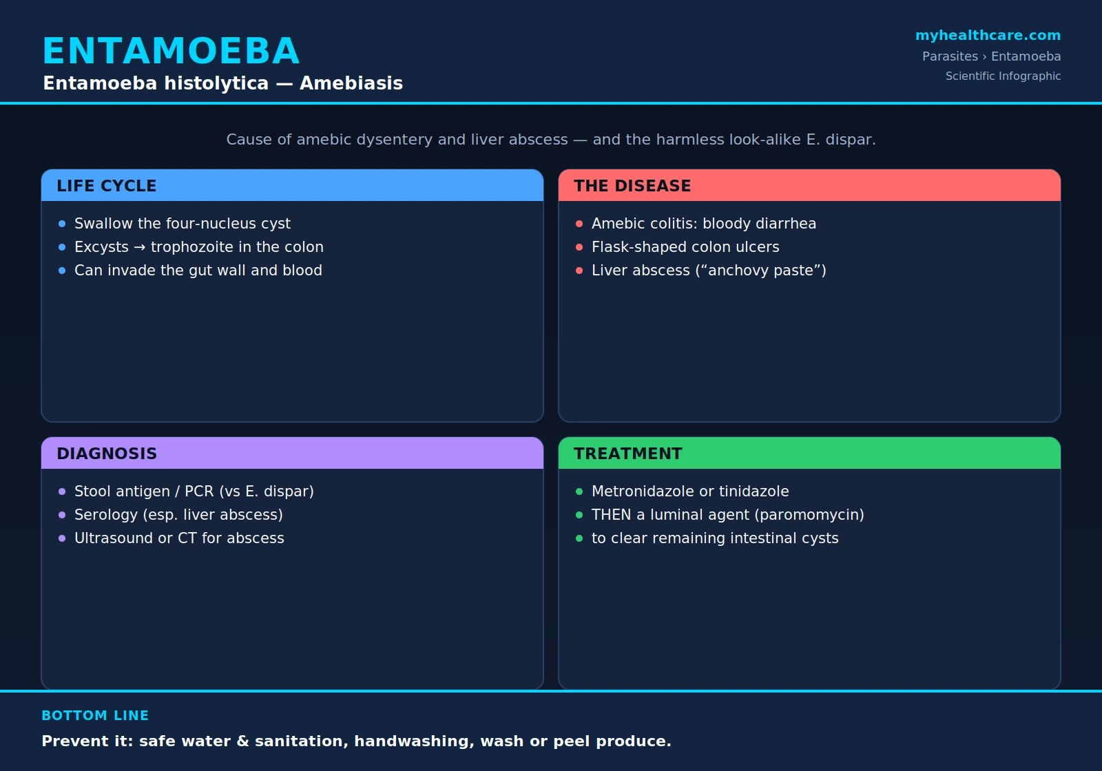
Entamoeba histolytica is a single-celled protozoan parasite and the cause of amebiasis — an infection of the large intestine that can range from a silent, symptom-free state to severe bloody dysentery and life-threatening abscesses of the liver. Worldwide it is one of the leading parasitic causes of death, killing tens of thousands of people each year, almost all of them in regions where clean water and reliable sanitation are scarce. A crucial and easily overlooked point runs through everything that follows: E. histolytica has a nearly identical, far more common, and completely harmless twin called Entamoeba dispar. The two cannot be told apart under an ordinary microscope, yet only E. histolytica invades human tissue and causes disease. This page explains what the parasite is, how it travels from person to person, the symptoms it produces, how modern laboratories distinguish the dangerous species from its benign look-alike, and how amebiasis is treated and prevented.
Table of Contents
- What Is Entamoeba histolytica?
- Life Cycle
- How It Spreads
- Symptoms
- Diagnosis
- Treatment
- Prevention
- Key Research Papers
- Featured Videos
1. What Is Entamoeba histolytica?
Entamoeba histolytica is a microscopic, single-celled protozoan parasite that lives in the human large intestine and is the agent of the disease called amebiasis (also spelled amoebiasis). Its species name is fitting: histolytica means "tissue-dissolving," a reference to the parasite's defining and dangerous ability to break down and invade the lining of the gut. Humans are its main host, and the infection is acquired by swallowing the parasite in food or water contaminated with infected stool.
The single most important fact about this organism is that it has a near-perfect impostor. Entamoeba dispar is a separate species that looks identical under the light microscope, colonizes the same part of the gut, and is many times more common — yet it is entirely harmless and never invades tissue. For most of the twentieth century the two were lumped together as a single species, which badly overstated how often "E. histolytica" actually caused disease. We now know that the great majority of people carrying microscopic "amebae" are in fact carrying E. dispar and need no treatment at all. Distinguishing the two requires tests that detect the parasite's specific proteins or DNA — a distinction that is central to the Diagnosis section below, because it determines who truly needs therapy.
Globally, amebiasis matters a great deal. It is among the most important parasitic causes of death, responsible for tens of thousands of deaths each year, and the burden falls overwhelmingly on parts of the world — in the tropics and subtropics, and wherever sanitation is poor — where contaminated water and food are common. The infection is also seen in travelers returning from these regions and in immigrants from them.
2. Life Cycle
The life cycle of E. histolytica is a straightforward fecal-oral loop — the parasite leaves one person's stool, survives in the environment, and is swallowed by the next person. It exists in two forms, and understanding the difference between them is the key to understanding both how the infection spreads and how it is treated.
- The cyst is the hardy, dormant, transmissible form. A mature cyst contains four nuclei and is enclosed in a tough wall that lets it survive for weeks outside the body in water, moist soil, and food, and resist stomach acid when swallowed. The cyst is the form that passes from person to person.
- The trophozoite is the active, feeding, dividing form that lives in the gut. It is fragile, does not survive long outside the body, and is the form that can invade tissue.
The cycle proceeds in a logical sequence:
- Ingestion. A person swallows mature cysts in food or water contaminated with infected feces.
- Excystation. The cysts pass safely through the stomach and, in the small intestine, each releases active trophozoites (a process called excystation).
- Colonization. The trophozoites travel to the large intestine (colon), where they live on the surface of the gut lining, feed, and multiply by simple division.
- Encystation and shedding. As trophozoites move down the bowel, many convert back into cysts, which are passed in the stool to continue the cycle in the next person. Because cysts — not the delicate trophozoites — are what survive in the environment, a person who passes formed stool full of cysts is the main source of new infections.
In most people the story ends with harmless colonization. But in a minority, the trophozoites turn invasive. They penetrate the wall of the large intestine, producing the characteristic ulcers of amebic colitis, and from there a few may enter the bloodstream and be carried to the liver — and, far more rarely, beyond it. This invasive turn is what transforms a silent carrier state into the serious disease described in the Symptoms section.
3. How It Spreads
Amebiasis spreads by the fecal-oral route: cysts shed in one person's stool are swallowed by another. In practice this happens through several overlapping pathways, all of which trace back to feces reaching the mouth.
- Contaminated food and water. Drinking water or eating food — especially raw fruits and vegetables — tainted with cyst-bearing feces is the dominant route. This is why the infection clusters where water supplies and sewage disposal are inadequate.
- Poor sanitation and hygiene. Inadequate sewage handling, the use of human waste as fertilizer, and unwashed hands after using the toilet all move cysts from stool into the food and water supply. Food handlers who are infected carriers can contaminate meals.
- Direct person-to-person contact that transfers fecal material, including certain sexual practices. Men who have sex with men are recognized as a group at increased risk of transmission through oral-anal contact.
Certain circumstances raise the odds of exposure. Travel to or residence in regions where amebiasis is common — much of the tropics and subtropics, and other areas with poor sanitation — is a leading risk factor, and amebiasis is a recognized cause of illness in returning travelers. Risk is also higher in institutional settings with crowding and limited hygiene. Although infection is far more frequent where sanitation is poor, the cyst's durability means that imported cases occur anywhere in the world.
4. Symptoms
The clinical picture of amebiasis is remarkably broad, ranging from no symptoms at all to a fatal abscess. It helps to think of the disease in three categories.
Asymptomatic infection. The majority of people infected with E. histolytica have no symptoms. The parasite lives quietly in the colon, the person feels well, and the only evidence of infection is the passage of cysts in the stool. Such carriers can still infect others and can, in principle, go on to develop invasive disease later.
Intestinal disease (amebic colitis / amebic dysentery). When the parasite invades the gut wall, it produces amebic colitis. Unlike the abrupt onset of many bacterial diarrheas, amebic colitis classically comes on gradually over one to several weeks. Typical features include:
- Bloody, mucus-containing diarrhea — the hallmark of amebic dysentery.
- Abdominal pain and cramping.
- Tenesmus — a painful, persistent sensation of needing to pass stool even when the bowel is empty.
- Weight loss and, in some patients, only low-grade fever (high fever is comparatively uncommon in uncomplicated colitis).
The signature pathological lesion is the "flask-shaped" ulcer: the parasite breaks through the thin surface lining and then spreads sideways in the looser tissue beneath, producing an ulcer that is narrow at the top and wide at the base, like a flask. In severe cases the colitis can become fulminant, with extensive bowel damage and the danger of perforation — a risk that is notably increased if the condition is mistaken for inflammatory bowel disease and treated with corticosteroids.
Extra-intestinal disease (amebic liver abscess). The most common form of amebiasis outside the gut is an amebic liver abscess, which forms when trophozoites travel from the colon to the liver through the bloodstream. It typically causes:
- Pain in the right upper part of the abdomen, where the liver sits, sometimes referred to the right shoulder.
- Fever and sweats, often more prominent than in intestinal disease.
- An enlarged, tender liver, with malaise and weight loss.
A characteristic feature is the abscess's contents, classically likened to "anchovy paste" — a reddish-brown, semi-liquid material made of necrotic liver tissue. Importantly, many people with an amebic liver abscess have no diarrhea at the time and may not recall ever having had it, which can make the diagnosis less obvious. Rarely, the parasite reaches other organs, including the lungs (sometimes by direct extension from a liver abscess) and, very uncommonly, the brain.
5. Diagnosis
Diagnosing amebiasis accurately is not simply a matter of "finding the ameba" — it is a matter of proving that the ameba present is the dangerous E. histolytica rather than its harmless twin E. dispar. Because the two are indistinguishable under a standard microscope, ordinary stool microscopy cannot make this critical call, and a positive microscope reading alone may lead to unnecessary treatment. Modern diagnosis therefore relies on more specific tools, chosen according to whether intestinal or liver disease is suspected.
- Stool antigen detection. Tests that detect E. histolytica-specific proteins in stool can distinguish it from E. dispar, making them far more meaningful than microscopy alone for identifying true infection.
- Molecular testing (PCR). Polymerase chain reaction detects the parasite's DNA and is highly sensitive and specific. It reliably separates E. histolytica from E. dispar (and the related E. moshkovskii) and is regarded as the most accurate approach for species identification.
- Serology (antibody blood tests). Tests for antibodies against the parasite are especially useful for amebic liver abscess, where the stool may be unrevealing; most patients with invasive disease mount a detectable antibody response. A limitation in highly endemic areas is that past infection can also produce antibodies, so results are interpreted alongside the clinical picture.
- Colonoscopy with biopsy. When intestinal disease is suspected but stool tests are inconclusive, direct examination of the colon can reveal the characteristic ulcers, and biopsy of the ulcer edge can show invading trophozoites.
- Imaging. Ultrasound or CT of the abdomen identifies a liver abscess, defines its size and location, and helps guide drainage if that becomes necessary. Imaging shows the abscess but does not by itself prove the cause, so it is paired with serology or other testing.
In short, the laboratory's job is twofold: confirm that a parasite is present and, just as importantly, confirm that it is the species capable of causing harm. That distinction is what separates a patient who needs treatment from one who does not.
6. Treatment
Treatment of amebiasis follows a logical two-step principle that mirrors the parasite's two forms: first kill the invasive trophozoites in the tissues, then clear the cysts that remain in the gut so the infection cannot smolder on or be passed to others. The information here is presented as commonly reported in the medical literature and should be directed by a clinician. This page does not provide medical advice; specific drugs, doses, and decisions about drainage are matters for a treating physician.
- Step 1 — a tissue-active drug. Invasive disease (amebic colitis or liver abscess) is first treated with a nitroimidazole — most commonly metronidazole or tinidazole. These drugs are effective against the trophozoites that have invaded the bowel wall and liver, but they do not reliably eliminate cysts sitting in the lumen (the open channel) of the gut.
- Step 2 — a luminal agent. Because the nitroimidazole leaves cysts behind, it is followed by a luminal (gut-acting) agent — such as paromomycin or diloxanide furoate — to clear the remaining intestinal cysts. This second step is what prevents relapse and stops the person from continuing to shed cysts. Luminal therapy is also used on its own to treat asymptomatic carriers of true E. histolytica in order to eradicate the parasite.
Drainage of a liver abscess is generally not required, because most abscesses respond well to drug therapy alone. However, drainage (typically image-guided needle aspiration) is sometimes needed — for example, for very large abscesses, those that do not respond to medication, or those at risk of rupturing. The decision is individualized and made by the medical team.
7. Prevention
Because amebiasis is acquired by swallowing cysts from contaminated food and water, prevention centers on breaking that fecal-oral chain. The same simple measures protect both communities and individual travelers.
- Safe drinking water. Use water that is known to be safe — boiled, properly treated, or sealed bottled water. Amebic cysts are resistant to the levels of chlorine used in routine water treatment, so chlorination alone cannot be relied upon; boiling is dependable.
- Good sanitation and sewage disposal. Proper handling of human waste keeps cysts out of the water and food supply and is the foundation of community-level prevention.
- Thorough handwashing with soap and water, especially after using the toilet and before preparing or eating food, interrupts person-to-person and food-handler transmission.
- Careful food choices. Wash and/or peel raw fruits and vegetables with safe water, and favor food that is freshly cooked and served hot. When traveling in higher-risk areas, the familiar rule applies: boil it, cook it, peel it, or forget it.
- Avoid untreated water when traveling — including ice made from tap water and water swallowed while swimming — in regions where sanitation is uncertain.
No vaccine against amebiasis is available, so these everyday hygiene and water-safety practices remain the most effective protection.
Key Research Papers
Peer-reviewed reviews and primary studies on Entamoeba histolytica and amebiasis — covering global burden, the biology of tissue invasion, clinical disease, and the diagnostics that separate the pathogen from its harmless look-alike. Journal names appear as plain text; the year/volume/pages link opens the full citation via DOI.
- Stanley SL Jr. Amoebiasis. The Lancet. 2003;361(9362):1025–1034.
- Haque R, Huston CD, Hughes J, Houpt E, Petri WA Jr. Amebiasis. New England Journal of Medicine. 2003;348(16):1565–1573.
- Shirley DT, Farr L, Watanabe K, Moonah S. A Review of the Global Burden, New Diagnostics, and Current Therapeutics for Amebiasis. Open Forum Infectious Diseases. 2018;5(7):ofy161.
- Marie C, Petri WA Jr. Regulation of Virulence of Entamoeba histolytica. Annual Review of Microbiology. 2014;68:493–520.
- Ralston KS, Solga MD, Mackey-Lawrence NM, Somlata, Bhattacharya A, Petri WA Jr. Trogocytosis by Entamoeba histolytica Contributes to Cell Killing and Tissue Invasion. Nature. 2014;508(7497):526–530.
- Espinosa-Cantellano M, Martínez-Palomo A. Pathogenesis of Intestinal Amebiasis: From Molecules to Disease. Clinical Microbiology Reviews. 2000;13(2):318–331.
- Fotedar R, Stark D, Beebe N, Marriott D, Ellis J, Harkness J. Laboratory Diagnostic Techniques for Entamoeba Species. Clinical Microbiology Reviews. 2007;20(3):511–532.
- Fotedar R, Stark D, Beebe N, Marriott D, Ellis J, Harkness J. PCR Detection of Entamoeba histolytica, Entamoeba dispar, and Entamoeba moshkovskii in Stool Samples. Journal of Clinical Microbiology. 2007;45(3):1035–1037.
- Shirley DT, Moonah S. Fulminant Amebic Colitis after Corticosteroid Therapy: A Systematic Review. PLoS Neglected Tropical Diseases. 2016;10(7):e0004879.
- Shirley DT, Watanabe K, Moonah S. Significance of Amebiasis: 10 Reasons Why Neglecting Amebiasis Might Come Back to Bite Us in the Gut. PLoS Neglected Tropical Diseases. 2019;13(11):e0007744.
Live PubMed Searches
Each link opens a live PubMed query so results stay current as new papers are indexed.
- Entamoeba histolytica amebiasis
- Amebic liver abscess
- Amebic colitis and dysentery
- Entamoeba histolytica vs. dispar
- Stool antigen and PCR diagnosis
- Metronidazole and paromomycin treatment
- Gal/GalNAc lectin and tissue invasion
- Amebiasis epidemiology and global burden
Connections
- Entamoeba Symptoms Overview
- Amoebic Dysentery and Colitis
- Liver Abscess and Extraintestinal Disease
- Diagnosis — Stool, Antigen, and Serology
- Entamoeba Treatments Overview
- Metronidazole and Tissue Amebicides
- Liver Abscess Drainage
- Prevention and Water Safety
- Parasites
- Acanthamoeba
- Giardia
- Malaria
- Toxoplasma
- Cryptosporidium
- Ascaris (Roundworm)
- Pinworm
- Hookworm
- Tapeworm
- Schistosoma
- Infectious Disease
- Gastroenterology
- Nephrology & Hepatology
- HIV/AIDS
- All Conditions