Parasites — Protozoa, Worms, and Free-Living Amoebae
Toxoplasma
An extremely common parasite spread by cats and undercooked meat; risky in pregnancy.
Ascaris
The giant roundworm — among the most common human infections on Earth.
Hookworm
A blood-feeding worm and a leading cause of iron-deficiency anemia.
Pinworm
The most common worm infection in temperate countries, especially in children.
Giardia
A flagellated protozoan and the most common waterborne cause of diarrhea (“beaver fever”).
Malaria
The mosquito-borne Plasmodium parasite — one of the world’s deadliest infections.
Schistosoma
Freshwater blood flukes that cause schistosomiasis (bilharzia).
Cryptosporidium
A chlorine-resistant protozoan behind swimming-pool diarrhea outbreaks.
Entamoeba
Entamoeba histolytica, the cause of amebic dysentery and liver abscess.
Tapeworm
Beef and pork tapeworms — and the brain disease neurocysticercosis.
Acanthamoeba
The free-living amoeba behind contact-lens eye infections and rare brain disease.
Naegleria fowleri
The rare “brain-eating amoeba” of warm freshwater — and how a nose clip prevents it.
Whipworm
A common soil-transmitted worm (Trichuris trichiura) of the large intestine.
Cyclospora
A foodborne parasite behind produce-linked outbreaks of prolonged watery diarrhea.
Fasciola
The liver fluke — caught from raw watercress and contaminated water (not meat); treated with triclabendazole.
Onchocerca
River blindness — a blackfly-borne worm causing intense itching and, over years, blindness; controlled with ivermectin.
Wuchereria bancrofti
The lymphatic-filariasis worm behind elephantiasis — mosquito-borne, and the target of a global elimination program.
Blastocystis
One of the most common organisms in human stool — often harmless, with a genuinely debated role in gut symptoms.
Clonorchis
The Chinese liver fluke from raw freshwater fish — a chronic bile-duct infection and a recognized cause of bile-duct cancer.
Paragonimus
The lung fluke from raw freshwater crab or crayfish — a chronic cough often mistaken for tuberculosis.
Guinea Worm
The meter-long “fiery serpent” from unfiltered water — now nearly eradicated without any drug.
Loa loa
The African “eye worm” that migrates visibly across the eye — spread by day-biting deer flies.
Echinococcus
Tapeworm larvae that form slow-growing hydatid cysts in the liver and lungs — caught from dogs and livestock.
Anisakis
The herring/sushi worm from raw marine fish — sudden stomach pain, and an under-recognized cause of fish allergy.
Angiostrongylus
The rat lungworm — the leading cause of eosinophilic meningitis, from raw snails, slugs, or unwashed produce.
Gnathostoma
From raw freshwater fish — migratory skin swellings and, rarely, dangerous nerve or eye migration.
Diphyllobothrium
The broad fish tapeworm from raw freshwater fish — the largest human tapeworm, and a classic cause of vitamin B12 deficiency.
Dientamoeba fragilis
A common gut protozoan with a genuinely debated role in digestive symptoms — and easily missed on ordinary stool tests.
Table of Contents
- What Are Parasites?
- Free-Living Amoebae: An Unusual Threat
- How These Infections Reach People
- Why They Are Hard to Treat
- Diagnosis Basics
- Prevention Principles
- Key Research Papers
- Featured Videos
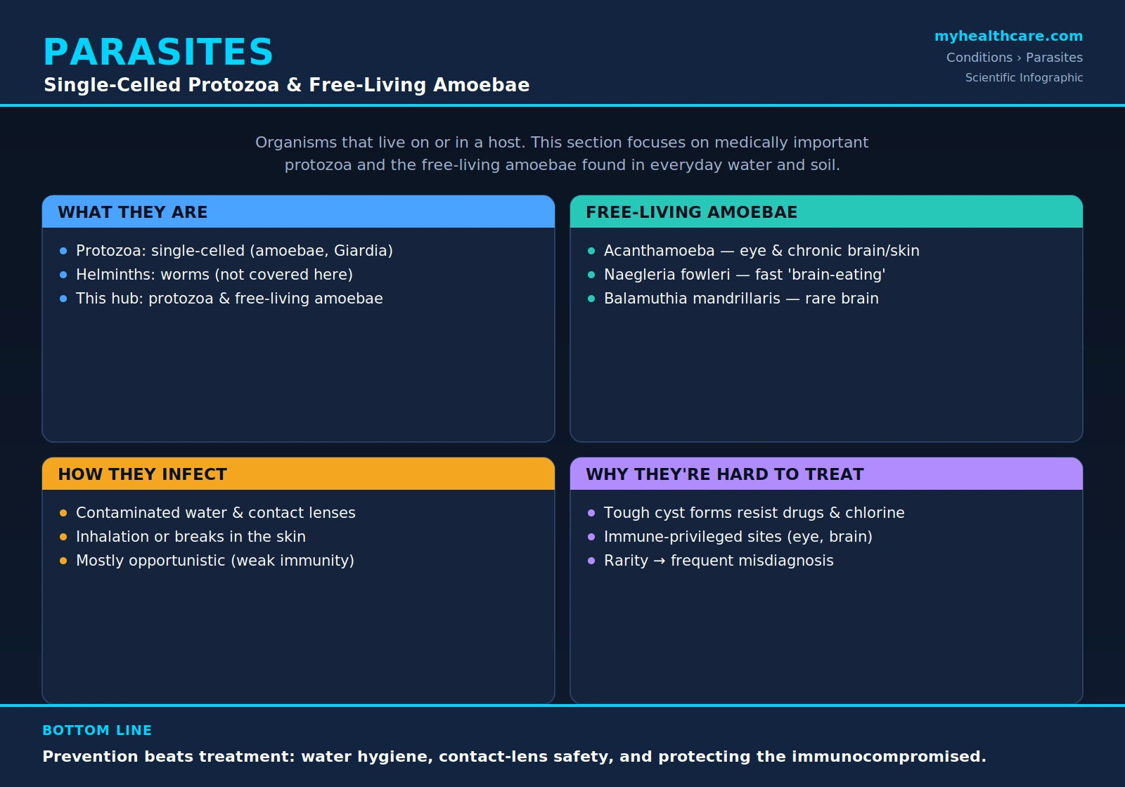
1. What Are Parasites?
A parasite is an organism that lives on or inside another living thing — its host — and benefits at the host's expense, drawing nutrients, shelter, or both while giving nothing useful in return. Human parasites span an enormous range of sizes and life cycles, from microscopic single cells to worms many centimeters long. For medical purposes they are usually divided into two broad groups: protozoa and helminths.
Protozoa are single-celled (unicellular) organisms. Despite being just one cell, they are biologically complex, able to move, feed, and in many cases multiply rapidly inside the human body. Familiar disease-causing protozoa include Plasmodium (the cause of malaria), Giardia, Entamoeba histolytica, and Toxoplasma. Helminths, by contrast, are multicellular parasitic worms — roundworms (nematodes), tapeworms (cestodes), and flukes (trematodes) — that are typically large enough to see with the naked eye.
This section of the site focuses on a specific and unusual corner of the protozoan world: medically important single-celled protozoa and the free-living amoebae. Unlike classic parasites such as malaria, which depend completely on a host to survive, the organisms covered here normally live independently in the environment and only occasionally end up infecting people. That difference matters, because it changes how the infections are caught, why they are so often missed, and how they are prevented.
2. Free-Living Amoebae: An Unusual Threat
Most parasites need a host to complete their life cycle. Free-living amoebae are different. They are amphizoic organisms — a term meaning they can lead two kinds of life. For the vast majority of the time they live freely in the environment, in warm fresh water, soil, dust, and even tap-water systems, feeding on bacteria. Only rarely, and opportunistically, do they invade a human and cause disease. They do not need us; infection is essentially an accident for both sides. Three genera account for nearly all serious human infections, and it helps to contrast them clearly.
Acanthamoeba
Acanthamoeba is the most commonly encountered of the three and the main subject of this section. It is found almost everywhere — in soil, fresh and brackish water, swimming pools, hot tubs, contact-lens solution, and domestic tap water. In people it causes two very different diseases. The first is Acanthamoeba keratitis, a painful infection of the cornea (the clear front window of the eye) strongly associated with contact-lens wear, that can threaten sight if not treated. The second, far rarer, is granulomatous amebic encephalitis (GAE), a slowly progressing brain infection that occurs mainly in people whose immune systems are weakened. Acanthamoeba can also cause skin and sinus infections.
Naegleria fowleri — the "brain-eating amoeba"
Naegleria fowleri is the organism behind the dramatic headlines about a "brain-eating amoeba." It thrives in warm fresh water — lakes, rivers, hot springs, and poorly maintained pools, especially in summer. It does not cause infection by being swallowed. Instead, when contaminated water is forced up the nose (during swimming, diving, or nasal rinsing), the amoeba can travel along the olfactory nerve into the brain and cause primary amebic meningoencephalitis (PAM). Unlike the slow course of Acanthamoeba brain disease, PAM is sudden and fast-moving, resembling acute bacterial meningitis, and is almost always fatal within days. It is extremely rare, but its speed and severity are what set it apart.
Balamuthia mandrillaris
Balamuthia mandrillaris lives in soil and dust. Like Acanthamoeba, it can cause a granulomatous amebic encephalitis, but — importantly — it can strike people with apparently normal immune systems, not only the immunocompromised. Balamuthia infections often begin as a slow-healing skin lesion before spreading, weeks to months later, to the brain. Like the other amoebic brain infections, it is rare, hard to diagnose, and carries a very high fatality rate.
The key contrast: Naegleria fowleri is the fast, fresh-water nasal threat; Acanthamoeba is the widespread environmental amoeba most famous for contact-lens eye infections; and Balamuthia mandrillaris is the soil-dwelling amoeba that can cause a slow, skin-then-brain illness even in healthy people.
3. How These Infections Reach People
Because free-living amoebae are everyday environmental organisms, almost everyone is exposed to them harmlessly throughout life. Infection requires the amoeba to get past the body's defenses through a specific route, and these routes define the risk.
- Water and the eye (contact lenses). This is the most important everyday risk. Acanthamoeba keratitis is closely tied to contact-lens use — particularly storing or rinsing lenses in tap water, swimming or showering while wearing lenses, and poor case hygiene. The amoeba clings to the lens and is pressed against the cornea, where a tiny scratch lets it take hold.
- Water up the nose. Naegleria fowleri enters through the nasal passages when warm fresh water is forced upward — while diving, jumping into lakes, or using non-sterile water in a neti pot or other nasal rinse. Swallowing the water is harmless; it is the nasal route to the brain that is dangerous.
- Inhalation of dust and soil contact. Acanthamoeba and Balamuthia cysts in soil and dust may be inhaled or enter through breaks in the skin. From there, in vulnerable people, the organism can spread through the bloodstream to the brain.
- Breaks in the skin. Cuts, abrasions, and chronic wounds exposed to contaminated soil or water can become an entry point, especially for Balamuthia, which often starts as a skin lesion.
A crucial point: these infections are opportunistic and not contagious. They are not passed from person to person in everyday contact. Each case results from an individual's own exposure to the environment, which is why prevention focuses on water and hygiene rather than on isolating sick people.
4. Why They Are Hard to Treat
Free-living amoebic infections are notoriously difficult to cure, and the reasons are built into the biology of the organisms and the rarity of the diseases.
The cyst stage. When conditions turn hostile — drying out, lack of food, exposure to drugs or disinfectants — these amoebae transform from their active, feeding trophozoite form into a dormant, thick-walled cyst. The cyst is extraordinarily tough: it resists desiccation, many common disinfectants, and a number of antimicrobial drugs. A treatment that kills the active trophozoites may leave cysts untouched, allowing the infection to relapse once therapy stops. Effective regimens therefore have to be prolonged and must include agents capable of penetrating or destroying cysts.
Immune-privileged sites. The amoebae tend to settle in places the immune system and many drugs reach poorly. The cornea has no blood vessels, so circulating immune cells and oral medicines arrive only weakly — topical drops, applied directly and frequently, do most of the work. The brain sits behind the blood–brain barrier, which blocks many drugs from reaching infected tissue at useful concentrations. These sanctuaries let the organism persist.
Rarity and misdiagnosis. Because amoebic infections are so uncommon, they are frequently mistaken for something else — Acanthamoeba keratitis for a herpes or bacterial eye infection, PAM for ordinary bacterial meningitis. By the time the correct diagnosis is made, valuable time has been lost, and for the brain infections that delay is often decisive. There are also no large, standardized clinical trials to define the single best therapy, so treatment relies on accumulated case experience and combinations of drugs.
5. Diagnosis Basics
Confirming a free-living amoebic infection takes a high index of suspicion and specialized testing. Several methods are used, often together.
- Microscopy. Direct examination of a sample — a corneal scraping, cerebrospinal fluid, or tissue — under the microscope can reveal amoebic trophozoites or cysts. Special stains help highlight them, and for the eye, an in-office imaging technique called confocal microscopy can sometimes show the organisms in the living cornea without removing tissue.
- Culture. The sample is placed on agar plates seeded with bacteria (a food source for the amoebae). If amoebae are present, they grow and leave visible tracks over a few days. Culture confirms a living organism but takes time.
- PCR (molecular testing). Polymerase chain reaction detects amoeba-specific DNA with high sensitivity and can identify the genus quickly. It has become a key tool for diagnosing keratitis and for the rapid identification needed in brain infections.
- Biopsy and imaging. For suspected brain or skin disease, a tissue biopsy examined by a pathologist may be needed to see the amoebae in the tissue, and brain imaging (CT or MRI) helps locate lesions and guide sampling.
Because no single test is perfect, doctors combine clinical clues — such as severe eye pain out of proportion to the exam in a contact-lens wearer, or rapid-onset meningitis after fresh-water swimming — with these laboratory methods to reach a diagnosis.
6. Prevention Principles
Since these infections are rare and hard to treat, prevention is the most powerful tool, and it centers on a few practical habits.
- Water hygiene. Avoid forcing untreated fresh water up the nose. For nasal rinsing (neti pots and similar devices), use only distilled, sterile, or previously boiled-and-cooled water — never straight tap water. Keep swimming pools and hot tubs properly chlorinated and maintained.
- Contact-lens safety. This single area prevents most Acanthamoeba keratitis. Never rinse or store lenses in tap water; use only fresh, sterile contact-lens disinfecting solution. Do not "top off" old solution. Remove lenses before swimming, showering, or using a hot tub. Wash and dry hands before handling lenses, replace the lens case regularly, and follow the prescribed replacement schedule.
- Protecting the immunocompromised. People with weakened immune systems — from HIV, organ transplants, chemotherapy, or long-term steroids — are most at risk for the amoebic brain infections. Extra care with soil, dust, and water exposure, prompt attention to slow-healing skin lesions, and early medical evaluation of unexplained neurological symptoms are sensible precautions for this group.
None of these steps require special equipment — they are mostly about clean water and careful lens habits — yet together they prevent the large majority of free-living amoebic infections.
Key Research Papers
Peer-reviewed studies and reviews on the free-living amoebae that infect humans — covering their general biology, Acanthamoeba keratitis and its risk factors, the rapidly fatal Naegleria fowleri brain infection, Balamuthia mandrillaris encephalitis, and treatment challenges. Each citation links to the full text via DOI.
- Visvesvara GS, Moura H, Schuster FL. Pathogenic and opportunistic free-living amoebae: Acanthamoeba spp., Balamuthia mandrillaris, Naegleria fowleri, and Sappinia diploidea. FEMS Immunology & Medical Microbiology. 2007;50(1):1–26.
- Marciano-Cabral F, Cabral G. Acanthamoeba spp. as agents of disease in humans. Clinical Microbiology Reviews. 2003;16(2):273–307.
- Khan NA. Acanthamoeba: biology and increasing importance in human health. FEMS Microbiology Reviews. 2006;30(4):564–595.
- Siddiqui R, Khan NA. Biology and pathogenesis of Acanthamoeba. Parasites & Vectors. 2012;5:6.
- John DT. Primary amebic meningoencephalitis and the biology of Naegleria fowleri. Annual Review of Microbiology. 1982;36(1):101–123.
- Kiderlen AF, Laube U. Balamuthia mandrillaris, an opportunistic agent of granulomatous amebic encephalitis, infects the brain via the olfactory nerve pathway. Parasitology Research. 2004;94(1):49–52.
- Carnt N, Minassian DC, Dart JKG. Acanthamoeba keratitis risk factors for daily wear contact lens users: a case-control study. Ophthalmology. 2023;130(1):48–55.
- Sharma S, Garg P, Rao GN. Patient characteristics, diagnosis, and treatment of non-contact lens related Acanthamoeba keratitis. British Journal of Ophthalmology. 2000;84(10):1103–1108.
- Seal D, Kirkness C, Bennett H, Peterson M. Acanthamoeba keratitis in Scotland: risk factors for contact lens wearers. Contact Lens and Anterior Eye. 1999;22(2):58–68.
- Larkin DFP, Kilvington S, Dart JKG. Treatment of Acanthamoeba keratitis with polyhexamethylene biguanide. Ophthalmology. 1992;99(2):185–191.
Live PubMed Searches
Each link opens a live PubMed query so results stay current as new papers are indexed.
- Free-living amoebae human infection review
- Acanthamoeba keratitis contact lens
- Naegleria fowleri primary amebic meningoencephalitis
- Balamuthia mandrillaris granulomatous amebic encephalitis
- Acanthamoeba cyst treatment resistance
- Amebic keratitis PCR diagnosis
- Naegleria nasal rinsing neti pot
- Granulomatous amebic encephalitis immunocompromised
Connections
- Toxoplasma
- Ascaris (Roundworm)
- Hookworm
- Pinworm
- Giardia
- Malaria
- Schistosoma
- Cryptosporidium
- Entamoeba histolytica
- Tapeworm
- Acanthamoeba
- Naegleria fowleri
- Whipworm (Trichuris)
- Cyclospora
- Fasciola (Liver Fluke)
- Onchocerca (River Blindness)
- Wuchereria (Filariasis)
- Blastocystis
- All Conditions
- Infectious Disease
- Ophthalmology