Unexplained Weight Loss (Involuntary Weight Loss)
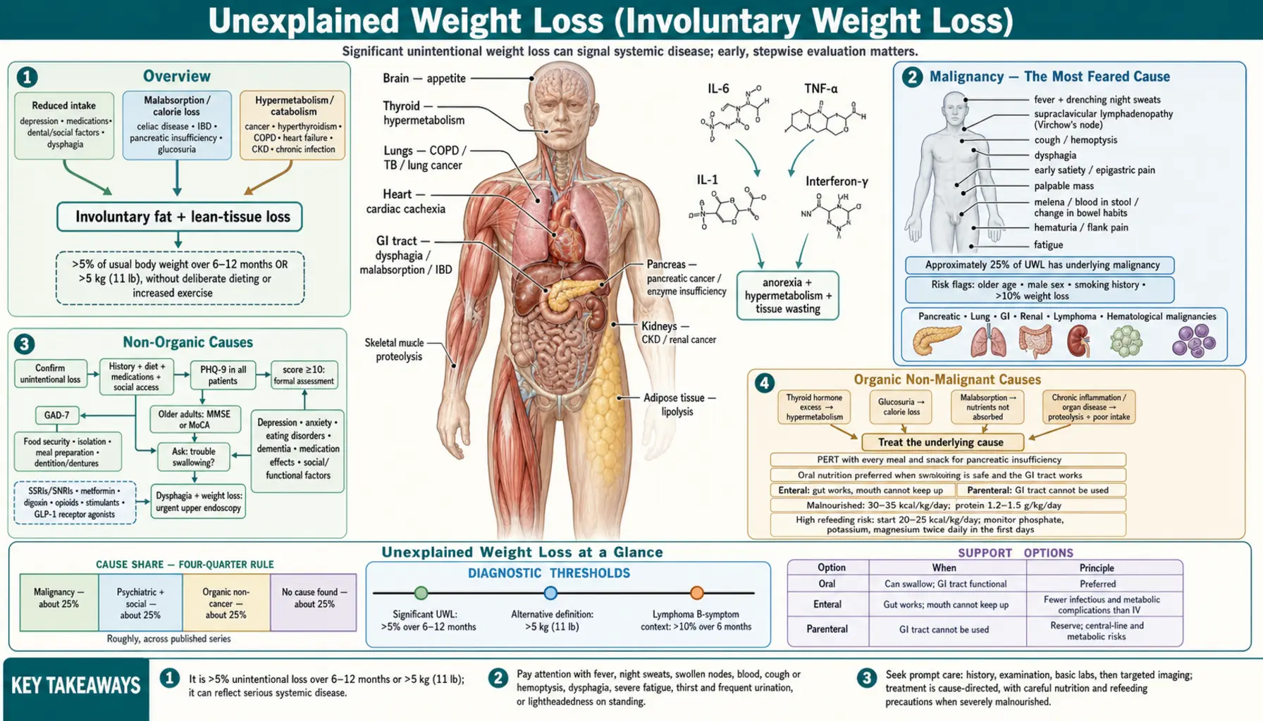
Table of Contents
- Overview
- Red Flags and the MEALS ON WHEELS Framework
- Malignancy — The Most Feared Cause
- Non-Organic Causes
- Anorexia Nervosa and Refeeding Syndrome
- Organic Non-Malignant Causes
- Endocrine Causes
- Malabsorption and GI Causes
- Evaluation Protocol
- Management
- When to Seek Medical Care
- Connections
- Key Research Papers
- Featured Videos
Overview
Unexplained weight loss (UWL), also called involuntary weight loss (IWL), is significant unintentional loss of body weight — generally defined as more than 5% of usual body weight over 6–12 months, or more than 5 kg (11 lbs) without deliberate dieting or increased exercise. This is a highly clinically significant finding: approximately 25% of patients with UWL have underlying malignancy, and even in those without cancer, the weight loss indicates significant systemic pathology requiring evaluation.
The evaluation of UWL is one of the most systematic in medicine, proceeding from history and physical examination to basic labs to targeted imaging. The differential diagnosis is broad — spanning malignancy, psychiatric illness, endocrine disorders, gastrointestinal disease, chronic infection, heart failure, and social factors — yet a careful, stepwise approach identifies the cause in approximately 75% of patients.
Unintentional weight loss is not a benign finding. Even when malignancy is excluded, patients with UWL have significantly higher mortality than weight-stable controls over the following one to three years. Identifying and treating the underlying cause as early as possible improves outcomes.
Red Flags and the MEALS ON WHEELS Framework
Some features, when they accompany weight loss, raise the probability of malignancy enough to justify expedited imaging and endoscopy rather than watchful waiting: older age, male sex, a smoking history, night sweats, lymphadenopathy, a palpable mass, hemoptysis, melena (black tarry stool), dysphagia, and the constitutional B symptoms of fever, drenching night sweats, and weight loss together.
In elderly patients the MEALS ON WHEELS mnemonic is the most practical checklist of causes, because it front-loads the social, dental, and medication problems that a cancer-focused workup tends to skip:
- M — Medications (many drugs suppress appetite or cause nausea).
- E — Emotional disturbance and depression.
- A — Anorexia (cancer, depression, age-related).
- L — Late-life paranoia (including delusions about food being tampered with).
- S — Swallowing disorders.
- O — Oral problems: poor dentition, mouth pain, ill-fitting dentures.
- N — No money: poverty and food insecurity.
- W — Wandering and other dementia-related behaviors.
- H — Hyperthyroidism and other endocrine disease.
- E — Enteric problems: malabsorption, inflammatory bowel disease.
- E — Eating problems, including the mechanical difficulty of Parkinson's disease.
- L — Low-salt and low-cholesterol diets, an iatrogenic restriction that makes food unpalatable.
- S — Social isolation and community problems.
A second framing worth carrying into the consultation is the four-quarter rule: across published series the causes of significant involuntary weight loss divide into roughly equal quarters — malignancy about 25%, psychiatric and social causes about 25%, organic non-cancer disease about 25%, and no cause found about 25%. Series differ mainly by setting: geriatric and primary-care cohorts return a higher psychosocial share, while referral and hospital cohorts return more cancer. The practical lesson is the same either way — a workup that only looks for a tumor will miss three-quarters of the diagnoses.
Malignancy — The Most Feared Cause
Approximately 25% of patients presenting with unexplained weight loss have an underlying malignancy. Any cancer can cause cachexia through cytokine-mediated mechanisms.
Cancer Cachexia Mechanism
IL-6, TNF-α (historically called cachectin), and IL-1 drive anorexia, hypermetabolism, proteolysis, and lipolysis simultaneously. Interferon-γ contributes to the same inflammatory program. The result is net tissue wasting even when caloric intake is adequate. The cancer cachexia index (CCI) combines skeletal muscle density with neutrophil-to-lymphocyte ratio to quantify severity and predict survival.
Cancer cachexia is not simple starvation, and the distinction changes management. In starvation the body defends lean tissue and burns fat; in cachexia, protein catabolism predominates over fat loss, so skeletal muscle disappears early. Because the inflammatory drive persists regardless of intake, cachexia cannot be reversed by feeding alone — nutritional support buys time but only treatment of the tumor interrupts the mechanism.
Most Commonly Associated Cancers
- Pancreatic cancer — produces the most severe cachexia, affecting up to 90% of patients at some point in the disease; weight loss is often the presenting symptom before abdominal pain or jaundice develops, and the tumor is frequently painless until late. New-onset diabetes mellitus in a person over 50 accompanied by weight loss should be treated as possible pancreatic cancer until proven otherwise. Check CA 19-9 and CT abdomen/pelvis.
- Lung cancer — especially small cell; paraneoplastic effects (ectopic ACTH, SIADH) compound the weight loss. Check chest CT in all smokers or those with respiratory symptoms.
- GI cancers — esophageal (dysphagia + weight loss), gastric (early satiety + epigastric pain), colorectal (blood in stool, change in bowel habits, iron-deficiency anemia). Upper and lower endoscopy are key diagnostic tools; CEA and CA 19-9 add supporting information but are not screening tests.
- Renal cell carcinoma — the classic triad is flank pain, hematuria, and a palpable mass, and weight loss is common; only a minority of patients have the full triad, so unexplained weight loss with microscopic hematuria deserves cross-sectional imaging of the kidneys.
- Lymphoma — B symptoms — night sweats, fever, and weight loss of more than 10% over six months — are a classic Hodgkin's and non-Hodgkin's lymphoma presentation. Check LDH, CBC, and CT chest-abdomen-pelvis; PET-CT for staging.
- Hematological malignancies — leukemia, multiple myeloma; CBC abnormalities (cytopenias, blasts, paraprotein on SPEP) are usually present.
When to Suspect Malignancy
- Weight loss as the primary complaint, particularly in patients over age 50.
- Weight loss exceeding 10% of usual body weight.
- Associated fatigue, night sweats, or unexplained fevers.
- Any unexplained lymphadenopathy, especially supraclavicular (Virchow's node).
- Elevated LDH, ESR, or uric acid without other explanation.
- Hypercalcemia without obvious benign cause.
Non-Organic Causes
Non-organic (psychosocial) causes account for approximately 40% of UWL cases in some series — making them collectively the most common cause overall. These causes are frequently underappreciated because clinicians and patients alike focus on ruling out cancer.
Depression
Anhedonia and loss of interest in eating drive reduced caloric intake. Severe depression can cause 10–20% body weight loss over months. Associated features include fatigue, insomnia, poor concentration, and psychomotor slowing. A PHQ-9 depression screen should be performed in all patients with UWL regardless of their primary complaint; a score of 10 or above warrants formal assessment. Depression is the single most common psychiatric cause of involuntary weight loss, and it is a genuinely reversible one — effective antidepressant treatment usually restores both appetite and weight.
Anxiety and Eating Disorders
Anxiety drives chronic nausea and poor appetite. Anorexia nervosa causes severe restriction and is classically described in younger women but occurs at any age, including older adults. Avoidant/restrictive food intake disorder (ARFID) is increasingly recognized across age groups. Somatization — persistent bodily preoccupation with nausea, bloating, or fear of choking — and late-life paranoia, in which an older person becomes convinced food is spoiled or tampered with, both reduce intake without any organic lesion to find. Anorexia nervosa is covered in detail in the next section.
Social and Functional Factors
These factors are the most overlooked causes of UWL, particularly in elderly patients:
- Poverty and food insecurity — inadequate access to food is directly responsible for weight loss.
- Social isolation — people who live alone often do not cook full meals; the social pleasure of shared eating is lost.
- Functional impairment — inability to shop, stand, or prepare meals due to pain, mobility limitations, or arthritis.
- Dental problems — poor dentition or ill-fitting dentures lead patients to avoid solid foods, dramatically reducing caloric density.
Always ask directly: "Are you able to buy the food you need? Is anyone helping you with meals?"
Cognitive Impairment and Dementia
Patients with dementia often forget to eat or lose the ability to recognize hunger. Late-stage dementia causes dysphagia and behavioral changes (food refusal, agitation at mealtimes) that further reduce intake. Assess cognition with the MMSE or MoCA in all elderly patients with UWL.
Medications
A complete medication review is mandatory. Common culprits include:
- Chemotherapy (nausea, mucositis, dysgeusia).
- SSRIs and SNRIs (early nausea and anorexia, especially in the first four to six weeks).
- Metformin (nausea, diarrhea, reduced appetite).
- Digoxin (nausea and anorexia are classic toxicity signs — check levels).
- Opioids (nausea, constipation, reduced appetite).
- Topiramate and zonisamide (appetite suppression is a known effect, and is sometimes used therapeutically in epilepsy).
- Stimulants — amphetamines and methylphenidate directly suppress appetite.
- Antibiotics (dysgeusia, the metallic or absent taste that makes food unappealing, plus nausea).
- GLP-1 receptor agonists such as semaglutide and liraglutide (intentional in many patients, but weight loss may be alarming to those not expecting it, and is often undesired in the frail elderly).
Dysphagia
Structural causes (esophageal cancer, peptic stricture, achalasia, eosinophilic esophagitis) or functional causes (neurological: Parkinson's disease, stroke, ALS) make eating painful or frightening. The mechanism here is mechanical restriction of intake rather than malabsorption — the food never gets far enough to be digested. Always ask "Do you have any trouble swallowing?" in UWL evaluation. Dysphagia with weight loss warrants urgent upper endoscopy.
Anorexia Nervosa and Refeeding Syndrome
Anorexia nervosa deserves separate treatment because it is the one cause of involuntary weight loss where the treatment itself can kill the patient. It is defined by restriction of intake, disturbance of body image, and an intense fear of weight gain. The female-to-male ratio is roughly 10:1 and onset clusters in adolescence and young adulthood. DSM-5 recognizes two subtypes: a restricting type (dietary restriction plus excessive exercise) and a binge-eating/purging type.
Medical Complications
- Cardiovascular — bradycardia, QTc prolongation with the attendant risk of fatal arrhythmia, and orthostatic hypotension.
- Electrolyte — hypokalemia, especially in the purging subtype, and hypophosphatemia.
- Skeletal — osteoporosis from prolonged estrogen deficiency compounded by undernutrition; fracture risk persists after weight is restored.
Refeeding Syndrome
After prolonged starvation, the body has shifted to fat and protein metabolism and total-body phosphate, potassium, and magnesium are depleted even when serum levels look normal. Reintroducing carbohydrate triggers an insulin surge that drives glucose — and phosphate, potassium, and magnesium with it — into cells. Serum levels then crash, and the result can be cardiac arrhythmia, respiratory failure, seizures, and death within the first few days of feeding. Thiamine demand also rises sharply as carbohydrate metabolism resumes.
Prevention is a deliberately slow caloric ramp with laboratory surveillance, not a nutritional catch-up. In patients at high refeeding risk, begin around 20–25 kcal/kg/day, advance by roughly 500 kcal every two to three days as tolerated, check phosphate, potassium, and magnesium twice daily during the first days, and replete before advancing rather than after.
Specific Management
Medical stabilization comes first — cardiac monitoring and electrolyte correction before any psychological work. Outpatient treatment suits medically stable patients; inpatient admission is indicated for a BMI below 15, acute medical complications, or failure of outpatient therapy. Family-based therapy (the Maudsley approach) has the strongest evidence base in adolescents, and cognitive behavioral therapy is first-line for adults.
Organic Non-Malignant Causes
Chronic Infections
- HIV/AIDS — weight loss occurs in all stages of HIV infection. HIV wasting syndrome (BMI below 20 with more than 10% weight loss) is a defining AIDS illness. Modern antiretroviral therapy has largely eliminated HIV wasting in treated patients; UWL in a person with HIV who is on ART should prompt evaluation for ART adherence, resistance, opportunistic infection, and malignancy. Test HIV antigen/antibody in all UWL patients with risk factors; CD4 count and viral load then guide how much of the weight loss is attributable to uncontrolled HIV itself.
- Tuberculosis — historically called consumption precisely because the wasting was its most visible sign. Constitutional symptoms including night sweats, fever, and weight loss combined with a productive cough are classic, and hemoptysis may appear as disease advances. Exposure history and immigration history are important clues. Diagnose with TST or IGRA plus chest imaging (upper-lobe infiltrates and cavitation on plain film; CT for detail); sputum AFB smear and culture if pulmonary TB is suspected.
- Chronic hepatitis B and C — progressive liver dysfunction reduces synthetic capacity, causing hypoalbuminemia, fatigue, and weight loss. Associated features include jaundice, ascites, peripheral edema in advanced disease.
- Endocarditis — prolonged fever with weight loss and a new or changing heart murmur. Look for embolic phenomena on examination: Janeway lesions (painless palm and sole macules), Osler nodes (tender fingertip nodules), and splinter hemorrhages under the nails. Blood cultures (minimum three sets) are the cornerstone of diagnosis.
- Chronic parasitic infection — intestinal helminths and other chronic parasites are a real cause of wasting in returning travelers, recent immigrants, and anyone with a relevant exposure history; stool studies for ova and parasites belong in that workup.
Heart Failure
Cardiac cachexia is driven by chronically elevated resting energy expenditure, neurohormonal activation (renin-angiotensin-aldosterone system, catecholamines), intestinal edema causing malabsorption, and reduced appetite secondary to dyspnea. Associated features include dyspnea on exertion, orthopnea, and peripheral edema. NT-proBNP and echocardiogram are key diagnostic tests. Weight loss in heart failure is not merely cosmetic — it is an independent marker of poor prognosis, and it can be masked on the scale by simultaneous fluid retention, so lean tissue may be melting away while the recorded weight holds steady.
COPD
The high mechanical work of breathing represents a chronically elevated energy expenditure. Eating exacerbates dyspnea in severe COPD, so patients eat less. Hyperinflated lungs flatten the diaphragm and press on the stomach, producing early satiety after only a few mouthfuls. Impaired gas exchange and hypoxemia further reduce appetite. The resulting pulmonary cachexia is associated with worse exercise tolerance and higher mortality independent of airflow obstruction.
Chronic Kidney Disease
Uremic anorexia and nausea reduce caloric intake. Hypermetabolism from chronic inflammation and metabolic acidosis accelerates proteolysis, breaking down muscle mass even in the face of adequate dietary protein. Dialysis adds a further loss, since amino acids and protein are removed across the membrane at every session. Nutritional assessment — including albumin, prealbumin, and dietary recall — is essential in dialysis patients.
Rheumatological Conditions
Rheumatoid arthritis, systemic lupus erythematosus, vasculitis, and other chronic inflammatory diseases drive cytokine-mediated cachexia. Medication side effects compound the problem: methotrexate causes nausea and oral mucositis; hydroxychloroquine causes GI symptoms; corticosteroids initially stimulate appetite but cause muscle wasting with chronic use.
Hypercalcemia
The classic mnemonic "bones, stones, groans, psychic moans, and abdominal groans" captures hypercalcemia's multisystem effects: nausea, vomiting, constipation, anorexia, polyuria, and confusion. Nausea and anorexia are often the most prominent symptoms at moderately elevated calcium levels (12–14 mg/dL). Causes include primary hyperparathyroidism (most common outpatient cause), malignancy-associated hypercalcemia (PTHrP secretion, osteolytic bone metastases), and vitamin D toxicity. Always correct serum calcium for albumin level.
Addison's Disease (Adrenal Insufficiency)
Weight loss with fatigue, postural hypotension, hyperpigmentation of skin creases and buccal mucosa (primary adrenal insufficiency), hyponatremia, and hyperkalemia should raise immediate suspicion for Addison's disease. This diagnosis must not be missed — adrenal crisis is life-threatening. Diagnose with 8AM serum cortisol: below 3 mcg/dL is insufficient; above 18 mcg/dL is sufficient; indeterminate values (3–18) require an ACTH stimulation test. On that test, a cortisol rise of less than about 9 mcg/dL above baseline after synthetic ACTH indicates adrenal insufficiency.
Endocrine Causes
Hyperthyroidism
Thyroid hormone excess causes hypermetabolism and appetite dysregulation. Weight loss occurs despite increased or normal appetite — hyperphagia does not fully compensate for the elevated metabolic rate. Associated features include palpitations, heat intolerance, tremor, anxiety, and diarrhea. TSH is the single best screening test and should be checked in every patient with UWL. If TSH is suppressed, follow with free T4, free T3, and radioactive iodine uptake scan to determine the cause (Graves' disease, toxic multinodular goiter, toxic adenoma). Age shifts the odds: Graves' disease is the usual cause in younger women, while toxic multinodular goiter predominates in older patients — who may also present with so-called apathetic hyperthyroidism, in which weight loss and atrial fibrillation appear without the tremor and agitation.
Uncontrolled Diabetes Mellitus
Osmotic diuresis and glucosuria result in calories lost in the urine even with adequate food intake. The classic presentation — polydipsia, polyuria, polyphagia, and weight loss (the "three Ps plus weight loss") — is type 1 diabetes mellitus until proven otherwise. Insulin deficiency adds protein catabolism and fat breakdown on top of the urinary calorie loss, so weight can fall very fast — sometimes the first presentation is diabetic ketoacidosis. This picture also occurs in very poorly controlled type 2 diabetes, and ketosis-prone type 2 diabetes (described particularly in African American and Hispanic patients) can mimic type 1 closely at onset. Diagnose with fasting plasma glucose and HbA1c; check for ketones if the patient is unwell.
Pheochromocytoma
Catecholamine excess causes hypermetabolism and decreased appetite alongside episodic hypertension, severe headache, palpitations, and diaphoresis. Pheochromocytoma is rare but treatable and should be considered when episodic symptoms accompany weight loss. Plasma free metanephrines have sensitivity exceeding 95% for pheochromocytoma; 24-hour urine metanephrines are the alternative biochemical test. Once biochemistry is positive, CT or MRI localizes the adrenal mass, and an MIBG scan finds extra-adrenal (paraganglioma) tumours that cross-sectional imaging of the adrenals alone would miss.
Hypogonadism
Testosterone deficiency in men causes loss of muscle mass, reduced appetite, and fatigue. In severe or prolonged hypogonadism, the loss of lean body mass can be clinically significant. Diagnose with LH, FSH, and total testosterone (morning specimen).
Malabsorption and GI Causes
The clue that separates malabsorption from every other cause on this page is weight loss despite adequate or even increased oral intake. The patient is eating; the calories are not being absorbed. Bloating, flatulence, and frequent loose or bulky stools usually accompany it, and when fat is the nutrient being lost the stools take on the characteristic appearance described under pancreatic insufficiency below.
Celiac Disease
Gluten triggers an autoimmune T-cell response in the small intestinal mucosa, causing villous atrophy and malabsorption of fat, carbohydrates, protein, iron, calcium, and fat-soluble vitamins. Symptoms include bloating, diarrhea, flatulence, and weight loss; iron-deficiency anemia is a common non-GI manifestation. Presentation is heterogeneous, and many adults arrive with extraintestinal disease rather than classical diarrhea — iron-deficiency anemia, osteoporosis, or peripheral neuropathy may be the only sign. Dermatitis herpetiformis is the skin form. Screen with anti-tissue transglutaminase IgA (anti-tTG IgA) plus total IgA (to detect IgA deficiency, which causes a false-negative tTG result). Confirm with duodenal biopsy showing villous atrophy, crypt hyperplasia, and intraepithelial lymphocytosis.
Inflammatory Bowel Disease
Both Crohn's disease and ulcerative colitis cause weight loss through different mechanisms. In Crohn's disease the loss is triple-sourced: patients eat less because eating provokes pain, absorb less across inflamed mucosa, and expend more because of systemic inflammation. Crohn's affects any part of the GI tract from mouth to anus; skip lesions, transmural inflammation, perianal disease, and fistulae characterize it; malabsorption is especially severe when the terminal ileum is involved (B12 deficiency, fat malabsorption). Ulcerative colitis causes bloody diarrhea and protein-losing enteropathy. Fecal calprotectin is a useful non-invasive inflammatory marker; colonoscopy with biopsies confirms the diagnosis.
Exocrine Pancreatic Insufficiency
When pancreatic lipase secretion falls below 10% of normal, fat malabsorption occurs, causing steatorrhea (oily, foul-smelling, floating stools) and deficiencies of fat-soluble vitamins A, D, E, and K. Causes include chronic pancreatitis (most common, often alcoholic or genetic), cystic fibrosis, and post-pancreatectomy states. Diagnose with fecal elastase-1 (below 200 mcg/g indicates EPI). Treat with pancreatic enzyme replacement therapy (PERT) taken with every meal and snack.
Small Intestinal Bacterial Overgrowth (SIBO)
Bacterial fermentation of carbohydrates in the small bowel produces gas (bloating, flatulence) and consumes B12, causing deficiency. Malabsorption of fat and carbohydrates follows. Bacterial competition for nutrients and low-grade mucosal injury compound the malabsorption. Diagnose with a glucose or lactulose breath test measuring hydrogen and methane, or empirically treat with rifaximin — a non-absorbable antibiotic that acts within the gut lumen — if clinical suspicion is high.
Mesenteric Ischemia
Chronic mesenteric ischemia causes "intestinal angina": postprandial abdominal pain develops 30–60 minutes after eating, conditioning patients to fear food and dramatically reduce intake. The result is progressive weight loss in a patient who may appear to be eating normally by self-report. This diagnosis should be considered in older patients with atherosclerotic disease and unexplained weight loss. CT angiography of the mesenteric vessels is the preferred imaging study.
Evaluation Protocol
Step 1 — History
The history is the most powerful tool in UWL evaluation:
- Rate of weight loss: percentage of usual body weight lost over what time period (weigh the patient today; compare to a documented prior weight if possible).
- Intentional vs. unintentional: "Were you trying to lose weight?" This is the first and most important question.
- Detailed dietary history: appetite (reduced, normal, or increased); ability to buy and prepare food; dysphagia; early satiety; nausea and vomiting; diarrhea or steatorrhea; abdominal pain.
- Associated symptoms: fever, night sweats, fatigue, cough, hemoptysis, blood in stool or urine, bone pain, headaches, palpitations, heat or cold intolerance.
- Complete medication and supplement review.
- Social history: PHQ-9 depression screen and GAD-7 anxiety screen, recent bereavement or loss of a spouse who did the cooking, alcohol and substance use, social support structure, food access ("Are you able to buy enough food?")
- Cancer history and family history of malignancy.
Step 2 — Physical Examination
- Measure actual weight and calculate percentage change from prior documented weight.
- Nutritional assessment: temporal wasting, thenar eminence muscle bulk, calf circumference, mid-arm circumference.
- Lymphadenopathy: cervical, supraclavicular (Virchow's node signals GI, lung, or breast malignancy), axillary, inguinal.
- Thyroid: goiter, nodule, tenderness.
- Breast examination and prostate examination (men over 50).
- Abdominal examination: masses, organomegaly, tenderness, ascites.
- Rectal examination and fecal occult blood test.
- Oral cavity: dentition and denture fit, oral thrush (immunosuppression), mucositis or ulcers (chemotherapy, Crohn's disease), dry mouth.
- Skin: jaundice (liver or biliary disease), acanthosis nigricans (insulin resistance or GI malignancy), hyperpigmentation of skin creases (Addison's disease), pallor (anemia), and rashes suggesting inflammatory bowel disease (erythema nodosum, pyoderma gangrenosum) or connective tissue disease.
- Neurological: brief cognitive screen (MMSE or MoCA) in elderly patients.
Step 3 — Initial Laboratory Evaluation
- CBC with differential — cytopenias, blasts, lymphocytosis, elevated WBC.
- Comprehensive metabolic panel — LFTs, creatinine, BUN, glucose, electrolytes (hyponatremia and hyperkalemia suggest Addison's disease), calcium.
- TSH — mandatory in every patient with UWL.
- Urinalysis with urine microscopy.
- Fecal occult blood test.
- HIV antigen/antibody.
- ESR and CRP — nonspecific but sensitive inflammatory markers.
- LDH — elevated in lymphoma, hemolysis, and many malignancies.
- Albumin and prealbumin — nutritional assessment and liver synthetic function.
- Anti-tTG IgA plus total IgA — celiac disease screen.
- PSA — in men over 50.
- HbA1c — uncontrolled diabetes.
- Lipase — if pancreatic disease suspected.
Step 4 — Imaging
- Chest X-ray — hilar adenopathy, lung mass, pleural effusion, TB upper-lobe infiltrate.
- CT chest-abdomen-pelvis with IV contrast — indicated when initial labs are negative but clinical suspicion for malignancy remains high, or when a patient is over 50 with otherwise unexplained UWL. Single most productive imaging study in UWL evaluation.
Step 5 — Targeted Evaluation Based on Findings
- Colonoscopy — age-appropriate colorectal cancer screening or symptomatic indications (blood in stool, change in bowel habits).
- Upper endoscopy — dysphagia, early satiety, epigastric pain, or upper GI symptoms.
- Mammography and cervical cancer screening — women who are overdue for screening.
- 8AM cortisol — if adrenal insufficiency suspected (fatigue, hypotension, hyponatremia, hyperpigmentation).
- Plasma free metanephrines — if episodic symptoms (pheochromocytoma).
- PET/CT — when CT is negative but clinical suspicion for malignancy remains high; sensitivity approximately 90% for metabolically active malignancy; detects occult primary tumors.
- Bone marrow biopsy — when lymphoma or myeloma is suspected and CT is non-diagnostic.
- Fecal elastase — steatorrhea or pancreatic disease.
How Probability Shifts With Age
The same 5% weight loss means different things at different ages, and the workup should be weighted accordingly.
- Under 50 — psychiatric causes (depression, eating disorders), hyperthyroidism, inflammatory bowel disease, and malabsorption syndromes are disproportionately common. Cancer is possible but far less likely, so exhaustive imaging before the basic labs and a depression screen is usually the wrong order.
- Over 65 — malignancy, depression, cardiac and pulmonary disease, and social or functional factors (poverty, dementia, poor dentition, isolation) all rise together, and more than one of them is often operating at once.
When the baseline evaluation in an older patient comes back negative, that is genuinely reassuring but not final: follow-up series find malignancy declaring itself at roughly 2–4% per year over the next two years. Re-weigh the patient at intervals rather than closing the case.
Prognosis of UWL
Patients with UWL have significantly higher mortality than weight-stable controls over the following one to three years, even when malignancy is excluded. One-year mortality is approximately 20–30% in hospitalized patients with UWL. Identifying the underlying cause within six months of presentation allows treatment that may reverse weight loss. Idiopathic UWL (no cause found after complete evaluation) carries a relatively more benign prognosis, though follow-up surveillance is warranted because occult malignancy may subsequently declare itself.
Management
Management of UWL is entirely cause-directed. Treating the underlying condition is the highest priority — weight regain follows treatment of hyperthyroidism, celiac disease, heart failure, depression, uncontrolled diabetes, and most infections.
Nutritional Support
- High-calorie, high-protein diet: target 30–35 kcal/kg/day and 1.2–1.5 g protein/kg/day in malnourished patients.
- Oral nutritional supplements (Ensure, Boost, Fortisip) as bridging support while the underlying cause is treated; each serving delivers roughly 250–400 kcal, so two or three a day between meals closes a substantial deficit without displacing food.
- A registered dietitian referral is worth making early — to fix the macronutrient balance, identify specific deficiencies, and modify texture for anyone with swallowing difficulty.
- Oral interventions are preferred over enteral (tube feeding) or parenteral (IV) nutrition when the patient can swallow safely and the GI tract is functional.
- Refeeding syndrome: reintroducing calories too quickly in a severely malnourished patient can be lethal. The mechanism and the safe caloric ramp are set out under Anorexia Nervosa and Refeeding Syndrome above, and the same precautions apply to severe undernutrition from any cause — cancer, alcohol dependence, or simple destitution — not only to eating disorders.
- Micronutrient repletion: multivitamin; B12 if deficient; iron if deficient; vitamin D if deficient.
Enteral and Parenteral Nutrition
When the gut works but the mouth cannot keep up, enteral nutrition is the next step and is preferred over the intravenous route because it keeps the intestinal mucosa fed and carries far fewer infectious and metabolic complications. A nasogastric tube suits short-term use; a percutaneous endoscopic gastrostomy (PEG) tube is considered when support is expected to run beyond four to six weeks — head and neck cancer during chemoradiation and progressive neurological dysphagia are the usual indications.
Parenteral nutrition is reserved for a gastrointestinal tract that cannot be used at all: short bowel syndrome, bowel obstruction, severe mucositis, or a high-output fistula. It carries real risks — central line infection, hyperglycemia, and metabolic and hepatic complications — and should not be started when enteral feeding is feasible.
Appetite Stimulation in Cancer Cachexia
- Megestrol acetate (Megace, 160–800 mg/day orally) — a synthetic progestin approved for HIV wasting and cancer cachexia; increases appetite and body weight but the gain is mostly fat rather than lean muscle. Risks include thromboembolism, adrenal suppression, edema, and delirium, so it is used cautiously in elderly or hypercoagulable patients.
- Dexamethasone (4–8 mg/day orally) — short-term appetite stimulation; not suitable for prolonged use due to muscle wasting and immunosuppression.
- Mirtazapine — an antidepressant whose potent H1 antihistamine activity stimulates appetite; useful when depression co-occurs with anorexia. Sedation is dose-dependent and can be turned to advantage when insomnia is also present.
- Dronabinol — a synthetic cannabinoid with modest evidence for improving appetite in HIV wasting and cancer cachexia; CNS effects (euphoria, confusion, dizziness) limit its use in the elderly.
- Oxandrolone — an anabolic steroid used mainly in HIV-associated wasting to preserve and restore lean body mass; hepatotoxicity and androgenic effects restrict its use elsewhere.
- Omega-3 fatty acids (EPA/DHA) — modest evidence for attenuating muscle wasting in cancer cachexia.
- Anamorelin — ghrelin receptor agonist approved in Japan; increases lean body mass and appetite in cancer cachexia; under regulatory review in the US and EU.
Dysphagia Management
When dysphagia contributes to weight loss, a speech-language pathology evaluation is essential. Interventions include texture modification (minced and moist, pureed, or liquid diets as appropriate), thickened liquids, swallowing exercises, and positioning strategies. In progressive neurological disease, a proactive conversation about goals of care and the role of tube feeding is important.
When to Seek Medical Care
See a doctor promptly if you notice any of the following:
- Weight loss of more than 5% of your body weight over six months without trying (for example, losing 9 pounds if you normally weigh 175 pounds).
- Weight loss accompanied by fever, night sweats, or swollen lymph nodes in the neck, armpit, or groin.
- Weight loss with blood in stool or urine.
- Weight loss with a new cough, coughing up blood, or difficulty breathing.
- Weight loss with difficulty swallowing or pain when eating.
- Weight loss with severe fatigue, profound loss of appetite, or persistent nausea.
- Weight loss with increased thirst and frequent urination (may signal uncontrolled diabetes).
- Weight loss with lightheadedness upon standing, skin darkening, or excessive salt craving (may signal adrenal insufficiency).
Unexplained weight loss is one of the most important alarm symptoms in medicine. It should never be attributed to "aging" or "stress" without a thorough evaluation. Early identification of the cause dramatically improves treatment outcomes.
Connections
- All Symptoms
- Weight Loss (Remedies) — the opposite question: intentional weight loss, covering satiety, protein and fiber, and how the popular diets actually work. This page is about weight you did not mean to lose.
- Fatigue
- Loss of Appetite
- Night Sweats
- Swollen Lymph Nodes
- Lymphoma
- Thyroid Disorders
- Hyperthyroidism
- Diabetes
- Type 1 Diabetes
- Depression
- Eating Disorders
- Celiac Disease
- Inflammatory Bowel Disease
- Tuberculosis
- Heart Failure
- COPD
- Chronic Kidney Disease
- Iron
- Vitamin B12
Key Research Papers
Weight loss as a sign of disease is one of the oldest observations in medicine — Hippocrates described the wasting of phthisis, the disease now called tuberculosis. The word cachexia (Greek kakos, bad, and hexis, condition) entered systematic clinical use in the 19th century. The MEALS ON WHEELS mnemonic was popularized in the geriatric literature by Morley and Kraenzle's 1994 nursing-home work, and the modern mechanistic era began in the 1990s and 2000s when the cytokine biology of cachexia — IL-6 and TNF-α in particular — was worked out in human patients, culminating in the 2011 international consensus definition of cancer cachexia.
Key Research Papers
- Marton KI, Sox HC, Krupp JR. Involuntary weight loss: diagnostic and prognostic significance. Annals of Internal Medicine. 1981;95(5):568-574. — Search PubMed
- Lankisch P, Gerzmann M, Gerzmann JF, Lehnick D. Unintentional weight loss: diagnosis and prognosis. The first prospective follow-up study from a secondary referral centre. Journal of Internal Medicine. 2001;249(1):41-46. — Search PubMed
- Hernandez JL, Matorras P, Riancho JA, Gonzalez-Macias J. Involuntary weight loss without specific symptoms: a clinical prediction score for malignant neoplasm. QJM. 2003;96(9):649-655. — PubMed PMID: 12925720
- Metalidis C, Knockaert DC, Bobbaers H, Vanderschueren S. Involuntary weight loss. Does a negative baseline evaluation provide adequate reassurance? European Journal of Internal Medicine. 2008;19(5):345-349. — PubMed PMID: 18549937
- Fearon K, Strasser F, Anker SD, et al. Definition and classification of cancer cachexia: an international consensus. Lancet Oncology. 2011;12(5):489-495. — Search PubMed
- Prado CM, Baracos VE, McCargar LJ, et al. Sarcopenia as a determinant of chemotherapy toxicity and time to tumor progression in metastatic breast cancer patients receiving capecitabine treatment. Clinical Cancer Research. 2009;15(8):2920-2926. — PubMed PMID: 19351764
- Prado CM, Lieffers JR, McCargar LJ, et al. Prevalence and clinical implications of sarcopenic obesity in patients with solid tumours of the respiratory and gastrointestinal tracts. Lancet Oncology. 2008;9(7):629-635. — PubMed PMID: 18539529
- Morley JE. Anorexia of aging: physiologic and pathologic. American Journal of Clinical Nutrition. 1997;66(4):760-773. — Search PubMed
- Stinton LM, Shaffer EA. Practical management of unexplained weight loss in the ambulatory care setting. Canadian Family Physician. 2012;58(5):543-547. — Search PubMed
- Bosch X, Monclus E, Escoda O, et al. Unintentional weight loss: Clinical characteristics and outcomes in a prospective cohort of 2677 patients. PLOS ONE. 2017;12(4):e0175125. — Search PubMed
- Huffman GB. Evaluating and treating unintentional weight loss in the elderly. American Family Physician. 2002;65(4):640-650. — Search PubMed
- Alibhai SM, Greenwood C, Payette H. An approach to the management of unintentional weight loss in elderly people. CMAJ. 2005;172(6):773-780. — Search PubMed
- Huhmann MB, Cunningham RS. Importance of nutritional screening in treatment of cancer-related weight loss. Lancet Oncology. 2005;6(5):334-343. — Search PubMed
- Laviano A, Meguid MM, Inui A, Muscaritoli M, Rossi-Fanelli F. Therapy insight: cancer anorexia-cachexia syndrome — when all you can eat is yourself. Nature Clinical Practice Oncology. 2005;2(3):158-165. — Search PubMed
PubMed Topic Searches
- PubMed: Unexplained weight loss, malignancy evaluation — systematic reviews
- PubMed: Cancer cachexia cytokines — TNF and IL-6 mechanism
- PubMed: Involuntary weight loss in the elderly — causes and prognosis
- PubMed: Unintentional weight loss — causes
- PubMed: Anorexia nervosa — treatment
- PubMed: Anorexia nervosa — medical complications and refeeding syndrome
- PubMed: Malabsorption syndromes and weight loss
- PubMed: Appetite stimulants in cachexia