Gilman and Rodbell: The Switch Inside the Cell, and Why a Third of Our Drugs Work
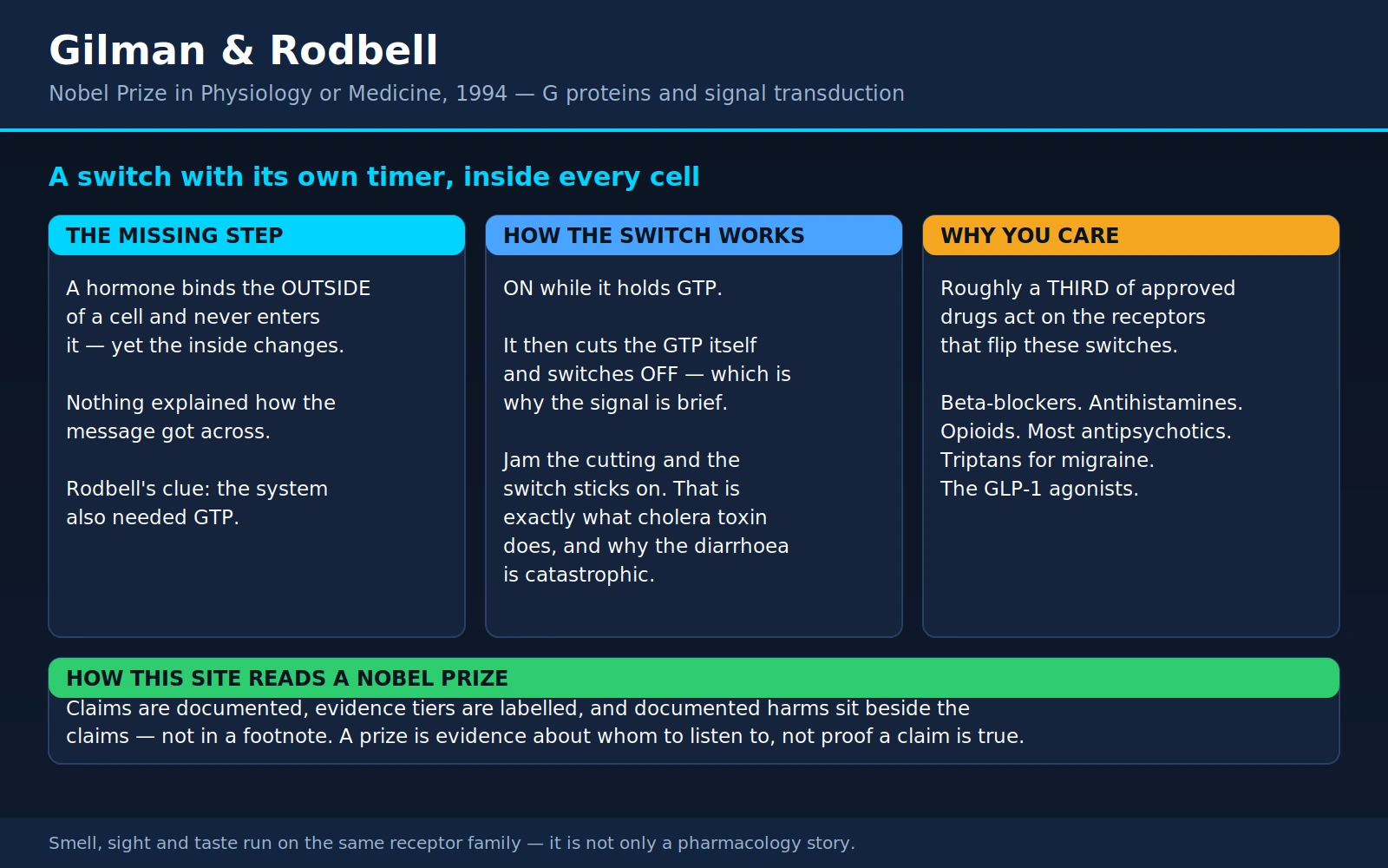
The 1994 Nobel Prize in Physiology or Medicine went to Alfred G. Gilman and Martin Rodbell "for their discovery of G-proteins and the role of these proteins in signal transduction in cells." That sentence is almost designed to be skipped. It sounds like interior biochemistry — a detail for specialists, three letters and a hyphen.
It is, in fact, one of the most useful things anyone has ever worked out about how medicines function. Open the average household medicine cabinet and count: the blood-pressure tablet, the asthma inhaler, the antihistamine, the heartburn tablet, the migraine pill, the antipsychotic, the strong painkiller, the newest weight-loss injection. Nearly all of them work through the machinery these two men described. Roughly a third of all approved drugs act on one family of receptors, and those receptors do nothing at all until a G protein — the thing Rodbell inferred and Gilman held in a test tube — passes the message on.
This page explains what that switch is, how it was found, why it explains cholera and a rare bone disease and a great many prescriptions, and what it means for the words on a patient information leaflet.
Table of Contents
- The Missing Step
- Rodbell's Three-Part Idea — and the GTP Clue
- Gilman's Proof: Holding the Missing Piece
- What a G Protein Actually Does
- GPCRs: The Biggest Receptor Family, and the Most Drugged
- The Drugs, by Family
- When the Switch Jams
- Biased Agonism: The Idea That Promised More Than It Delivered
- Why "Receptor" Language Misleads Patients
- Smell, Sight and Taste
- The Prize, and Who Was Not On It
- Where Mainstream Medicine Agrees — and What Remains Debated
- Key Research Papers
- Connections
- Featured Videos
1. The Missing Step
Start with the problem as it stood in the 1960s, because the problem is what makes the answer beautiful.
Adrenaline reaches a heart cell and the cell speeds up. Glucagon reaches a liver cell and the cell releases sugar. Histamine reaches a stomach cell and the cell pumps acid. In every case something outside changes something inside — and in every case the hormone never gets in. Adrenaline is a water-soluble molecule; it cannot slip through the greasy interior of a cell membrane, and experiments with hormones chemically tethered to beads far too large to enter had already shown that a hormone stuck permanently on the outside still works. The message crosses the wall without the messenger crossing it.
Half of this puzzle had already been solved, brilliantly. Earl Sutherland and his colleagues discovered cyclic AMP in the late 1950s and built from it the idea of the second messenger: the hormone is the first messenger, and it never enters; inside the cell a small molecule is manufactured on demand and it is that molecule which fans out and does the work — switching on the enzymes that break down glycogen, and much else besides. Sutherland received the Nobel Prize in Physiology or Medicine in 1971 for exactly this. It explained why so many different hormones produce overlapping effects, and why the effects are so fast.
But it left a hole in the middle of the story, and the hole was obvious to everyone working on it. The enzyme that makes cyclic AMP — adenylyl cyclase — sits in the membrane facing inward. The receptor sits in the membrane facing outward. How does the receptor tell the enzyme? For a decade the default assumption was the tidiest one: perhaps the receptor and the enzyme are two ends of a single protein, and binding at the outer end simply flexes the inner end into activity. One molecule, one hinge, no mystery.
That assumption was wrong, and the way it turned out to be wrong is the whole discovery. There is a third thing in between, and it does something no hinge can do: it decides how long the message lasts.
2. Rodbell's Three-Part Idea — and the GTP Clue
Martin Rodbell (1925–1998) was working at the National Institutes of Health in Bethesda, studying how the hormone glucagon stimulates cyclic-AMP production in membranes prepared from rat liver. He was a careful membrane biochemist with an unusual habit of mind: he thought about biological signalling in the vocabulary of communications engineering, and he insisted the system had to have parts with jobs.
His framework named three of them:
- a discriminator — what we now call the receptor. Its job is to tell things apart: to bind glucagon and not adrenaline, to recognise one signal out of the thousands washing past the cell surface.
- a transducer — the missing middle. Its job is to convert one kind of event (a molecule docking on the outside) into another kind of event (activity on the inside).
- an amplifier — the enzyme, adenylyl cyclase. Its job is to turn one small event into a large one, producing many molecules of cyclic AMP for each activated receptor.
The word transducer is borrowed on purpose. A microphone is a transducer: it does not amplify sound and it does not select sound, it changes sound into electricity so that something else can. Rodbell's claim was that biology needed a component whose entire function was that conversion — and that this component was a separate molecule, not part of either neighbour.
Then came the observation that turned a framework into a discovery. In a series of papers in the Journal of Biological Chemistry in 1971, Rodbell and his colleagues Lutz Birnbaumer, Stephen Pohl and Henri Krans reported that glucagon could not stimulate adenylyl cyclase unless GTP was present. Not ATP — GTP. The fifth paper in the series carries the finding in its title: An obligatory role of guanylnucleotides in glucagon action. The fourth paper reported the mirror-image result on the other side of the membrane: guanine nucleotides also changed how glucagon itself bound to the receptor.
Why was needing GTP such a loud clue? Because GTP had no business being there. The reaction under study converts ATP into cyclic AMP; ATP is the raw material, and its requirement is trivially expected. GTP is a different nucleotide entirely — chemically similar, biologically distinct — and it is neither the substrate nor a product of the reaction. Yet without it the hormone did nothing. A molecule that is not consumed by the chemistry, but without which the chemistry will not respond to the signal, is not fuel. It is information. Something in that membrane must be binding GTP and using the act of binding it as a switch.
That is the reasoning in a sentence: an unexplained requirement is a hidden component. The GTP requirement was the transducer announcing its own existence without being seen. Rodbell had found the fingerprint. Nobody had yet found the finger.
3. Gilman's Proof: Holding the Missing Piece
Alfred Goodman Gilman (1941–2015) was, by an accident of naming that reads like fiction, destined for pharmacology. His father Alfred Gilman co-wrote The Pharmacological Basis of Therapeutics with Louis S. Goodman — the textbook every medical student still calls "Goodman & Gilman" — and named his son after his co-author. That son grew up to win the Nobel Prize in pharmacology's central question.
Gilman's contribution was not to guess better. It was to isolate the thing, and the difference between those two matters enormously.
The trick was a broken cell line. In 1975, Henry Bourne, Philip Coffino and Gordon Tomkins at the University of California, San Francisco selected a variant of the S49 mouse lymphoma line that had lost the ability to make cyclic AMP in response to any stimulator — a clone they described as deficient in adenylyl cyclase, and which the field came to call cyc−. It was a cell with intact receptors on its surface and no response inside. Gilman's laboratory, then at the University of Virginia, recognised what that was: not a dead end, but an assay. If you can find the substance whose addition makes a cyc− cell respond again, you have found the missing component — and you will know you have found it, because the cell will tell you.
Two 1977 experiments framed the answer. In one, Gilman and Elliott Ross showed that heating an extract of normal S49 membranes destroyed its cyclase activity, and that mixing the heat-killed extract with an extract from the cyc− cells restored it — each supplying what the other lacked, which meant at least two proteins were required. In the other, they took membrane extracts from mouse L cells (which have adenylyl cyclase but no β-adrenergic receptors) and added them to cyc− membranes (which have β-adrenergic receptors but no working cyclase). The reconstituted mixture responded to catecholamines exactly as a normal cell does — a hormone-sensitive system rebuilt out of two halves, neither of which worked alone.
Then, in 1980, came the paper that closed the argument. Gilman's group — John Northup, Paul Sternweis, Murray Smigel, Lee Schleifer, Elliott Ross and Gilman — reported the purification of the regulatory component of adenylyl cyclase from rabbit liver membranes, "essentially to homogeneity," enriched about two-thousand-fold through six chromatographic steps. The purified protein turned out to be a complex of three polypeptides. Added back to cyc− membranes, it restored guanine-nucleotide-, fluoride- and hormone-stimulated activity. Added to the other broken variant — one in which receptor and enzyme were present but uncoupled — it restored the coupling.
Why purification rather than inference is the whole point. Until 1980, the transducer was a deduction. It was the best explanation for a set of kinetic oddities and genetic defects, and it might well have been right — but "the data are best explained by an unseen component" is a claim about a model, not about the world. Purification changes the category of the claim. Once the protein is in a tube you can weigh it, count its subunits, ask what it binds, sequence it, raise antibodies against it, and eventually clone its gene. Above all you can perform the one test that no amount of inference can substitute for: put it back and watch the dead cell come alive. That demonstrates sufficiency, not correlation. It is the difference between deducing that a house has a fuse box and finding the fuse box.
The purified protein was named for what it did: a G protein, for guanine-nucleotide-binding protein. Gilman spent the following years showing there was not one of them but a family — a stimulatory one (Gs), an inhibitory one (Gi), and others — and his 1987 review in the Annual Review of Biochemistry, titled simply G proteins: transducers of receptor-generated signals, gave the field its settled vocabulary. Rodbell's word had become a molecule.
4. What a G Protein Actually Does
Here is the mechanism in plain language. It is worth a few minutes because almost everything else on this page falls out of it.
A G protein is a team of three subunits, called α, β and γ, sitting on the inner face of the cell membrane. The α subunit is the interesting one: it has a pocket that holds a guanine nucleotide. In the resting state that pocket holds GDP, and the three subunits are stuck together as a unit. Nothing is happening.
Then a hormone lands on the receptor outside. The receptor changes shape, and its inner face — now presented differently — prises the α subunit's pocket open. GDP falls out; GTP, which is abundant inside every cell, drops in. That single nucleotide swap is the switch being thrown. GTP-loaded α lets go of βγ, and both freed pieces go off to do things: α may switch adenylyl cyclase on, or off, or activate a different enzyme entirely, while βγ opens ion channels of its own.
And now the part that makes it a genuinely clever piece of engineering. The α subunit is not just a GTP holder — it is a GTP cutter. It slowly hydrolyses the GTP it is holding, snipping off one phosphate to leave GDP behind. The moment it does, it reverts to its resting shape, re-binds βγ, and the signal stops.
So the best way to picture a G protein is a light switch with a timer built into it — the sort found in a stairwell, where you press the button, the light comes on, and a mechanism inside the switch turns it off again after a set interval. Nobody has to come back and switch it off. The switch has an off-switch inside it, and the interval is a property of the switch itself.
Three consequences follow directly, and each one shows up in medicine:
- Signals are transient by design. The duration of the response is set by how fast the α subunit cuts its own GTP — typically seconds. A cell does not need a separate shut-off system, and a hormone does not need to be actively removed for its message to end. This self-timing is why your heart rate comes down after a fright rather than staying up.
- Signals are amplified. One activated receptor does not switch on one G protein; during the seconds it stays active it can throw dozens of them, and each activated G protein drives an enzyme that produces many molecules of cyclic AMP. A handful of hormone molecules outside becomes a substantial chemical event inside. This is also why drugs acting here can work at very low doses.
- If the cutting stops, the switch jams ON. Anything that disables the α subunit's ability to hydrolyse GTP — a mutation, or a bacterial toxin — leaves the signal permanently on with no hormone present at all. Section 7 is entirely about what happens then, and it is where the mechanism stops being abstract.
Two refinements worth knowing. First, "G protein" is a family, not an individual: Gs stimulates adenylyl cyclase and raises cyclic AMP, Gi inhibits it and lowers cyclic AMP, Gq takes a different route entirely and releases calcium inside the cell. The same receptor architecture can therefore produce opposite effects depending on which G protein it is wired to — which is exactly why adrenaline speeds the heart through one adrenergic receptor subtype and relaxes the airways through another. Second, the timer is adjustable: a family of proteins discovered in the 1990s, the regulators of G-protein signalling (RGS proteins), accelerate the α subunit's GTP-cutting and so shorten the signal. Cells tune their own switch speeds.
5. GPCRs: The Biggest Receptor Family, and the Most Drugged
The receptors that talk to G proteins share a distinctive shape: a single protein chain that threads back and forth through the cell membrane seven times, leaving a pocket on the outside where the signal molecule binds and loops on the inside where the G protein docks. They are called G-protein-coupled receptors, or GPCRs, and sometimes seven-transmembrane (7TM) receptors.
They are the largest family of membrane receptors in the human genome. A 2003 genome-wide analysis by Fredriksson and colleagues identified more than 800 human GPCR sequences, of which 342 were unique functional non-olfactory receptors — and that split is the key to reading the number honestly. Roughly half the family is devoted to smell: a 2004 analysis of the human genome by Malnic, Godfrey and Buck counted 339 intact olfactory receptor genes alongside 297 that have decayed into pseudogenes. So the useful summary is: approximately 800 GPCR genes in all, of which about 350–400 respond to hormones, neurotransmitters and other internal signals, and most of the rest smell things. Treat every one of these figures as approximate; they shift slightly with each genome annotation and with where you draw the family boundary.
Why are they so unusually druggable? Four reasons, and they compound:
- The target is on the outside. A drug does not have to get inside the cell to reach a GPCR's binding pocket — it only has to reach the bloodstream and the tissue fluid. That removes the single hardest obstacle in drug design, and it is why so many GPCR drugs can be swallowed.
- The pocket already binds small molecules. The natural signals — adrenaline, histamine, acetylcholine, serotonin, dopamine — are tiny. A pocket shaped to hold a tiny molecule is a pocket a medicinal chemist can fill with a synthetic tiny molecule that either mimics or blocks the original.
- There are hundreds of subtly different versions. Selectivity is at least achievable: a drug can be tuned to hit the histamine H1 receptor and largely spare H2, or the β2 receptor in the airways more than the β1 in the heart. Not perfectly — section 12 is honest about where this fails — but achievably.
- The amplification is free. Because one receptor throws many G-protein switches, occupying a modest fraction of receptors can produce a full clinical effect. Less drug is needed than the arithmetic of "one molecule per target" would suggest.
And now the number this page exists to deliver. Two independent surveys, published a year apart, using different databases and different counting rules, arrived at the same headline:
- Hauser and colleagues (2017), in Nature Reviews Drug Discovery, reported that 475 drugs — approximately 34% of all drugs approved by the US Food and Drug Administration — act at 108 unique GPCRs.
- Sriram and Insel (2018), in Molecular Pharmacology, curated their own list and concluded that as of November 2017, 134 GPCRs were targets for drugs approved in the United States or European Union, and estimated that ~700 approved drugs target GPCRs, implying that approximately 35% of approved drugs target GPCRs. They also noted that GPCRs themselves account for roughly 12% of all protein targets of approved drugs — making them the largest single family of drug targets.
The drug counts differ substantially — 475 versus about 700 — because the two teams drew on different registries and counted combination products, salts and formulations differently. The fraction, remarkably, does not: both land on about a third. That convergence from different methods is what makes "roughly a third of approved drugs act on GPCRs" a claim worth carrying around.
Read it precisely, though. It is a count of approved molecular entities, not of prescriptions written, not of pills swallowed, and not of pharmaceutical revenue. And it is a fraction of the whole approved pharmacopoeia, including drugs almost nobody takes. What it tells you is where the pharmacological action has been concentrated for fifty years — and it tells you why Rodbell's transducer and Gilman's purified protein are not a footnote.
6. The Drugs, by Family
This is the practical payoff. What follows is not exhaustive; it is a walk through the medicine cabinet, and the point is breadth. Somewhere in this list is something you or someone in your household has taken.
- Beta-blockers — propranolol, metoprolol, atenolol, bisoprolol, carvedilol. They block β-adrenergic receptors, the GPCRs through which adrenaline speeds and strengthens the heartbeat. Used for high blood pressure, angina, arrhythmias, heart failure, tremor, and performance anxiety. Propranolol, the first of them, came from James Black.
- Beta-agonists — salbutamol (albuterol), salmeterol, formoterol. The same receptor family, pushed the other way: the blue rescue inhaler in asthma is a β2-receptor agonist relaxing airway smooth muscle within minutes.
- Alpha-blockers and alpha-agonists — doxazosin and tamsulosin block α1 receptors (blood pressure, prostate symptoms); clonidine activates α2 receptors. Same neurotransmitter, different receptor subtypes, opposite manipulations.
- Antihistamines — cetirizine, loratadine, fexofenadine, diphenhydramine. These act at the histamine H1 receptor and are the mainstay of hay fever, urticaria and allergic reactions. Pharmacologically most of them are inverse agonists rather than simple blockers — a distinction that matters, and section 9 explains why.
- Acid-reducing H2 blockers — famotidine, cimetidine, and formerly ranitidine, acting at the histamine H2 receptor on the stomach's acid-producing parietal cells. (Ranitidine was withdrawn from most markets around 2019–2020 over NDMA contamination, not over its pharmacology; famotidine remains widely used.) James Black designed both propranolol and cimetidine — one man, two receptor families, two revolutions in prescribing.
- Opioids — morphine, oxycodone, codeine, fentanyl, buprenorphine, and the antidote naloxone. All act at the μ-opioid receptor, a GPCR coupled to Gi. See opioids for chronic pain and opioid overdose.
- Antipsychotics — haloperidol, risperidone, olanzapine, quetiapine, aripiprazole. Their common thread is action at the dopamine D2 receptor, usually alongside serotonin receptors. See schizophrenia.
- Triptans and the newer migraine drugs — sumatriptan and its relatives act at serotonin 5-HT1B/1D receptors; lasmiditan at 5-HT1F; and the "gepants" (rimegepant, ubrogepant) block the CGRP receptor. All GPCRs. Every modern acute migraine drug class works through this family.
- Muscarinic drugs — acetylcholine's GPCR receptors, M1 through M5. Blocked by ipratropium and tiotropium in the airways, by oxybutynin and solifenacin in the overactive bladder, by scopolamine for motion sickness, and by atropine in emergencies. Activated by pilocarpine for dry mouth and glaucoma.
- Angiotensin-receptor blockers (ARBs) — losartan, valsartan, candesartan, irbesartan. They block the AT1 receptor for angiotensin II. (Their cousins the ACE inhibitors are a useful contrast: those block an enzyme, not a receptor.)
- GLP-1 receptor agonists — semaglutide, liraglutide, dulaglutide, exenatide, and the dual agonist tirzepatide. The GLP-1 receptor is a GPCR — a class B receptor that, as a 2017 cryo-electron-microscopy structure showed directly, signals primarily through the stimulatory G protein Gs: the same transducer Gilman purified from rabbit liver. The most-discussed drug class of the moment for obesity and type 2 diabetes sits squarely inside the 1994 Nobel Prize. See GLP-1 receptor agonists.
- And a scattering more — latanoprost for glaucoma (prostaglandin FP receptor), aprepitant for chemotherapy nausea (NK1 receptor), oxytocin in labour, cannabinoid drugs (CB1/CB2), sphingosine-1-phosphate modulators such as fingolimod in multiple sclerosis, and the anticoagulant vorapaxar (PAR-1). All GPCRs.
What is not on this list is just as instructive. Statins block an enzyme. Proton-pump inhibitors block a pump. SSRIs block a transporter. Insulin acts on a receptor that is its own enzyme. Paracetamol and ibuprofen act on cyclo-oxygenase enzymes. Levothyroxine acts on a receptor inside the cell nucleus. Ondansetron, despite being a serotonin drug, acts on a 5-HT3 receptor that is an ion channel, not a GPCR — a genuine exception in a family where nearly every other serotonin receptor is one. So the claim is not that all drugs are GPCR drugs. The claim is narrower and more impressive: out of every biological mechanism medicine has learned to manipulate, this single family accounts for about a third of the pharmacopoeia.
7. When the Switch Jams
A self-timing switch has one obvious failure mode: the timer breaks and the light stays on. Three examples show what that looks like in a human being, and the first is one of the most clarifying stories in all of medicine.
Cholera: the switch welded on
Cholera is caused by Vibrio cholerae, swallowed in contaminated water. Here is the striking thing: the bacterium does not invade. It does not destroy the intestinal lining, it does not enter the bloodstream, and on inspection the gut of a cholera victim looks remarkably intact. Everything the disease does, it does with one secreted protein.
That protein is cholera toxin. It is built like a flower: five B subunits forming a ring that grips a particular sugar-lipid (the GM1 ganglioside) on the surface of intestinal cells, and one A subunit which that grip delivers into the cell. Once inside, the active fragment of the A subunit does exactly one chemical thing — and it is the thing this whole page has been building towards.
It attaches an ADP-ribose group onto the α subunit of Gs, at a single arginine residue: arginine 201. That was shown in 1978 by Cassel and Pfeuffer, and independently by Gill and Meren, who tracked the modification to the GTP-binding component of the adenylyl cyclase system — work published within months of Gilman's own reconstitution experiments, in a field that was suddenly converging from several directions at once.
The consequence of decorating that one arginine is precise and catastrophic: the α subunit can no longer cut its own GTP. The timer is destroyed. Gs stays in the GTP-bound, ON conformation permanently, with no hormone anywhere near the cell. Adenylyl cyclase runs without stopping. Cyclic AMP inside the intestinal cell climbs to levels it was never designed to reach, protein kinase A phosphorylates the CFTR chloride channel and holds it open, and chloride pours out of the cell into the gut lumen. Sodium follows the chloride, water follows the salt by osmosis, and absorption in the opposite direction is simultaneously shut down.
The clinical result is the disease as physicians have described it for two centuries: painless, odourless, cloudy "rice-water" stool, produced in volumes that in severe cases can approach a litre an hour. Nobody dies of cholera because their intestine was damaged. They die of dehydration — of losing more salt and water than a circulation can spare, in hours.
Understanding the mechanism explains the treatment, which is the part that makes this a genuinely beautiful example. Cholera toxin jams the chloride-secreting machinery, but it leaves untouched the entirely separate transporter that absorbs sodium together with glucose. Give the patient water containing salt and sugar in the right proportions and that intact transporter pulls sodium in, water follows it, and absorption can outrun the secretion. That is oral rehydration solution — a sachet of glucose and salts costing pennies, which turns a disease with a historically reported case-fatality of around 50% in untreated severe illness into one where mortality with prompt, adequate rehydration falls below 1%. A single G-protein switch, jammed by a bacterial enzyme, and a treatment that works precisely because it routes around the jam.
Pertussis toxin: the same trick, the opposite direction
Bordetella pertussis, the cause of whooping cough, secretes a toxin that also ADP-ribosylates a G-protein α subunit — but it targets Gi, the inhibitory one, and modifies it at a different site with the opposite effect: the modified Gi can no longer be activated by its receptors at all. The brake is disconnected rather than the accelerator jammed, and cyclic AMP rises for the mirror-image reason. Katada and Ui demonstrated the biochemistry in 1982, working with what they then called "islet-activating protein" and tracking its ADP-ribosylation of a 41,000-dalton membrane protein.
An honest note on this one: pertussis toxin is a superb laboratory reagent — blocking a response with it remains the standard test for whether that response runs through Gi — but its precise contribution to the clinical syndrome of whooping cough is more tangled than cholera toxin's contribution to cholera. The biochemistry is settled; the pathophysiology is not as clean.
GNAS mutations: the same residue, inherited by a cell line
Now the elegant twist. The gene encoding the Gs α subunit is called GNAS. What happens if a mutation does to arginine 201 what cholera toxin does to it chemically?
Exactly what you would predict. In 1989, Landis and colleagues reported that a subset of growth-hormone-secreting human pituitary tumours carry somatic mutations that inhibit the GTP-cutting activity of Gsα, leaving adenylyl cyclase permanently stimulated and bypassing the cell's normal requirement for a trophic hormone — the "gsp" oncogene. The affected cells behave as if a growth signal were always present, and the patient develops acromegaly.
In 1991, Weinstein, Shenker, Spiegel and colleagues at the NIH found the same class of mutation behind McCune-Albright syndrome, a puzzling condition combining patchy bone lesions (polyostotic fibrous dysplasia), café-au-lait skin patches and precocious puberty with overactivity of several endocrine glands. Analysing tissue from four patients, they found an activating mutation in exon 8 of the Gsα gene in every one — histidine substituted for arginine at position 201 in two patients, and a different substitution at the same position in the others. The mutation arises after fertilisation, so the patient is a mosaic: some cell lineages carry the jammed switch and some do not, which is precisely why the disease is patchy — bone lesions here and not there, pigmented patches with jagged borders, one gland overactive and its neighbour normal.
The unifying logic is worth stating once, plainly. A cell whose Gs switch cannot turn itself off behaves as though its hormone were permanently present. In a pituitary somatotroph that means unrelenting growth hormone. In a bone-forming precursor, disorganised fibrous bone. In an ovary, puberty at three years old. In a thyroid cell, an autonomously functioning nodule. Different tissues, different diseases, one broken timer. (Mutations running the other way — ones that inactivate GNAS — cause the opposite family of conditions, in which tissues fail to respond to hormones that are present in normal amounts.)
That the exact residue a bacterial toxin evolved to attack is also the residue that cancer-causing mutations hit is not coincidence. Arginine 201 sits at the heart of the GTP-cutting machinery. There are only so many ways to break a switch, and evolution and mutation found the same one.
8. Biased Agonism: The Idea That Promised More Than It Delivered
This section is here for two reasons: it is where the field's frontier actually is, and it is a case study in how a genuinely interesting scientific idea can be oversold to patients before the evidence arrives.
The idea. A GPCR does not have just one "on" shape. Different ligands can stabilise subtly different active conformations, and those conformations can engage downstream partners to different degrees — notably the G protein on one hand and β-arrestin on the other. (β-arrestin's original job is to shut receptors down and pull them off the cell surface, but it turns out to initiate signalling of its own.) A drug that activates one route preferentially is called a biased agonist. If wanted and unwanted effects of a drug travel down different routes, then in principle you could design a drug that takes only the route you want.
The application that captured everyone's imagination. Opioids relieve pain and suppress breathing, and the second of those kills people. Early work in mice lacking β-arrestin2 suggested that morphine's analgesia was preserved or enhanced while respiratory depression and constipation were reduced — implying that analgesia ran through the G protein and the lethal side effect through β-arrestin. Separate the two and you would have a strong painkiller that does not stop people breathing. It is difficult to overstate how attractive that was: it would be one of the most consequential drugs of the century.
What happened. Oliceridine (TRV130, marketed as Olinvyk) was designed explicitly on that rationale — a G-protein-biased μ-opioid agonist — and taken through phase III trials. In APOLLO-1, in patients with moderate-to-severe pain after bunionectomy, oliceridine produced clear analgesia: responder rates of 50%, 62% and 65.8% across three dose regimens against 15.2% for placebo, with the two higher regimens non-inferior to morphine. It was approved by the FDA in 2020 for intravenous use in supervised settings.
But read the safety result in the same trial carefully, because this is where the story turns. The trial's composite measure of respiratory safety burden increased dose-dependently across the oliceridine regimens, and none of them was statistically different from morphine. The drug worked. The specific promise — analgesia without the breathing risk — was not demonstrated in the pivotal trial.
Then the underlying rationale itself came under serious challenge, twice.
- In 2020, Gillis and colleagues systematically re-evaluated the leading candidate biased opioids — oliceridine, PZM21 and SR-17018 — in assays designed to avoid the small signal windows that had confounded earlier bias measurements. They found all three had low intrinsic efficacy in every pathway, and a strong correlation between measures of receptor activation, G-protein coupling and β-arrestin recruitment. Their conclusion was deflating in the most useful way: what looked like bias may largely have been partial agonism. These were not differently-shaped keys. They were weaker keys — and a weaker opioid has a better side-effect profile at equianalgesic dose for a much more ordinary reason.
- Also in 2020, a consortium of three independent laboratories in different countries re-tested the founding observation itself. They administered morphine and fentanyl to β-arrestin2 knockout mice and found dose-dependent respiratory depression and constipation indistinguishable from wild-type animals. As the authors noted, the entire decade-long programme had rested on evidence "from a single study." That study did not replicate.
Where this leaves things. Biased agonism is real as receptor biology — structural work can now visualise distinct active conformations, and nobody disputes that ligands stabilise them differently. It remains a legitimate and active research programme across many receptors, not just opioid ones. But as a clinical strategy for separating opioid analgesia from respiratory depression, it is unsettled at best and, on the current evidence, unsupported. Oliceridine is an approved intravenous opioid; it carries the same class warnings as other opioids, and there is no good basis for a patient to regard it as a safe opioid.
9. Why "Receptor" Language Misleads Patients
Once you know that a receptor is a switch with a timer and a G protein behind it, the ordinary shorthand of drug information — "blocks histamine," "boosts dopamine" — starts to look actively unhelpful. Five distinctions do real work in everyday medicine.
- Agonist. Turns the receptor on, mimicking the natural signal. Salbutamol at the β2 receptor; morphine at the μ-opioid receptor; semaglutide at the GLP-1 receptor. An agonist adds a signal that was not there.
- Antagonist. Occupies the binding site without turning it on, and so prevents the natural signal from getting in. Propranolol, naloxone, losartan, most classical "blockers." Crucially, an antagonist does nothing on its own. Its entire effect depends on how much natural signalling there was to block — which is why propranolol barely changes a resting heart rate but blunts the surge during exercise or fright, and why naloxone does nothing at all to a person who has taken no opioid.
- Partial agonist. Turns the receptor on, but only part-way, no matter how much you give — there is a ceiling built into the molecule. The clinically useful consequence is that a partial agonist behaves like an agonist when the natural signal is low and like an antagonist when the natural signal is high, because it displaces something stronger. Buprenorphine works this way at the opioid receptor, which is central to how it is used in dependence treatment; aripiprazole works this way at the dopamine D2 receptor.
- Inverse agonist. Many receptors are not silent when empty — they have a low level of constitutive activity, a background hum. An antagonist silences the hum's response to signal; an inverse agonist pushes activity below the resting baseline. Most H1 antihistamines are inverse agonists, which is part of why they can help conditions where histamine release is not the whole story.
- Tolerance, downregulation and rebound. Cells adapt to sustained input. Persistent stimulation causes the receptor to be phosphorylated, bound by β-arrestin, uncoupled from its G protein and pulled inside the cell, and with prolonged exposure the cell simply makes fewer of them. Persistent blockade does the reverse: the cell makes more receptors and couples them more tightly, becoming supersensitive.
Two practical consequences follow, and both are the kind of thing worth knowing before it happens to you.
Why some drugs stop working. The decongestant nasal spray that gives instant relief and then, after four or five days, seems to make the blockage worse is the classic demonstration: sustained α-agonist stimulation downregulates the response, and stopping produces rebound congestion. The same logic underlies opioid tolerance — escalating dose requirements are often the receptor system adapting, not the pain getting worse and not the patient being difficult.
Why beta-blockers are never stopped abruptly. Weeks of β-blockade leave the heart with more β-receptors than it started with, and more sensitive ones. Stop the drug overnight and that supersensitised system meets the full force of ordinary circulating adrenaline. The result — well described in prescribing information and standard practice — is rebound tachycardia, rising blood pressure, and in people with coronary artery disease a real risk of angina or a cardiac event. This is why β-blockers are tapered over days to weeks, and why "I felt fine so I stopped taking it" is precisely the wrong instinct. The same rebound logic applies to clonidine, where abrupt withdrawal can produce a sharp hypertensive surge.
None of this is visible in "blocks adrenaline." All of it follows from the switch.
10. Smell, Sight and Taste
One more thing about this receptor family, and it is the part that tends to delight people who expected pharmacology to be dry: your senses run on it.
Smell. In 1991, Linda Buck and Richard Axel reported a large, previously unknown multigene family expressed in the nose, and proposed that it encoded odorant receptors. They were right, and it earned them the 2004 Nobel Prize. Olfactory receptors are GPCRs — several hundred distinct ones in humans, the largest gene family we have — and each one couples to a specialised G protein (Golf, a close relative of Gilman's Gs) which switches on adenylyl cyclase, raises cyclic AMP, opens an ion channel and fires the neuron. The smell of coffee, of rain on hot pavement, of a particular person, reaches your brain through the exact machinery Rodbell inferred from rat liver membranes and Gilman purified from rabbit liver.
Sight. Rhodopsin, the pigment in the rod cells of the retina, is a GPCR — and an unusual one, because its "ligand" is already bolted inside it: a molecule of retinal, derived from vitamin A, which changes shape when it absorbs a photon. That shape change activates rhodopsin, which activates its G protein transducin, which switches on an enzyme that destroys cyclic GMP, which closes ion channels and changes the cell's electrical state. Note the pleasing inversion: light turns the photoreceptor off, not on. The amplification is so extreme that a rod cell can respond to a single photon — the built-in gain of the G-protein cascade, taken to its physical limit. When the first crystal structure of any GPCR was finally solved in 2000, it was rhodopsin, for the practical reason that it is the one you can obtain in bulk, from cow retinas.
Taste. Sweet, umami and bitter are detected by GPCRs (the T1R and T2R receptor families), coupled to yet another transducin relative, gustducin. Salty and sour are not — those are ion channels. So three of the five basic tastes travel through the Rodbell–Gilman machinery and two take a different road entirely, which is a good reminder that biology rarely standardises on one solution. (Bitter receptors also turn up in tissues that have nothing to do with tasting, including airway smooth muscle; what that means physiologically is an active research question rather than a settled fact.)
The reach of this family is the argument for its importance. The same molecular design that lets a drug lower your blood pressure is what lets you see, smell and taste — and it was worked out, in the first instance, by two men measuring an enzyme in liver membranes.
11. The Prize, and Who Was Not On It
The 1994 Nobel Prize in Physiology or Medicine was shared by Alfred G. Gilman, then at the University of Texas Southwestern Medical Center in Dallas, and Martin Rodbell, by then scientific director at the National Institute of Environmental Health Sciences in North Carolina.
They were not collaborators, and this is unusual enough to be worth stating. Rodbell's decisive experiments were done at the NIH around 1969–1971; Gilman's were done at the University of Virginia and later in Texas between 1977 and 1980. They worked on different hormones, in different tissues, with different techniques, roughly a decade apart. What united them was a single object — one inferred it must exist, the other put it in a bottle.
Every prize of this kind raises the question of who was left out, and here there are two omissions worth recording, both verifiable from the published record rather than from gossip.
The co-authors. The Nobel statutes cap a prize at three recipients. The 1971 papers that established the GTP requirement carry four names: Rodbell, Lutz Birnbaumer, Stephen Pohl and Henri Krans. Birnbaumer was a co-author on both of the pivotal papers and went on to a long and distinguished career in G-protein biology; he was not among the laureates, and it was he who wrote Rodbell's obituary in Science in 1999.
The receptor. Rodbell's scheme had three parts, and the prize honoured the middle one. The discriminator — the receptor itself — was purified and later cloned by Robert Lefkowitz and his colleagues, work that established the seven-transmembrane architecture and opened the entire GPCR field. Lefkowitz was not among the 1994 laureates. He shared the 2012 Nobel Prize in Chemistry with Brian Kobilka "for studies of G-protein-coupled receptors" — eighteen years later, in a different category. There is a certain grace in the sequel: Lefkowitz himself wrote the appreciation of Rodbell and Gilman published in Trends in Pharmacological Sciences in December 1994, and he wrote Gilman's obituary in Nature in 2016.
Read together, the awards form a chain rather than a set of isolated honours: 1971 to Sutherland for the second messenger, 1994 to Rodbell and Gilman for the transducer, 2012 to Lefkowitz and Kobilka for the receptor. Three prizes, forty-one years, one pathway — assembled backwards from the inside out, which is how much of biology actually gets solved.
Rodbell died in 1998, aged 73. Gilman died in 2015, aged 74.
12. Where Mainstream Medicine Agrees — and What Remains Debated
Settled — not controversial at any level
- Heterotrimeric G proteins exist and work as described. The αβγ architecture, the GDP↔GTP cycle, the intrinsic GTPase that times the signal out — all of it is textbook, structurally visualised, and taught identically in every pharmacology course in the world.
- GPCRs are the largest human receptor family and the largest single family of drug targets, accounting for roughly a third of approved drugs on two independent counts.
- Cholera toxin acts by ADP-ribosylating Gsα and disabling its GTPase, and oral rehydration is the treatment that follows from that mechanism.
- Activating GNAS mutations at arginine 201 cause McCune-Albright syndrome and a subset of growth-hormone-secreting pituitary adenomas.
- Receptor desensitisation, downregulation and withdrawal rebound are real and clinically consequential — the reason beta-blockers are tapered and decongestant sprays are time-limited.
Genuinely unsettled
- Whether biased agonism can deliver a clinical therapeutic window. See section 8. The idea is real biology; its translation into safer drugs is, on present evidence, unproven and its founding animal result failed to replicate.
- What to do with the orphans. Sriram and Insel noted that around 100 of the roughly 360 human non-sensory GPCRs are orphan receptors — no known natural ligand. They are plainly there for a reason, and each is a potential drug target nobody can yet aim at.
- Subtype selectivity has genuine limits. The five muscarinic receptor subtypes have near-identical binding pockets, which is exactly why muscarinic drugs cause dry mouth, blurred vision, constipation and confusion along with their intended effect. Allosteric modulators — drugs binding a site away from the natural one, where the subtypes differ more — are the leading proposed fix, and a promising one, but very few have reached approval.
- The precise numbers move. 34% or 35%; 475 drugs or 700; 800 GPCR genes or somewhat more. These depend on which database, which year and which counting rules. Carry "about a third" and "roughly 800, half of them for smell," and do not carry the decimal places.
Not supported by evidence
Because this site exists partly to sort real biology from things sold using its vocabulary: there is no supplement, herb, diet or device that has been shown to "repair," "restore," "optimise" or "reset" G-protein signalling, and there is no clinical test that measures an individual patient's "G-protein function." G-protein biology is genuinely central to how medicine works, which unfortunately makes it excellent marketing material. If a product's claim rests on the phrase "cellular signalling," ask which receptor, which G protein, which trial — and expect no answer.
13. Key Research Papers
- Rodbell M, Birnbaumer L, Pohl SL, Krans HM. The glucagon-sensitive adenyl cyclase system in plasma membranes of rat liver. V. An obligatory role of guanylnucleotides in glucagon action. J Biol Chem 1971;246(6):1877-82 — the GTP requirement; the observation that implied a transducer.
- Rodbell M, Krans HM, Pohl SL, Birnbaumer L. The glucagon-sensitive adenyl cyclase system in plasma membranes of rat liver. IV. Effects of guanylnucleotides on binding of 125I-glucagon. J Biol Chem 1971;246(6):1872-6 — the companion paper: guanine nucleotides also alter hormone binding at the receptor.
- Bourne HR, Coffino P, Tomkins GM. Selection of a variant lymphoma cell deficient in adenylate cyclase. Science 1975;187(4178):750-2 — the S49 cyc− line, the broken cell that became Gilman's assay.
- Ross EM, Gilman AG. Reconstitution of catecholamine-sensitive adenylate cyclase activity: interactions of solubilized components with receptor-replete membranes. Proc Natl Acad Sci U S A 1977;74(9):3715-9 — a hormone-responsive system rebuilt from two non-responsive halves.
- Northup JK, Sternweis PC, Smigel MD, Schleifer LS, Ross EM, Gilman AG. Purification of the regulatory component of adenylate cyclase. Proc Natl Acad Sci U S A 1980;77(11):6516-20 — the transducer purified essentially to homogeneity; three polypeptides; restores response when added back to cyc− membranes.
- Gilman AG. G proteins: transducers of receptor-generated signals. Annu Rev Biochem 1987;56:615-49 — the review that gave the field its vocabulary.
- Rodbell M. Nobel Lecture. Signal transduction: evolution of an idea. Biosci Rep 1995;15(3):117-33
- Gilman AG. Nobel Lecture. G proteins and regulation of adenylyl cyclase. Biosci Rep 1995;15(2):65-97
- Cassel D, Pfeuffer T. Mechanism of cholera toxin action: covalent modification of the guanyl nucleotide-binding protein of the adenylate cyclase system. Proc Natl Acad Sci U S A 1978;75(6):2669-73
- Gill DM, Meren R. ADP-ribosylation of membrane proteins catalyzed by cholera toxin: basis of the activation of adenylate cyclase. Proc Natl Acad Sci U S A 1978;75(7):3050-4 — the independent, near-simultaneous demonstration.
- Levis MJ, Bourne HR. Activation of the alpha subunit of Gs in intact cells alters its abundance, rate of degradation, and membrane avidity. J Cell Biol 1992;119(5):1297-307 — states the identity directly: replacing arginine-201 activates Gsα by slowing its GTPase, "like the cholera toxin-catalyzed modification."
- Katada T, Ui M. Direct modification of the membrane adenylate cyclase system by islet-activating protein due to ADP-ribosylation of a membrane protein. Proc Natl Acad Sci U S A 1982;79(10):3129-33 — pertussis toxin's target.
- Landis CA, Masters SB, Spada A, Pace AM, Bourne HR, Vallar L. GTPase inhibiting mutations activate the alpha chain of Gs and stimulate adenylyl cyclase in human pituitary tumours. Nature 1989;340(6236):692-6 — the gsp oncogene.
- Weinstein LS, Shenker A, Gejman PV, Merino MJ, Friedman E, Spiegel AM. Activating mutations of the stimulatory G protein in the McCune-Albright syndrome. N Engl J Med 1991;325(24):1688-95 — exon 8 mutations at arginine 201 in all four patients studied.
- Fredriksson R, Lagerström MC, Lundin LG, Schiöth HB. The G-protein-coupled receptors in the human genome form five main families. Mol Pharmacol 2003;63(6):1256-72 — more than 800 human GPCR sequences; 342 unique functional non-olfactory receptors.
- Hauser AS, Attwood MM, Rask-Andersen M, Schiöth HB, Gloriam DE. Trends in GPCR drug discovery: new agents, targets and indications. Nat Rev Drug Discov 2017;16(12):829-842 — 475 drugs, ~34% of all FDA-approved drugs, acting at 108 unique GPCRs.
- Sriram K, Insel PA. G protein-coupled receptors as targets for approved drugs: how many targets and how many drugs? Mol Pharmacol 2018;93(4):251-258 — 134 GPCRs targeted by approved drugs; ~700 drugs, ~35% of approvals. The independent check on the number above.
- Zhang Y, Sun B, Feng D, et al. Cryo-EM structure of the activated GLP-1 receptor in complex with a G protein. Nature 2017;546(7657):248-253 — the GLP-1 receptor is a class B GPCR signalling primarily through Gs, shown in atomic detail.
- Wilding JPH, Batterham RL, Calanna S, et al; STEP 1 Study Group. Once-weekly semaglutide in adults with overweight or obesity. N Engl J Med 2021;384(11):989-1002 — 1,961 adults; mean weight change −14.9% with semaglutide 2.4 mg versus −2.4% with placebo at 68 weeks (treatment difference −12.4 percentage points).
- Viscusi ER, Skobieranda F, Soergel DG, Cook E, Burt DA, Singla N. APOLLO-1: a randomized placebo and active-controlled phase III study investigating oliceridine (TRV130), a G protein-biased ligand at the μ-opioid receptor. J Pain Res 2019;12:927-943 — effective analgesia; respiratory safety burden rose with dose and was not statistically different from morphine.
- Gillis A, Gondin AB, Kliewer A, et al. Low intrinsic efficacy for G protein activation can explain the improved side effect profiles of new opioid agonists. Sci Signal 2020;13(625):eaaz3140 — oliceridine, PZM21 and SR-17018 had low intrinsic efficacy in all pathways; G-protein and β-arrestin measures correlated strongly. Directly contests the bias rationale of the paper above.
- Kliewer A, Gillis A, Hill R, et al. Morphine-induced respiratory depression is independent of β-arrestin2 signalling. Br J Pharmacol 2020;177(13):2923-2931 — a three-laboratory consortium; morphine and fentanyl depressed breathing in β-arrestin2 knockout mice indistinguishably from wild type.
- Rasmussen SG, DeVree BT, Zou Y, et al. Crystal structure of the β2 adrenergic receptor-Gs protein complex. Nature 2011;477(7366):549-55 — receptor and G protein caught together; the structure behind the 2012 Chemistry Nobel.
- Palczewski K, Kumasaka T, Hori T, et al. Crystal structure of rhodopsin: a G protein-coupled receptor. Science 2000;289(5480):739-45 — the first GPCR structure of any kind.
- Buck L, Axel R. A novel multigene family may encode odorant receptors: a molecular basis for odor recognition. Cell 1991;65(1):175-87
- Malnic B, Godfrey PA, Buck LB. The human olfactory receptor gene family. Proc Natl Acad Sci U S A 2004;101(8):2584-9 — 339 intact olfactory receptor genes and 297 pseudogenes in the human genome.
- Chandrashekar J, Hoon MA, Ryba NJ, Zuker CS. The receptors and cells for mammalian taste. Nature 2006;444(7117):288-94
- Lefkowitz RJ. Alfred Goodman Gilman (1941-2015). Nature 2016;529(7586):284 — written by the man who received the receptor half of the story's Nobel eighteen years later.
Live PubMed Searches
- GPCR drug targets
- Heterotrimeric G protein GTPase cycle
- Cholera toxin ADP-ribosylation of Gs alpha
- GNAS activating mutations and McCune-Albright
- Biased agonism at the mu-opioid receptor
14. Connections
- All Notable Doctors
- Nobel Prize in Physiology or Medicine — the full roll, 1901 to the present
- Earl Sutherland — cyclic AMP and the second messenger, the 1971 prize this page picks up from
- Katz, von Euler & Axelrod — how a neurotransmitter is released, arrives and is cleared away. Their story ends at the receptor; this page is what happens in the next few milliseconds, on the other side of the membrane
- Black, Elion & Hitchings — James Black built propranolol and cimetidine by reasoning about receptors before anyone knew what was behind them
- Axel & Buck — the odorant receptors, the largest GPCR family in the genome and the largest gene family we have
- Carlsson, Greengard & Kandel — what happens downstream of the G protein in the brain: phosphorylation cascades, memory and dopamine
- Rosalyn Yalow — radioimmunoassay, the technique that made it possible to measure hormones and count receptor binding at all
- Furchgott, Ignarro & Murad — nitric oxide and cyclic GMP: a second messenger system running in parallel, and the pharmacology of sildenafil
- Cholera and Vibrio cholerae — the disease whose entire pathology is one jammed G-protein switch
- GLP-1 Receptor Agonists — semaglutide and relatives, acting on a class B GPCR coupled to Gs
- Asthma — the β2-agonist inhaler, and why the rescue inhaler works in minutes
- High Blood Pressure — beta-blockers, alpha-blockers and ARBs: three GPCR drug families in one condition
- Migraine — triptans, gepants and ditans, all of them GPCR drugs
- Prizes That Aged Badly — the companion page for Nobel decisions the century did not vindicate. This one is not on it