Wald, Hartline & Granit: Vitamin A, and the Chemistry of Seeing
Three men shared the 1967 Nobel Prize in Physiology or Medicine "for their discoveries concerning the primary physiological and chemical visual processes in the eye." Between them they answered a question that had been open since antiquity: what actually happens when light hits you?
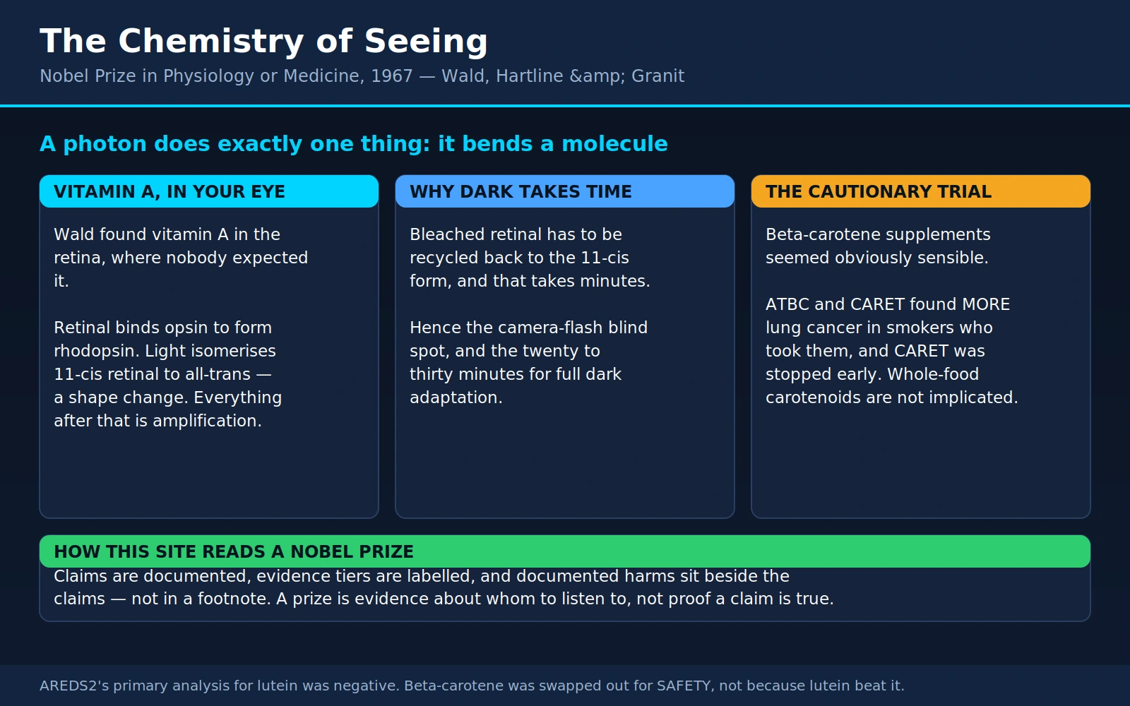
The answer turned out to be startlingly simple at its core. A photon strikes a molecule in your retina, and that molecule bends. That is the whole of it. One bond twists, one molecule changes shape, and everything else — the electrical signal, the nerve impulse, the image, the recognition of a face — is amplification and interpretation stacked on top of that single mechanical event. And the molecule that bends is made from vitamin A.
Table of Contents
- The Prize and the Three Men
- Wald's Discovery: Vitamin A in the Retina
- The Visual Cycle, and Why It Matters Practically
- Night Blindness and the World's Largest Preventable-Blindness Story
- Beta-Carotene, and the Trial That Ended the Enthusiasm
- Rods, Cones, and Colour
- Hartline: Lateral Inhibition and the Edge-Sharpening Eye
- Granit: The Electroretinogram and the Receptor Types
- What This Explains in Everyday Sight
- Eye-Health Claims, Tiered
- Where Mainstream Medicine Agrees / What Remains Debated
- Key Research Papers
- Connections
- Featured Videos
1. The Prize and the Three Men
The 1967 prize is unusual in that the three laureates barely worked together. They approached the same organ from three different directions — chemistry, single-cell electrophysiology, and whole-eye electrical recording — and their results interlock so neatly that in retrospect the shared award looks inevitable.
George Wald (1906–1997)
George Wald was born in New York City in 1906, the son of immigrant parents, and took his doctorate at Columbia in 1932 under Selig Hecht — a biophysicist who had spent his career measuring, with great precision, how little light a human being needs in order to see. That apprenticeship matters. Hecht taught Wald to think of vision as a quantitative photochemical problem rather than a vague biological one, and Hecht's own most famous experiment, published with Simon Shlaer and Maurice Pirenne in 1942, established that a dark-adapted human eye can register a flash carrying only a handful of light quanta — meaning a single rod cell must be able to respond to a single photon. Once you accept that, you are forced to ask what molecule is doing the catching.
Wald then spent a travelling fellowship year in Europe that reads like a deliberate assembly of the tools he would need: Berlin with Otto Warburg, who taught him the micro-methods of biochemistry; Zurich with Paul Karrer, who had determined the chemical structure of vitamin A; and Heidelberg with Otto Meyerhof. He joined Harvard in 1934 and stayed for the rest of his working life, becoming professor of biology in 1948.
Wald was also, by all accounts, a spectacular lecturer — his introductory biology course at Harvard was famous — and he was a fluent and vivid writer. That combination of laboratory rigour and public eloquence is what took him well beyond the laboratory in his last decades.
Haldan Keffer Hartline (1903–1983)
H. Keffer Hartline was born in Bloomsburg, Pennsylvania, took his MD at Johns Hopkins in 1927, and then did something slightly unusual for a newly qualified physician: he went and studied physics and mathematics. He worked at the Eldridge Reeves Johnson Foundation for Medical Physics at the University of Pennsylvania, returned to Johns Hopkins, and from 1953 was at the Rockefeller Institute (later Rockefeller University) in New York.
Hartline's gift was for the delicate, patient business of isolating a single nerve fibre and listening to it. In the 1930s, at a time when almost nobody could do it, he learned to dissect out one optic-nerve fibre from a frog and record its impulses in response to light. He then found an animal whose eye made the experiment far easier — the Atlantic horseshoe crab, Limulus polyphemus — and that choice of animal is a large part of why he won a Nobel Prize.
Ragnar Granit (1900–1991)
Ragnar Granit was born near Helsinki in 1900 into Finland's Swedish-speaking minority, took his MD at the University of Helsinki in 1927, and worked in Oxford with Charles Sherrington and at the University of Pennsylvania before returning to a chair in physiology at Helsinki. In 1940, in the aftermath of the Winter War, he moved to Sweden and the Karolinska Institute in Stockholm, where from 1945 he directed the Nobel Institute for Neurophysiology. He became a Swedish citizen in 1941.
Granit's method was the electroretinogram — the electrical signal generated by the eye as a whole in response to light — which he learned to take apart into its components, and then to complement with recordings from individual optic-nerve fibres. His work is the bridge between Wald's chemistry (which pigments exist) and everyday clinical medicine (a test you can still have done at a hospital eye clinic this afternoon).
Wald's second career, and an honest note about it
George Wald became publicly famous in a way that has little to do with retinal chemistry. On 4 March 1969 he gave a speech at the Massachusetts Institute of Technology titled A Generation in Search of a Future, a searing address on the Vietnam War, the draft, and nuclear weapons. It was reprinted, recorded, circulated on campuses, and read aloud at rallies; for a great many Americans, Wald was the Nobel laureate who spoke against the war, and the rhodopsin was a footnote. He campaigned for nuclear disarmament for the rest of his life and served on international human-rights tribunals.
It should be said plainly, because this site's readers are owed it: Wald also lent his name and his Nobel authority to positions well outside his expertise, particularly in his later years. He was a prominent opponent of recombinant DNA research during the Cambridge, Massachusetts moratorium debates of the mid-1970s, arguing for restrictions on a technology whose risks he was not in a position to assess and which went on to produce insulin, vaccines, and clotting factors; he opposed nuclear power on grounds that mixed genuine radiological concern with broader political argument; and he made confident public pronouncements on a range of scientific-policy questions where a prize in visual biochemistry conferred no special standing.
None of this diminishes the science. It is included because a recurring failure of public health reasoning is treating a laureate's opinion on any subject as expert testimony. Wald's chemistry was superb and it is settled. His opinions were opinions. Both things are true, and a reader is better served by being told so than by being handed a saint.
2. Wald's Discovery: Vitamin A in the Retina
In the early 1930s, working in Warburg's Berlin laboratory as a young postdoctoral researcher, Wald did something that sounds obvious now and was not obvious then. He took retinas, extracted them, and looked for vitamin A.
He found it. In quantity. And essentially nobody expected that.
At the time, vitamin A was understood as a growth factor — one of the "accessory food factors" whose existence Frederick Gowland Hopkins had demonstrated with his milk-supplemented rat experiments, and whose deficiency was known to cause both stunted growth and eye disease. It was a nutrition topic. Nobody had proposed that it was a structural component of the light-sensing apparatus itself. It was known that the retina contained a reddish-purple pigment — rhodopsin, or "visual purple," described by Franz Boll and Wilhelm Kühne in the 1870s — which was bleached to a pale yellow by light and slowly regenerated in the dark. But nobody knew what it was made of.
Wald's answer, assembled over the following years at Harvard and published largely in the Journal of General Physiology, was that rhodopsin is vitamin A plus a protein.
The chain, step by step
Here is the sequence Wald and his collaborators established, in plain language:
- Retinol — vitamin A in its alcohol form, the form you eat and the form your liver stores — is delivered to the retina.
- Retinol is oxidised to retinal (also called retinaldehyde), which is the same molecule with an aldehyde group where the alcohol group was. This is a small chemical change that makes the molecule far more chemically reactive.
- Retinal exists in different shapes. The one that matters is 11-cis-retinal — a version with a kink in its carbon chain at the eleventh position.
- 11-cis-retinal binds to a protein called opsin, tucking into a pocket in it and attaching through a chemical bond to a lysine residue. The combination is rhodopsin — visual purple. Each rod cell in your eye holds on the order of a hundred million of these molecules, stacked in a dense pile of membrane discs.
- A photon arrives and does exactly one thing: it isomerises 11-cis-retinal to all-trans-retinal. The kink straightens. The molecule changes shape. Nothing is broken, nothing is consumed, nothing is oxidised. A bond rotates.
That is the beginning of vision. That shape change, and only that shape change, is the point at which light becomes biology.
Everything after it is amplification
The straightened retinal no longer fits its pocket. The opsin protein, forced to accommodate a differently shaped passenger, changes its own shape through a rapid series of intermediate states — and the shape it settles into is an active enzyme-like form that can switch on other proteins.
The cascade that follows was largely worked out after the 1967 prize, by Wald's successors, but it is the direct completion of his account. One activated rhodopsin molecule switches on hundreds of molecules of a G protein called transducin; each of those activates an enzyme, phosphodiesterase, which chews up thousands of molecules of a small signalling molecule, cyclic GMP; the falling cGMP level closes ion channels in the cell membrane; and the cell's electrical state changes. The total gain is on the order of a hundred thousand to a million. That is why a single photon — an amount of energy far too small to matter to any ordinary chemical reaction — can produce a signal your brain can read.
One counter-intuitive detail worth knowing, because it surprises everyone: vertebrate photoreceptors are electrically active in the dark and go quieter in the light. In darkness a steady "dark current" flows through open channels; light closes those channels and the cell hyperpolarises. Your rods do not shout when photons arrive. They stop shouting.
3. The Visual Cycle, and Why It Matters Practically
Once retinal has straightened, that particular molecule is spent. It cannot catch another photon until something bends it back. This is the part of Wald's work with the most direct consequences for how your eyes behave, and it is called the visual cycle (or the retinoid cycle).
All-trans-retinal is released from the bleached opsin, reduced back to all-trans-retinol, and shipped out of the photoreceptor to a neighbouring layer of cells called the retinal pigment epithelium (RPE) — a single sheet of dark, hard-working cells lying immediately behind your photoreceptors. There it is esterified for storage, and then an enzyme called RPE65 performs the critical trick: it re-isomerises the molecule back to the 11-cis shape. The 11-cis-retinol is oxidised back to 11-cis-retinal, returned to the photoreceptor, and reloaded into a waiting opsin. The eye is ready again.
Three practical consequences follow directly, and each one is something you have personally experienced.
Camera flashes blind you briefly
A bright flash isomerises a large fraction of the rhodopsin in the illuminated patch of retina all at once. Until the visual cycle grinds that pigment back through the RPE and returns it, that patch is genuinely less sensitive — you are looking at a real chemical depletion, not a psychological aftereffect. The floating purple blob drifts with your gaze because it is your retina, not the room.
Full dark adaptation takes twenty to thirty minutes
Walking from bright sunlight into a dim cinema, you see almost nothing, then progressively more. Two processes are running. The cones adapt fast — most of their recovery is done within five to seven minutes. The rods adapt slowly, and their recovery is limited by the rate at which the visual cycle can resupply 11-cis-retinal. The classic dark-adaptation curve has a distinct kink in it, the "rod–cone break," where rod sensitivity overtakes cone sensitivity; from there the rods keep improving for another twenty minutes or more. By the end, your eye is roughly a hundred thousand times more sensitive than it was in daylight. There is no way to hurry this. It is enzyme kinetics.
It requires a continuing supply of vitamin A
The cycle is efficient but not perfect; retinoids are lost and must be replaced from the liver's stores, which are replenished from the diet. If the supply fails, rhodopsin regeneration slows. And because rods depend on rhodopsin far more critically than cones depend on their pigments, the first thing you lose is dim-light vision.
This is the hinge on which the next section turns. Wald's chemistry predicted, from first principles, that vitamin A deficiency should cause night blindness before it causes anything else. It does.
4. Night Blindness and the World's Largest Preventable-Blindness Story
Vitamin A deficiency remains one of the most consequential nutritional problems on Earth. The World Health Organization's estimate, cited in the current Cochrane review, is that roughly 190 million preschool children have low serum retinol levels. It is a leading cause of preventable childhood blindness, and it kills.
The progression, in order
Ocular vitamin A deficiency — xerophthalmia — follows a well-characterised sequence, graded by the WHO. It is worth setting out because the early stages are reversible in days and the late stages are not reversible at all.
- Night blindness (nyctalopia, WHO stage XN). The child stops moving around at dusk, bumps into things after sunset, will not leave the hut at night. In many affected communities there is a local word for it. This is the rhodopsin-regeneration failure Wald's chemistry predicts, and it reverses within a day or two of a vitamin A dose.
- Conjunctival xerosis (X1A). The white of the eye loses its wet sheen and looks dry, dull, and slightly wrinkled.
- Bitot's spots (X1B). Foamy, greyish-white triangular plaques on the conjunctiva, usually on the outer side of the eye. They are keratinised debris and desquamated cells. They look like dried soap suds and they are close to diagnostic.
- Corneal xerosis (X2). The cornea itself dries and becomes hazy. Still reversible.
- Corneal ulceration and keratomalacia (X3A, X3B). The cornea softens and melts. This can happen in hours, and is often precipitated by measles or severe diarrhoea in an already-depleted child. Whatever cornea is lost is lost.
- Corneal scarring (XS). Permanent blindness.
The cruelty of the sequence is that the reversible stages are quiet and the irreversible stage is fast. See our Vitamin A Deficiency section and its dedicated page on night blindness and eye damage for the clinical detail.
Supplementation reduces child deaths, not just blindness
This is the genuinely large effect, and it deserves to be stated with its evidence rather than asserted.
In 1986, Alfred Sommer and colleagues published a randomised community trial in northern Sumatra in The Lancet. Four hundred and fifty villages were randomised; 25,939 preschool children were followed for about a year; children over one year old received 200,000 IU vitamin A capsules twice, six months apart. Among children aged 12–71 months at baseline, mortality in the control villages was 7.3 per 1,000 against 4.9 per 1,000 where supplements were given — a 49% higher death rate in the controls. The finding was contentious at the time precisely because nobody had expected a vitamin capsule to cut child mortality by a third.
Three and a half decades of trials later, the current Cochrane review (Imdad and colleagues, 2022) pools 47 studies covering approximately 1.2 million children aged six months to five years. Its headline result for all-cause mortality is a risk ratio of 0.88 (95% CI 0.83 to 0.93) — a 12% reduction — graded as high-certainty evidence. Deaths from diarrhoea fell by a similar 12% (RR 0.88, 95% CI 0.79 to 0.98). High-certainty evidence in a Cochrane review of a million children is about as solid as public-health nutrition gets.
Twelve per cent is smaller than the third suggested by Sommer's Sumatra trial, and the reason is instructive. A very large Indian cluster-randomised trial, DEVTA, published in The Lancet in 2013, followed about a million preschool children and found a mortality ratio of 0.96 (95% CI 0.89 to 1.03, p=0.22) — no statistically significant benefit — while confirming that the supplements worked biochemically (mean plasma retinol rose, severe deficiency halved, Bitot's spots fell from 3.5% to 1.4%). DEVTA's own authors wrote that their result "contradicts the expectation from other trials that vitamin A supplementation would reduce child mortality by 20–30%, but cannot rule out some more modest effect." The Cochrane pooled estimate includes DEVTA, which is exactly why it lands at 12% rather than 30%. The honest summary is: the effect is real, it is smaller than the early enthusiasm, and it is still one of the best returns per dollar in global child health.
The counterweight: none of this applies to people who are not deficient
Everything above describes correcting a deficiency in a deficient population. Extending it to a well-nourished reader is a category error, and it is the single most common way vitamin A information goes wrong.
In populations that are not vitamin A deficient, supplementation does not improve vision. There is no reserve tank. A retina with a full supply of retinoids regenerates rhodopsin at the maximum rate the enzymes allow; adding more substrate does not speed up an enzyme that is already saturated. If your night vision is poor and you are well-nourished, the cause is something else — uncorrected refractive error, early cataract, retinal disease, or simply age — and vitamin A will not touch it.
Worse, high-dose preformed vitamin A (retinol and retinyl esters) is genuinely toxic. This is not a theoretical caution. See our Hypervitaminosis A section for the full picture; in brief:
- Acute hypervitaminosis A. A single very large dose — classically the polar-bear-liver story from Arctic exploration, more commonly a supplement error — causes headache, nausea, vomiting, blurred vision, dizziness, raised intracranial pressure, and later skin peeling.
- Chronic hypervitaminosis A. Sustained excess produces dry cracked skin and lips, hair loss, bone and joint pain with bony overgrowth, pseudotumor cerebri, and liver injury that can progress to fibrosis and cirrhosis. Penniston and Tanumihardjo's review in the American Journal of Clinical Nutrition is the standard reference.
- Teratogenicity. Retinoids are among the most reliably documented human teratogens. This is why isotretinoin — a vitamin A derivative used for severe acne — is dispensed only under a formal pregnancy-prevention programme (iPLEDGE in the United States, equivalent schemes elsewhere) requiring negative pregnancy tests and two forms of contraception. The programme is not bureaucratic caution; it exists because the drug causes severe craniofacial, cardiac, and central nervous system malformations. Preformed vitamin A supplements in pregnancy carry the same class of concern, which is why prenatal vitamins use beta-carotene or tightly capped retinol.
- Bone and hip fracture. Three independent observational studies point the same way. Melhus and colleagues (Annals of Internal Medicine, 1998) found that for every additional 1 mg/day of dietary retinol, hip fracture risk rose 68% (95% CI 18–140%), with intake above 1.5 mg/day roughly doubling risk (OR 2.1, 95% CI 1.1–4.0). Feskanich and colleagues in the Nurses' Health Study (JAMA, 2002; 72,337 women, 18 years) found the highest quintile of total vitamin A intake carried RR 1.48 (95% CI 1.05–2.07), driven by retinol (RR 1.89, 95% CI 1.33–2.68) — and, importantly, beta-carotene did not contribute significantly (RR 1.22, 95% CI 0.90–1.66). Michaëlsson and colleagues (New England Journal of Medicine, 2003) measured serum retinol in 2,322 men and followed them 30 years: hip fracture rate ratio 2.47 (95% CI 1.15–5.28) for the highest versus middle quintile, and again no association with serum beta-carotene. These are observational studies and causation is not settled — but three different designs, a plausible mechanism (retinoic acid stimulates bone-resorbing osteoclasts), and a consistent carotenoid-versus-retinol split is a serious signal.
The adult tolerable upper intake level for preformed vitamin A is 3,000 µg RAE (10,000 IU) per day. The recommended intake is 900 µg RAE for men and 700 µg RAE for women. Note that the upper limit applies to retinol, not to carotenoids — which is the subject of the next section, and the reason that distinction is not a technicality.
5. Beta-Carotene, and the Trial That Ended the Enthusiasm
This section is the practical heart of the page, and it is the clearest cautionary tale in modern nutrition science. It deserves to be told completely rather than summarised into a slogan.
Why the logic seemed impeccable
Beta-carotene is a provitamin A carotenoid: a plant pigment, orange, that your body can cleave into two molecules of retinal. Wald himself worked on carotenoids — his 1935 paper "Carotenoids and the visual cycle" is one of the founding documents of the field. Carotenoids are also antioxidants, quenching reactive oxygen species in a test tube with genuine efficiency.
By the 1980s, the reasoning ran like this, and each link in the chain was defensible on its own:
- People who eat more carotenoid-rich fruit and vegetables get less lung cancer. This was a robust, repeatedly replicated observational finding.
- People with higher blood levels of beta-carotene get less lung cancer. Also robust.
- Beta-carotene is an antioxidant, and oxidative damage to DNA is a plausible mechanism of carcinogenesis.
- Therefore giving beta-carotene supplements to people at high risk of lung cancer — smokers — should reduce their risk.
This was not fringe thinking. It was the mainstream consensus, endorsed by cancer institutes, and it was considered important enough to fund two enormous randomised trials.
ATBC (1994)
The Alpha-Tocopherol, Beta Carotene Cancer Prevention Study randomised 29,133 male smokers aged 50 to 69 in south-western Finland to alpha-tocopherol (50 mg/day), beta-carotene (20 mg/day), both, or placebo, and followed them five to eight years. Results published in the New England Journal of Medicine in April 1994:
- 876 new lung cancers occurred.
- Men who received beta-carotene had an 18% higher incidence of lung cancer (95% CI 3% to 36%) than those who did not.
- Total mortality was 8% higher (95% CI 1% to 16%) in the beta-carotene groups, driven by lung cancer and ischaemic heart disease deaths.
- Alpha-tocopherol produced no reduction in lung cancer, though fewer prostate cancers occurred; more haemorrhagic strokes occurred.
The trial's own conclusion was carefully worded: it "raises the possibility that these supplements may actually have harmful as well as beneficial effects." The initial reaction in the field was substantial disbelief. A single trial, however large, might be a statistical accident.
CARET (1996)
It was not an accident. The Beta-Carotene and Retinol Efficacy Trial was already running in the United States: 18,314 smokers, former smokers, and asbestos-exposed workers, randomised to 30 mg/day beta-carotene plus 25,000 IU/day retinol as retinyl palmitate, versus placebo. Results, published in the New England Journal of Medicine in May 1996 after a mean 4.0 years of follow-up:
- 388 new lung cancers.
- Relative risk of lung cancer in the active-treatment group: 1.28 (95% CI 1.04 to 1.57), P = 0.02.
- Relative risk of death from lung cancer: 1.46 (95% CI 1.07 to 2.00).
- Relative risk of death from any cause: 1.17 (95% CI 1.03 to 1.33).
- Relative risk of cardiovascular death: 1.26 (95% CI 0.99 to 1.61).
CARET was stopped 21 months early on these findings. A trial designed to demonstrate prevention was terminated because the intervention appeared to be causing the disease it was meant to prevent.
What the story actually teaches
Four things, and all four generalise far beyond beta-carotene.
An observational association is not a mechanism you can bottle. Carotenoid-rich diets protect against lung cancer. It does not follow that carotenoids are what does the protecting. Vegetables contain hundreds of compounds; beta-carotene may simply be the easiest marker of "this person eats vegetables," and the marker is not the medicine.
A plausible mechanism is not evidence of benefit. Antioxidant activity in a cuvette is a real chemical property. Whether it translates into less cancer in a living smoker's lung is an entirely separate empirical question, and the answer came back not merely "no" but "the opposite." Proposed explanations include beta-carotene acting as a pro-oxidant at high concentration in the oxygen-rich, oxidant-saturated environment of a smoker's lung, and disruption of retinoic-acid signalling in bronchial epithelium. Neither explanation was predicted in advance. Nobody saw this coming.
Dose and context change the sign of the effect. The trials used 20–30 mg/day of isolated beta-carotene, roughly ten times what a good diet supplies, delivered as a purified compound rather than in a food matrix, to a population with an extraordinary oxidative burden in the target organ. None of those conditions resemble eating vegetables.
Food carotenoids are not implicated. This must be said clearly, because the trials are sometimes misquoted as showing that carrots cause cancer. They do not show that and nothing shows that. There are two reasons the distinction is real rather than rhetorical. First, conversion of beta-carotene to vitamin A by the enzyme BCO1 in the intestinal wall is regulated — when your vitamin A status is adequate, conversion is downregulated, which is why you cannot give yourself hypervitaminosis A by eating carrots. (You can turn your palms orange. Carotenodermia is harmless and reverses.) Second, whole plant foods deliver carotenoids at ordinary doses, mixed with hundreds of other compounds, in a matrix that limits absorption. The observational evidence favouring carotenoid-rich diets was never overturned. What was overturned was the supplement.
The finding has had a permanent institutional consequence, described in section 9: it is the reason the standard eye-health supplement formulation was reformulated. See Carrots and Lutein for the food-level picture.
6. Rods, Cones, and Colour
Wald did not stop at rhodopsin. His later work at Harvard, much of it with Paul K. Brown, attacked the harder problem: the pigments of the cones, the photoreceptors responsible for daylight and colour vision.
This was technically brutal. Rods are numerous (roughly 120 million per human eye) and packed with pigment, so rhodopsin can be extracted in bulk. Cones number around 6 million, are concentrated in the central fovea, and hold far less pigment each. Brown and Wald solved it with microspectrophotometry — shining a beam of light narrow enough to pass through a single photoreceptor, and measuring which wavelengths it absorbed.
Their 1964 paper in Science, "Visual pigments in single rods and cones of the human retina," reported measurements on individual human photoreceptors and found what the theory had predicted for a century: three classes of cone, with absorption peaks in the blue, green, and yellow-green regions of the spectrum. A group at Johns Hopkins — Marks, Dobelle, and MacNichol — published closely comparable measurements from primate cones in Science within weeks. Independent confirmation arriving that fast is unusually good evidence.
Modern values put the peaks at roughly 420–430 nm (S, short / "blue"), 530–535 nm (M, medium / "green"), and 558–565 nm (L, long / "red"). Rhodopsin peaks near 500 nm. Brown and Wald's numbers were close to these.
Why colour vision is three-channel
A single photoreceptor cannot report colour. It reports how many photons it caught, and a bright dim-wavelength light and a dim bright-wavelength light look identical to it. This is the principle of univariance, and it is why a rod-only eye is colour-blind: in dim light, when only rods are working, you genuinely see in shades of grey.
Colour arises from comparison. With three cone types whose absorption curves overlap, any wavelength produces a distinctive ratio of S : M : L responses, and the ratio identifies the wavelength unambiguously. Three channels, plus the downstream opponent processing (red-versus-green, blue-versus-yellow, light-versus-dark) that Ewald Hering had inferred from pure psychology in the nineteenth century, is enough to generate the roughly one million distinguishable colours a normal human eye can discriminate.
Three is also why colour reproduction works. A television, a phone screen, and a printed photograph do not reproduce the spectrum of the original scene — they cannot. They produce a mixture of three primaries that happens to excite your three cone types in the same ratio the original scene would have. The image is a forgery aimed precisely at the receptors Wald measured.
Red-green colour blindness, and why it is a men's condition
In 1986, Jeremy Nathans, David Thomas, and David Hogness identified the genes encoding the three cone pigments, published in Science. The findings explain the epidemiology exactly.
The gene for the S (blue) pigment sits on chromosome 7 — an autosome, one copy from each parent. The genes for the L (red) and M (green) pigments sit adjacent to each other in a tandem array on the X chromosome, and they are about 98% identical in sequence.
Two consequences follow:
- Nearly identical neighbouring genes recombine badly. During the formation of eggs, the two X chromosomes line up and swap material. When two almost-identical genes sit side by side, the alignment can slip — producing chromosomes that have lost a gene, gained an extra copy, or ended up with a fused hybrid gene coding for a pigment with an intermediate spectral peak. This is why red-green defects are common while blue defects (tritanopia) are rare: it is not that the S pigment is more robust, it is that it is not sitting next to its own near-twin.
- Men have only one X. A woman with a faulty L or M gene on one X usually has a working copy on the other, and sees normally — she is a carrier. A man has no second copy. Whatever is on his single X is what he gets.
The result is the familiar epidemiology: red-green colour vision deficiency affects roughly 8% of men of Northern European ancestry and about 0.5% of women, with lower rates in some other populations. A boy inherits it from his mother's X chromosome, which is why it characteristically skips from a colour-blind grandfather to a grandson through an unaffected daughter.
Most affected people are anomalous trichromats — they still have three pigments, but one is shifted so that red and green responses overlap more than they should, compressing that part of colour space rather than eliminating it. True dichromats, missing a pigment entirely, are less common.
7. Hartline: Lateral Inhibition and the Edge-Sharpening Eye
Wald explained how a photoreceptor catches light. Hartline explained what the retina does with the answer — and the answer is that it does far more than pass it along.
Listening to one nerve fibre at a time
In the 1930s Hartline mastered the dissection of a single optic-nerve fibre from a frog's eye and recorded its impulses directly. Working this way through the late 1930s and early 1940s, publishing chiefly in the American Journal of Physiology — papers from that era and journal are not indexed in PubMed, which is why they carry no link here — he established two things that reshaped sensory neuroscience:
- Optic-nerve fibres come in classes. Some fire when a light comes on ("ON" fibres), some when it goes off ("OFF" fibres), and some at both transitions ("ON–OFF" fibres). The eye is not a photographic plate reporting brightness; it is already sorting events into categories.
- Each fibre has a receptive field. Hartline introduced this concept in 1938: the specific patch of retina where light can influence that particular fibre. "Receptive field" has since become one of the most productive ideas in all of neuroscience — it is the framework within which Stephen Kuffler described centre–surround organisation, and within which Hubel and Wiesel described orientation-selective cortical cells and won their own Nobel Prize in 1981.
The horseshoe crab
Frog retinas are difficult. The compound eye of the Atlantic horseshoe crab, Limulus polyphemus, is not. It is built of several hundred ommatidia — individual optical units, each with its own receptor cell and its own nerve fibre — large, tough, robustly separated, and possible to illuminate one at a time with a fine beam while recording from the fibre of a chosen unit. Hartline chose it deliberately, and it is one of the great model-organism choices in biology. (It also had a second life: the horseshoe crab's blue blood is the source of the Limulus amoebocyte lysate assay used to test injectable drugs for bacterial endotoxin. The animal has done a great deal for human medicine.)
The discovery: an illuminated receptor suppresses its neighbours
With Floyd Ratliff and Henry Wagner, in a series of Journal of General Physiology papers from 1956 through 1958, Hartline established the phenomenon that carries his name:
When you illuminate an ommatidium, the firing rate of its neighbours goes down.
The inhibition is:
- Mutual — each unit inhibits its neighbours and is inhibited by them, in proportion to how hard each is being driven.
- Graded with distance — strongest for near neighbours, falling off further away.
- Graded with intensity — a brightly lit unit inhibits harder than a dimly lit one.
- Quantifiable — Hartline and Ratliff wrote it as a set of simultaneous linear equations, one per receptor, each unit's output being its own excitation minus the summed weighted inhibition from all the others. The eye was doing arithmetic, and they wrote down the arithmetic.
Why this sharpens edges
Consider a boundary between a bright field and a dark field falling across the eye.
A receptor deep inside the bright region is surrounded on all sides by other brightly lit receptors, so it receives inhibition from every direction and its output is pulled down. A receptor sitting just on the bright side of the boundary has bright neighbours on one side only — its other neighbours are in the dark and inhibit it weakly — so it receives less total inhibition and fires more than its deep-field colleagues. Symmetrically, a receptor just on the dark side of the boundary is being inhibited by the bright receptors next to it, so it fires less than receptors deep in the dark.
The signal leaving the eye therefore shows a bright overshoot on the light side of the edge and a dark undershoot on the dark side. The edge has been exaggerated before the brain ever sees it. Uniform regions are compressed and boundaries are amplified — which is a very sensible thing for a visual system to do, since boundaries carry nearly all the information about where objects are and uniform expanses carry almost none.
Mach bands, the Hermann grid, and what you can see for yourself
Mach bands are the direct perceptual demonstration. Print a staircase of grey strips, each uniformly shaded and each a step darker than the last. Where two strips meet, you will see a bright line along the lighter strip's edge and a dark line along the darker strip's edge. Measure the ink and the bands are not there. They are your lateral inhibition, drawn on the world. Ernst Mach described them in the 1860s and correctly inferred that some neural interaction must be responsible — ninety years before Hartline recorded it happening.
The Hermann grid — a lattice of black squares separated by white streets, in which shadowy grey blobs appear at the intersections you are not looking at — is the other standard illustration, described by Ludimar Hermann in 1870. The textbook account attributes it to centre–surround lateral inhibition: a receptive field centred on an intersection has white in more of its surround than one centred in a street, so it is inhibited more and the intersection looks darker.
Here honesty requires a caveat, because this site would rather be accurate than tidy. The simple lateral-inhibition account of the Hermann grid has been seriously challenged. The strongest objection is easy to check yourself: if the grid's streets are made gently wavy instead of straight, the illusion largely disappears — and a purely circular centre–surround mechanism has no way to care whether a line is straight or curved. The illusion is now generally thought to involve orientation-selective mechanisms in visual cortex as well as, or instead of, retinal inhibition. Mach bands remain a good demonstration of lateral inhibition. The Hermann grid is a more complicated story than the textbooks say.
The general principle survives all of this intact, and it is the thing worth taking away: your visual system enhances contrast before the signal reaches your brain. What arrives at your cortex is not a photograph. It has already been edited, and the editing is done by neurons in your eye, according to rules Hartline wrote down while listening to a horseshoe crab.
8. Granit: The Electroretinogram and the Receptor Types
Granit's approach sat between Wald's test tube and Hartline's single fibre: he recorded the electrical response of the whole retina, and then learned to take that response apart.
Dissecting the electroretinogram
When light strikes an eye, the eye generates a measurable voltage — the electroretinogram (ERG). Willem Einthoven, better known for the electrocardiogram, had recorded it in 1908. But a whole-organ signal is a sum of contributions from many cell layers, and by itself it is nearly uninterpretable.
In a 1933 paper in the Journal of Physiology — "The components of the retinal action potential in mammals and their relation to the discharge in the optic nerve" — Granit separated the mammalian ERG into three components, which he named PI, PII, and PIII in the order they were abolished by deepening anaesthesia. Modern work has assigned them to their sources:
- PIII — the fastest, a negative deflection, arising from the photoreceptors themselves. It is the source of the clinical a-wave.
- PII — a positive deflection from the inner retina, principally the bipolar cells with a contribution from Müller glia. It is the source of the clinical b-wave.
- PI — a slow positive component from the retinal pigment epithelium, seen clinically as the c-wave.
Because a-wave and b-wave come from different layers, their ratio localises a disease. That single fact is why the ERG is still in clinical use nearly a century later.
Dominators and modulators
Granit then went further and recorded from individual optic-nerve fibres while varying the wavelength of the stimulus, mapping each fibre's spectral sensitivity curve. Working with Anna Munsterhjelm and with Carl Wrede, he published a matched pair of studies in the Journal of Physiology in 1937 — the electrical responses of dark-adapted and of light-adapted frogs' eyes to monochromatic stimuli.
He found two kinds of curve, and named them:
- Dominators — broad curves covering most of the visible spectrum with a single peak, corresponding to overall brightness sensing. The scotopic (dark-adapted) dominator peaked near 500 nm — matching the absorption spectrum of Wald's rhodopsin. The photopic (light-adapted) dominator peaked further toward the yellow-green.
- Modulators — narrow curves with peaks at different wavelengths across the spectrum, which he interpreted as the signature of separate receptor mechanisms with distinct spectral tuning.
Granit refined the scotopic dominator's correspondence with visual purple in later work with K. O. Donner. The importance of the modulators is that they were physiological evidence for multiple spectrally distinct receptor types recorded from living tissue — arriving from the electrode side of the field decades before Brown and Wald could measure the cone pigments directly with light. When the two independent lines of evidence converged in the 1960s, the case became overwhelming. That convergence is, in effect, what the 1967 prize was awarded for.
The ERG is a working clinical test today
This is not history. If you or a family member is investigated for a retinal disorder, you may well have a Granit descendant performed on you. Standards are set by the International Society for Clinical Electrophysiology of Vision (ISCEV), and the test involves dark-adapting for around twenty minutes (that visual-cycle timing again), then recording responses to standardised flashes with an electrode against the eye.
Current uses include:
- Retinitis pigmentosa and other rod–cone dystrophies. Rod responses are reduced or extinguished, often before changes are visible when looking into the eye. The ERG can confirm a diagnosis at a stage when the fundus still looks close to normal.
- Cone dystrophies and cone–rod dystrophies, including Stargardt disease, where the photopic responses fail first.
- Congenital stationary night blindness, which produces the characteristic "negative ERG" — a preserved a-wave with a reduced b-wave, meaning the photoreceptors are working but the signal is not getting through the inner retina. That is a Granit PIII-versus-PII reading, done at a hospital, this year.
- Retinal toxicity screening, notably multifocal ERG for hydroxychloroquine retinopathy.
- Endpoints in gene-therapy and drug trials, where an objective measure of retinal function is needed.
9. What This Explains in Everyday Sight
Dark adaptation, and why red light preserves it
Astronomers, submariners, aircrew, night-shift radiologists, and photographers working in darkrooms all use dim red illumination, and the reason is pure Wald chemistry.
Rhodopsin absorbs strongly around 500 nm (blue-green) and very weakly at long wavelengths. Deep red light around 630 nm and beyond therefore bleaches almost no rhodopsin — your rods stay loaded and stay dark-adapted. Your cones, particularly the L-cones peaking near 560 nm, still respond to red light well enough that you can read a chart or check an instrument. So red light lets you use daylight vision without spending your night vision. Step into a room with a white light for ten seconds and you have thrown away twenty minutes of adaptation.
Two practical footnotes. The effect requires the light to be dim as well as red: bright red light will bleach rhodopsin perfectly well. And it should be genuinely deep red, not orange — a "red" LED with a broad spectrum leaking into the yellow undoes much of the benefit.
Why night vision worsens with age
Several independent changes stack up, and knowing which is which helps you know what can be fixed:
- The pupil gets smaller. Senile miosis reduces the maximum dark-adapted pupil area substantially between young adulthood and old age, so less light enters. Nothing to be done about it.
- The lens yellows and scatters. An ageing lens absorbs short wavelengths and scatters light, reducing retinal illumination and increasing glare — which is why oncoming headlights become so much worse with age. Cataract is the same process continued, and cataract surgery genuinely restores night driving for many people.
- Dark adaptation itself slows. The rate-limiting step is the visual cycle — the RPE's handling and re-isomerisation of retinoids — and it slows with age, particularly where the RPE is stressed. Delayed dark adaptation is now recognised as one of the earliest functional markers of age-related macular degeneration, sometimes preceding visible drusen.
The point worth internalising: worsening night vision in a well-nourished older adult is a lens problem, a pupil problem, or an RPE problem. It is not a vitamin A supply problem, and treating it as one wastes time that should be spent at an eye clinic.
Retinitis pigmentosa and gene therapy: a direct descendant of the visual cycle
Retinitis pigmentosa is a family of inherited retinal dystrophies in which photoreceptors progressively die. It typically begins with night blindness — rods first — then constricting peripheral fields, then central vision. More than seventy genes are implicated.
One of them is RPE65: the enzyme in the retinal pigment epithelium that performs the re-isomerisation step, converting all-trans retinoid back to the 11-cis form. It is precisely the enzymatic step Wald's chemistry required to exist. Children with biallelic RPE65 mutations cannot regenerate 11-cis-retinal efficiently; they present with severe visual impairment and profound night blindness from early childhood (Leber congenital amaurosis type 2 / early-onset severe retinal dystrophy), and they go on losing vision.
Voretigene neparvovec (marketed as Luxturna) is a gene therapy for exactly this condition: a working copy of the RPE65 gene packaged into an adeno-associated virus vector and injected under the retina. The phase 3 trial, published by Stephen Russell and colleagues in The Lancet in 2017, randomised 31 participants 2:1 to treatment or control. The primary endpoint was change over one year in a multi-luminance mobility test — walking a marked obstacle course at progressively lower light levels, which measures something a patient actually cares about rather than a letter chart. Mean bilateral score change was 1.8 light levels in the treated group versus 0.2 in controls (difference 1.6, 95% CI 0.72–2.41, p=0.0013), and 13 of 20 treated participants, against none of the controls, passed the course at the dimmest level tested (1 lux) — the maximum improvement the test could register. No product-related serious adverse events occurred.
It was approved by the FDA in December 2017, the first directly administered gene therapy for an inherited disease approved in the United States. Realistic framing matters: it applies only to patients with confirmed biallelic RPE65 mutations, a small fraction of inherited retinal dystrophy; it does not restore normal sight; it does not regrow photoreceptors already lost, so it works best when treated early; the durability of benefit over decades is still being established; and it is extraordinarily expensive.
But the intellectual line is unbroken and worth naming. A young man in Berlin in the early 1930s extracted retinas and found vitamin A. That led to the visual cycle. The visual cycle required an isomerising enzyme. The enzyme was identified as RPE65. Mutations in RPE65 were found to cause childhood blindness. A gene therapy was built to replace it, and children walked an obstacle course in near-darkness who could not have done so before. That is what basic science looks like when you follow it for eighty years.
Macular degeneration supplements: what the trials actually show
Age-related macular degeneration is the leading cause of severe central vision loss in older adults in high-income countries. It is also the one eye condition where a nutritional supplement has real randomised-trial evidence — and where the evidence is routinely overstated in both directions.
AREDS (2001). The Age-Related Eye Disease Study randomised 3,640 participants aged 55–80 to a daily formulation of vitamin C 500 mg, vitamin E 400 IU, beta-carotene 15 mg, and zinc 80 mg (as oxide) with copper 2 mg, or components, or placebo, with average follow-up of 6.3 years. Antioxidants plus zinc reduced progression to advanced AMD, odds ratio 0.72 (99% CI 0.52–0.98); among the higher-risk participants the estimate strengthened to 0.66 (99% CI 0.47–0.91), and moderate visual acuity loss fell (OR 0.73, 99% CI 0.54–0.99).
Two limits on that result are frequently dropped and should not be. First, the benefit was confined to people who already had substantial AMD — extensive intermediate drusen, large drusen, or advanced disease in one eye. Participants with no AMD or only early changes had just a 1.3% five-year probability of progressing, and the trial showed no benefit for them; taking the formulation as general "eye vitamins" with healthy eyes is not supported. Second, the companion report from the same trial found no benefit for cataract — the same supplement, same participants, and no effect on lens opacity or cataract-related vision loss.
AREDS2 (2013) asked whether the formulation could be improved: could lutein and zeaxanthin, or omega-3 fatty acids, do better, and could beta-carotene be removed? It randomised 4,203 participants at risk of progression. The primary analysis was negative: lutein + zeaxanthin gave a hazard ratio of 0.90 (98.7% CI 0.76–1.07, P=.12), and DHA + EPA gave 0.97 (98.7% CI 0.82–1.16, P=.70). Neither reached significance. Omega-3 did nothing. Removing beta-carotene did not reduce the formulation's effectiveness.
And then the safety finding that determined everything afterwards: more lung cancers occurred in participants assigned beta-carotene — 23 (2.0%) versus 11 (0.9%), nominal P=.04 — overwhelmingly in former smokers. Fifteen years after ATBC and CARET, in a trial about eyes, in ex-smokers rather than current smokers, the same signal appeared.
That is why the modern AREDS2 formulation replaces beta-carotene with lutein and zeaxanthin. It is important to understand the actual reasoning, because the common shorthand ("AREDS2 showed lutein works better") misstates it. The primary analysis did not show lutein was better. The substitution was justified because lutein and zeaxanthin were at least as good, and beta-carotene carried a cancer signal that lutein did not. It was a safety swap between roughly equivalent options.
The ten-year follow-up, AREDS2 Report 28 (Chew and colleagues, 2022, 3,882 participants), then strengthened it in both directions. The lung cancer odds ratio at ten years was 1.82 (95% CI 1.06–3.12, P=.02) for beta-carotene and a non-significant 1.15 (95% CI 0.79–1.66) for lutein/zeaxanthin. And with longer follow-up, lutein/zeaxanthin did show a modest benefit for progression to late AMD: hazard ratio 0.91 (95% CI 0.84–0.99, P=.02) versus no lutein/zeaxanthin, and 0.85 (95% CI 0.73–0.98, P=.02) in a direct comparison against beta-carotene.
The practical summary: if you have intermediate AMD or advanced AMD in one eye, the AREDS2 formulation has real evidence and is worth discussing with your ophthalmologist. If you have healthy eyes, it has no demonstrated benefit. And nobody with any smoking history should be taking a beta-carotene-containing eye supplement — check the label, because old-formula products are still sold.
10. Eye-Health Claims, Tiered
Vision is a field crowded with supplement marketing, much of it invoking Wald's chemistry as a warrant. Here is where the evidence actually sits.
🟢 Carrots and night vision — true if you are deficient, false if you are not
The most famous eye-nutrition claim in the English-speaking world is partly a wartime deception, and the real story is better than either the myth or the debunking.
During the Second World War, British night-fighter crews began shooting down German bombers in darkness at rates that demanded an explanation. The actual explanation was airborne interception radar, a secret worth protecting. Among the cover stories put about was that RAF pilots — John "Cat's Eyes" Cunningham most famously — owed their night vision to a diet heavy in carrots. It also suited the Ministry of Food, which had a carrot surplus and a blackout to make tolerable. The story has been told and retold for eighty years and is now firmly lodged in popular belief.
But here is the part the debunkings usually miss. The claim is not arbitrary nonsense — it has a real kernel, and the kernel is exactly Wald's chemistry. Carrots are rich in beta-carotene; beta-carotene is converted to vitamin A; vitamin A is the substrate for rhodopsin; and vitamin A deficiency genuinely does cause night blindness as its very first symptom. A vitamin A–deficient person who starts eating carrots will, in fact, see better in the dark. That is not folklore; it is the reversal of stage XN xerophthalmia, and it is the basis of programmes that protect millions of children's sight.
What does not follow is the extrapolation. In someone whose vitamin A status is already adequate, eating more carrots does not improve night vision at all. The rhodopsin regeneration machinery is running at capacity; more substrate does not make a saturated enzyme faster. Carrots are an excellent food for many reasons. Night-vision enhancement in a well-fed adult is not one of them. See Carrots.
Tier: correcting a deficiency, strong evidence. Supplementing when replete, no evidence.
🟡 Lutein and zeaxanthin — genuine for advanced AMD, thin for screens
Lutein and zeaxanthin are xanthophyll carotenoids that concentrate in the macula, forming the yellow macular pigment. They absorb short-wavelength light and quench reactive oxygen species, so the mechanism is entirely plausible — but as section 5 should have made permanently clear, plausible mechanisms in this field have an unimpressive track record.
Where the evidence is real: in the AREDS2 population — people with substantial existing AMD — lutein/zeaxanthin at 10 mg and 2 mg respectively is part of a formulation with trial evidence, and the ten-year follow-up gives it a modest independent benefit (HR 0.91, 95% CI 0.84–0.99). It is also the correct and safer replacement for beta-carotene in anyone who has ever smoked.
Where the evidence is thin: lutein supplements for screen fatigue, "digital eye strain," or general eye health in people without AMD. That market is far larger than the AMD market, and it rests on extrapolation rather than trials. Dietary lutein from kale, spinach, and egg yolks is sensible, well-absorbed, and costs nothing extra if you eat those foods anyway.
Tier: good evidence within the AREDS2 indication; extrapolation beyond it.
🔴 "Blue light" supplements and blue-blocking lenses — not supported
The 2023 Cochrane review of blue-light-filtering spectacle lenses (Singh and colleagues) pooled 17 randomised trials. Its conclusions: blue-light filtering lenses may not reduce eye strain with computer use over short-term follow-up compared with ordinary lenses; there is probably little or no effect on best-corrected visual acuity; effects on sleep quality were indeterminate and mixed; and there was no randomised evidence at all bearing on macular health, contrast sensitivity, colour discrimination, or glare. The trials were small and mostly at high risk of bias.
Supplements marketed specifically as "blue light protection" rest on the same premise and have less evidence again. If your eyes ache at a screen, the effective interventions are the boring ones: correct your refraction, get a proper eye examination, fix the lighting and screen distance, and blink deliberately — screen use reduces blink rate and the resulting dry eye is the most common real cause of the symptom.
Tier: not supported for eye strain or eye health.
🟡 Bilberry — a persistent wartime myth, and a thin modern evidence base
Bilberry (Vaccinium myrtillus) carries its own aviator legend: that RAF pilots ate bilberry jam to sharpen night vision. Like the carrot story it is wartime folklore, and unlike the carrot story it does not even have a deficiency-correction kernel to stand on. Bilberry anthocyanins have been studied for rhodopsin regeneration and dark adaptation for decades, and controlled trials in healthy people have consistently failed to show a night-vision benefit.
The modern evidence is more modest and points elsewhere. A randomised, double-blind, placebo-controlled trial of a 12-week bilberry extract reported improved objective measures of ciliary muscle contraction — that is, focusing effort, not night vision — in participants with eye fatigue. That is a small, single, industry-relevant trial of a mechanism unrelated to the original claim, and it should be read as a hypothesis rather than a recommendation.
Tier: night vision, not supported. Accommodation and eye fatigue, preliminary only.
🔴 Eye exercises for refractive error — will not change your prescription
Myopia, hyperopia, and astigmatism are optical and anatomical. Myopia is principally an eyeball that is too long for its optics; no amount of exercise shortens an eyeball. Systems promising to eliminate glasses through eye exercises have been marketed for over a century (the Bates method dates to the 1920s) and have never been shown to alter refractive error.
A partial exception deserves mention for accuracy. The eye-acupoint exercises performed daily in Chinese schools have been studied for myopia control — slowing progression, not reversing refraction. A 2023 systematic review and meta-analysis found a 24% apparent reduction in myopia in unadjusted analysis (OR 0.76, 95% CI 0.62–0.89) which lost significance once covariates were adjusted for (OR 0.87, 95% CI 0.72–1.02), with modest protective effects surviving in some subgroups. The authors concluded the effect "may not be enough to prevent the progress of myopia in the long term." That is a fair reading: a small, uncertain, heavily confounded signal for progression, and nothing at all for correcting existing refractive error.
Separately, convergence insufficiency — difficulty pointing both eyes at a near target — is a genuine binocular-vision disorder that does respond to supervised vergence therapy. That is a real, evidence-backed use of eye exercises, and it is not the same thing as throwing away your glasses.
Tier: refractive error, not supported. Myopia progression, weak and confounded. Convergence insufficiency, supported — different condition.
11. Where Mainstream Medicine Agrees / What Remains Debated
Settled
- The photochemistry of vision. Rhodopsin is 11-cis-retinal bound to opsin; a photon isomerises it to all-trans; that isomerisation initiates the transduction cascade. This is textbook, taught worldwide, and has been confirmed by structural biology, spectroscopy, and genetics down to the femtosecond and the individual atom.
- The visual cycle and its rate-limiting role in dark adaptation. Confirmed enzymatically, genetically (RPE65, LRAT, RDH5 mutations all cause the predicted disease), and therapeutically.
- Vitamin A deficiency causes night blindness, xerophthalmia, and irreversible blindness, in that order.
- Vitamin A supplementation reduces all-cause child mortality in deficient populations — RR 0.88 (95% CI 0.83–0.93), high-certainty Cochrane evidence over 1.2 million children.
- Three cone types underlie human colour vision, encoded by genes on chromosome 7 (S) and the X chromosome (L and M), with the X-linked tandem arrangement explaining the sex distribution of red-green deficiency.
- Lateral inhibition sharpens edges in the retina. Recorded directly, formalised mathematically, and demonstrable perceptually with Mach bands.
- Beta-carotene supplements increase lung cancer risk in smokers. Two large independent randomised trials, one stopped early, plus a concordant signal in ex-smokers in AREDS2 sustained at ten years.
- High-dose preformed vitamin A is toxic and teratogenic. Uncontested; the isotretinoin pregnancy-prevention programmes exist because of it.
- The ERG is a valid clinical test for retinal dystrophies, with international recording standards.
Debated or unresolved
- The magnitude of the vitamin A mortality benefit. Sommer's Sumatra trial suggested about a third; DEVTA's million children found no significant effect; Cochrane's pooled figure is 12%. Whether the difference reflects changing background nutrition, differing baseline deficiency, or trial methodology is still argued.
- Whether high retinol intake causes hip fracture. Three good observational studies agree and the mechanism is plausible, but no randomised trial has tested it and some cohorts have not replicated the finding. It is a strong reason for caution, not a settled fact.
- Why beta-carotene harms smokers. The pro-oxidant and retinoic-acid-signalling hypotheses are both plausible and neither is proven. This matters, because without knowing the mechanism we cannot be confident which other carotenoids at which doses are safe in that population.
- Whether lutein and zeaxanthin genuinely slow AMD independently. AREDS2's primary analysis said no; the ten-year follow-up and secondary analyses say modestly yes. Long-term observational follow-up of a randomised trial is a weaker design than the trial itself.
- The Hermann grid. The classical retinal lateral-inhibition explanation does not survive the curved-line variant; how much of the illusion is cortical remains open. Mach bands are not in dispute.
- Whether earlier or repeat gene therapy improves durability in RPE65 dystrophy, and whether the approach extends usefully to the far more common retinal dystrophy genes.
- How much of age-related dark-adaptation delay is treatable. Delayed dark adaptation is an early AMD marker; whether it can be modified, and whether doing so alters disease course, is under active study.
12. Key Research Papers
Two notes on sourcing. First, several of Wald's foundational publications — including his 1933 and 1934 letters to Nature reporting vitamin A in the retina, and Hartline's American Journal of Physiology papers of 1938 and 1940 introducing the receptive field and the ON/OFF/ON–OFF fibre classes — are not indexed in PubMed and therefore carry no link here. They are described in the text with journal and year so a reader can find them in a library or archive. Second, Wald's 1967 Nobel lecture appeared in print in 1968 in two journals; both records are given below rather than picking one and implying the other does not exist.
- Wald G. Vitamin A in eye tissues. Journal of General Physiology, 1935;18(6):905–915. The measurement that started it: vitamin A present in retinal tissue, where nobody had looked for it.
- Wald G. Carotenoids and the visual cycle. Journal of General Physiology, 1935;19(2):351–371. The paper that names and frames the visual cycle, connecting dietary carotenoids to retinal photochemistry.
- Hecht S, Shlaer S, Pirenne MH. Energy, quanta, and vision. Journal of General Physiology, 1942;25(6):819–840. Wald's mentor establishing the human absolute threshold — the result that forces the conclusion that a single rod responds to a single photon.
- Wald G. The molecular basis of visual excitation. Nature, 1968;219(5156):800–807. Wald's Nobel lecture, delivered December 1967. A companion version appeared as "Molecular basis of visual excitation," Science, 1968;162(3850):230–239.
- Brown PK, Wald G. Visual pigments in single rods and cones of the human retina: direct measurements reveal mechanisms of human night and color vision. Science, 1964;144(3614):45–52. Microspectrophotometry of individual human photoreceptors; the three cone pigments measured directly.
- Nathans J, Thomas D, Hogness DS. Molecular genetics of human color vision: the genes encoding blue, green, and red pigments. Science, 1986;232(4747):193–202. The genes behind Wald's pigments, and the tandem X-linked arrangement that explains red-green colour blindness.
- Hartline HK, Ratliff F. Inhibitory interaction of receptor units in the eye of Limulus. Journal of General Physiology, 1957;40(3):357–376. The core lateral-inhibition result: illuminated receptors suppress their neighbours, mutually and in proportion.
- Hartline HK, Ratliff F. Spatial summation of inhibitory influences in the eye of Limulus, and the mutual interaction of receptor units. Journal of General Physiology, 1958;41(5):1049–1066. The quantitative formulation — the simultaneous equations describing what the retina computes.
- Granit R. The components of the retinal action potential in mammals and their relation to the discharge in the optic nerve. Journal of Physiology, 1933;77(3):207–239. The dissection of the electroretinogram into PI, PII, and PIII, which underlies every clinical ERG performed today.
- Granit R, Munsterhjelm A. The electrical responses of dark-adapted frogs' eyes to monochromatic stimuli. Journal of Physiology, 1937;88(4):436–458. Spectral sensitivity measured electrically; the dominator and modulator curves that implied multiple receptor types.
- Sommer A, Tarwotjo I, Djunaedi E, West KP Jr, Loeden AA, Tilden R, Mele L. Impact of vitamin A supplementation on childhood mortality: a randomised controlled community trial. The Lancet, 1986;1(8491):1169–1173. 450 villages in northern Sumatra; control mortality 49% higher than in supplemented villages.
- Imdad A, Mayo-Wilson E, Haykal MR, Regan A, Sidhu J, Smith A, Bhutta ZA. Vitamin A supplementation for preventing morbidity and mortality in children from six months to five years of age. Cochrane Database of Systematic Reviews, 2022;3(3):CD008524. 47 studies, ~1.2 million children; all-cause mortality RR 0.88 (95% CI 0.83–0.93), high-certainty evidence.
- Awasthi S, Peto R, Read S, Clark S, Pande V, Bundy D; DEVTA team. Vitamin A supplementation every 6 months with retinol in 1 million pre-school children in north India: DEVTA, a cluster-randomised trial. The Lancet, 2013;381(9876):1469–1477. The large null trial — mortality ratio 0.96 (95% CI 0.89–1.03) — that pulled the pooled estimate down. Included for balance, not despite it.
- The Alpha-Tocopherol, Beta Carotene Cancer Prevention Study Group. The effect of vitamin E and beta carotene on the incidence of lung cancer and other cancers in male smokers. New England Journal of Medicine, 1994;330(15):1029–1035. 29,133 Finnish male smokers; lung cancer incidence 18% higher with beta-carotene (95% CI 3–36%).
- Omenn GS, Goodman GE, Thornquist MD, Balmes J, Cullen MR, Glass A, et al. Effects of a combination of beta carotene and vitamin A on lung cancer and cardiovascular disease. New England Journal of Medicine, 1996;334(18):1150–1155. CARET: 18,314 participants; lung cancer RR 1.28 (95% CI 1.04–1.57); trial stopped 21 months early.
- Age-Related Eye Disease Study Research Group. A randomized, placebo-controlled, clinical trial of high-dose supplementation with vitamins C and E, beta carotene, and zinc for age-related macular degeneration and vision loss: AREDS report no. 8. Archives of Ophthalmology, 2001;119(10):1417–1436. Antioxidants plus zinc, advanced AMD odds ratio 0.72 (99% CI 0.52–0.98); benefit confined to higher-risk participants.
- Age-Related Eye Disease Study 2 Research Group. Lutein + zeaxanthin and omega-3 fatty acids for age-related macular degeneration: the AREDS2 randomized clinical trial. JAMA, 2013;309(19):2005–2015. Primary analysis negative for both additions; more lung cancers with beta-carotene (2.0% vs 0.9%), mostly former smokers — the basis for the reformulation.
- Chew EY, Clemons TE, Agrón E, Domalpally A, Keenan TDL, Vitale S, et al. Long-term outcomes of adding lutein/zeaxanthin and ω-3 fatty acids to the AREDS supplements on age-related macular degeneration progression: AREDS2 report 28. JAMA Ophthalmology, 2022;140(7):692–698. Ten-year data: beta-carotene lung cancer OR 1.82 (95% CI 1.06–3.12); lutein/zeaxanthin late-AMD HR 0.91 (95% CI 0.84–0.99).
- Penniston KL, Tanumihardjo SA. The acute and chronic toxic effects of vitamin A. American Journal of Clinical Nutrition, 2006;83(2):191–201. The standard review of hypervitaminosis A. (Note: a letter with an identical title appeared in the same journal later that year; this is the review article.)
- Michaëlsson K, Lithell H, Vessby B, Melhus H. Serum retinol levels and the risk of fracture. New England Journal of Medicine, 2003;348(4):287–294. 2,322 men followed 30 years; hip fracture rate ratio 2.47 (95% CI 1.15–5.28) in the highest retinol quintile, with no association for beta-carotene.
- Russell S, Bennett J, Wellman JA, Chung DC, Yu ZF, Tillman A, et al. Efficacy and safety of voretigene neparvovec (AAV2-hRPE65v2) in patients with RPE65-mediated inherited retinal dystrophy: a randomised, controlled, open-label, phase 3 trial. The Lancet, 2017;390(10097):849–860. Gene therapy aimed at the visual cycle's isomerase; mobility-test improvement 1.8 versus 0.2 light levels (difference 1.6, 95% CI 0.72–2.41, p=0.0013).
- Singh S, Keller PR, Busija L, McMillan P, Makrai E, Lawrenson JG, Hull CC, Downie LE. Blue-light filtering spectacle lenses for visual performance, sleep, and macular health in adults. Cochrane Database of Systematic Reviews, 2023;8(8):CD013244. Seventeen trials; no demonstrated benefit for eye strain or visual acuity, and no randomised evidence on macular health at all.
Live PubMed Searches
- Rhodopsin, visual cycle, and retinal
- Vitamin A deficiency and night blindness in children
- Beta-carotene supplementation and lung cancer
- AREDS2 and macular degeneration
- Voretigene neparvovec and RPE65
13. Connections
- Notable Doctors — the full index of physicians and scientists profiled on this site.
- Nobel Prize in Physiology or Medicine — the complete roll of laureates, 1901 to the present, in which the 1967 prize sits.
- Frederick Gowland Hopkins — the accessory food factors that became the vitamins; without his concept, "vitamin A in the retina" would not have been a sentence anyone could say.
- Christiaan Eijkman — beriberi, brown rice, and the deficiency-disease idea that made nutritional blindness intelligible.
- Albert Szent-Györgyi — vitamin C, and the same 1930s European biochemistry that trained Wald.
- Cajal & Golgi — the neuron doctrine, worked out substantially by drawing retinal neurons; Cajal's diagrams of retinal cell layers are the anatomical map that Hartline and Granit later recorded from.
- Julius & Patapoutian — the receptors for heat, chili, menthol, and touch; the same problem as vision, solved for other senses.
- Vitamin A — the nutrient at the centre of this story: sources, forms, requirements, and interactions.
- Vitamin A Deficiency — symptoms, causes, and recovery, including the night blindness and eye damage progression described above.
- Hypervitaminosis A — the toxicity side, including birth defects in pregnancy and liver damage.
- Ophthalmology — the full eye-disease section of this site.
- Age-Related Macular Degeneration — where the AREDS and AREDS2 evidence is applied in practice.
- Retinitis Pigmentosa — the inherited dystrophies diagnosed by Granit's ERG and, for RPE65, now treatable.
- Lutein — the macular carotenoid that replaced beta-carotene in the AREDS2 formulation.
- Bilberry — the other wartime night-vision legend, assessed on its evidence.
- Carrots — beta-carotene in the food matrix, where the trials' warnings do not apply.
- Zinc — a component of the AREDS formulation and a cofactor for the retinol dehydrogenases of the visual cycle.