TSH Test (Thyroid-Stimulating Hormone): Reference Range and Interpretation
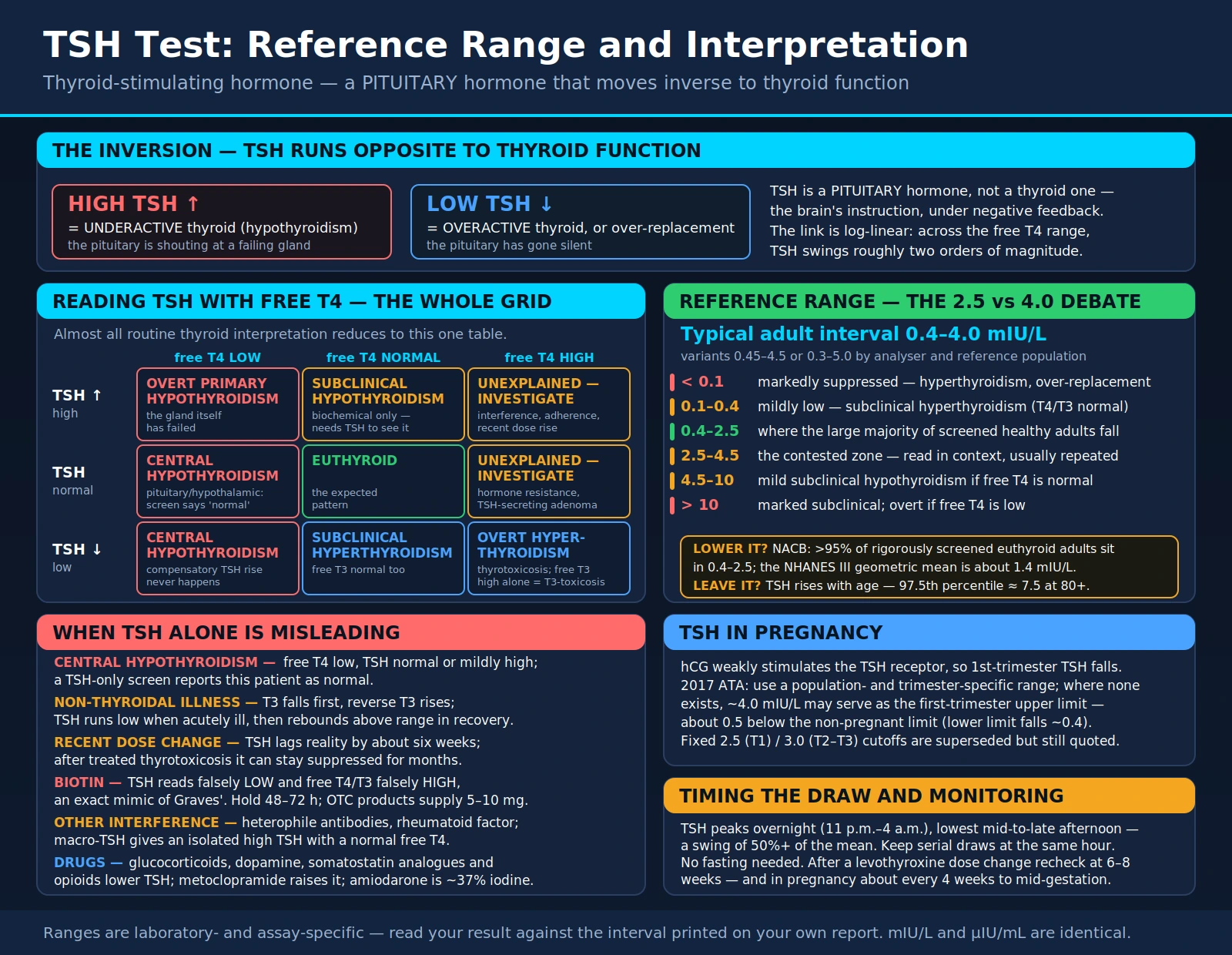
Thyroid-stimulating hormone (TSH, also called thyrotropin) is the single most frequently ordered endocrine blood test in medicine. It is a pituitary hormone — not a thyroid hormone — and that one fact explains almost everything readers find confusing about their results. TSH is the brain's instruction to the thyroid gland, and the brain adjusts that instruction based on how much thyroid hormone it is already detecting. When the thyroid is underperforming, the pituitary shouts louder and TSH goes up. When the thyroid is overperforming, the pituitary falls silent and TSH goes down. TSH therefore moves in the opposite direction to thyroid function, which is why a high number on a lab report so often means an underactive gland. This page explains the feedback loop that produces that inversion, why the upper end of the reference range is genuinely contested among endocrinologists, how TSH is read alongside free T4, and the specific situations in which a TSH result — on its own — will mislead you.
Interactive Visualization How Your Thyroid Is Controlled Run the TSH feedback loop, convert T4 into T3, and set your metabolic rate — then switch on Hashimoto's, Graves', or iodine deficiency and watch the labs move. Launch →Table of Contents
- What TSH Is and Why It Is Ordered First
- The Feedback Loop: Why TSH Runs Backwards
- The Reference Range and the 2.5-vs-4.0 Debate
- Timing the Draw: Diurnal Variation and Preparation
- Reading TSH Together With Free T4
- Subclinical Hypothyroidism and Hyperthyroidism
- TSH in Pregnancy
- When TSH Alone Is Misleading
- Monitoring Levothyroxine and the 6–8 Week Rule
- Companion Tests and What Each Adds
- Key Research Papers
- Connections
- Featured Videos
What TSH Is and Why It Is Ordered First
TSH is a glycoprotein hormone secreted by specialised cells — thyrotrophs — in the anterior pituitary gland. It travels to the thyroid, the butterfly-shaped gland straddling the windpipe, and binds the TSH receptor on thyroid follicular cells. That binding drives essentially every step of hormone production: it pulls iodide into the gland through the sodium-iodide symporter, drives thyroid peroxidase to attach iodine onto the thyroglobulin scaffold, and triggers release of finished hormone into the circulation. Sustained stimulation also makes the gland physically grow, which is why an iodine-starved thyroid under relentless TSH drive enlarges into a goiter.
The thyroid's output is roughly 80–90% thyroxine (T4) and only a small fraction triiodothyronine (T3). T4 is best thought of as a circulating reserve — a prohormone with a half-life of about seven days. The biologically potent hormone is T3, and most of the body's T3 is generated not by the thyroid but inside target tissues, where deiodinase enzymes strip a single iodine atom off T4 to activate it. A separate deiodinase routes T4 down an inactivating path to reverse T3 instead. This tissue-level conversion is one reason a single blood test cannot capture everything about thyroid status.
TSH became the first-line screening test because it is amplified. Owing to the logarithmic relationship described in the next section, a change in thyroid hormone too small to push free T4 outside its reference interval will already have moved TSH substantially. TSH is therefore the earliest quantitative signal that the thyroid is drifting, and modern third-generation immunoassays measure it down to about 0.01–0.02 mIU/L — low enough to distinguish a partially suppressed TSH from a fully suppressed one.
Thyroid dysfunction is common enough to justify that screening volume. In the NHANES III survey, 4.6% of participants had hypothyroidism — 4.3% of it subclinical, meaning detectable only by TSH — and 1.3% had hyperthyroidism, of which 0.7% was subclinical. Prevalence is several times higher in women and rises steadily with age. Clinicians order a TSH for fatigue, unexplained weight change, cold or heat intolerance, hair thinning, bowel changes, menstrual irregularity, subfertility, palpitations, tremor, low mood, cognitive fog, high cholesterol, a palpable goiter or nodule, a family history of thyroid or autoimmune disease, before and during pregnancy, and to monitor anyone already taking thyroid hormone or antithyroid medication.
One small unit note that trips people up: TSH is reported in mIU/L (milli-international units per litre) or µIU/mL (micro-international units per millilitre). These are numerically identical — a TSH of 2.1 mIU/L and 2.1 µIU/mL are the same result, so there is nothing to convert when comparing reports from different laboratories.
The Feedback Loop: Why TSH Runs Backwards
The hypothalamic-pituitary-thyroid axis is a thermostat. The hypothalamus releases thyrotropin-releasing hormone (TRH), which tells the pituitary to release TSH, which tells the thyroid to release T4 and T3. Circulating thyroid hormone then travels back to the pituitary and hypothalamus and suppresses further TRH and TSH release. This is classic negative feedback, and it is the entire reason TSH reads backwards.
Work through it slowly, because this is the point most readers get wrong:
- An underactive thyroid raises TSH. If the gland is damaged, surgically removed, iodine-deficient, or being destroyed by autoimmune thyroiditis, it delivers less T4. The pituitary detects the shortfall and compensates by pushing out more TSH to flog the failing gland harder. A high TSH is therefore the signature of an underactive thyroid — hypothyroidism.
- An overactive thyroid lowers TSH. If the gland is churning out excess hormone — from Graves' disease, an autonomous nodule, thyroiditis dumping stored hormone, or too large a dose of thyroid medication — the pituitary detects the surplus and shuts its own output down. A low or undetectable TSH is the signature of an overactive thyroid, or of over-replacement.
Patients frequently reverse this and read a TSH of 8 as "high thyroid." It is the opposite. A useful mental image: TSH is the volume of the shouting, not the amount of work being done. A loud instruction means the worker is not responding; silence means the worker is already doing too much.
Two refinements make the axis far more sensitive than a simple thermostat.
The pituitary makes its own T3. The thyrotroph does not respond mainly to circulating T3. It expresses type 2 deiodinase, which converts incoming T4 into T3 locally, inside the cell, and it is that locally generated T3 that switches off TSH gene transcription. The consequence is that pituitary TSH output is tuned almost specifically to circulating T4 — which is precisely why TSH and free T4 are the pairing that matters diagnostically, and why free T3 adds relatively little to a routine screen.
The relationship is log-linear, not proportional. A straight-line fall in free T4 produces an exponential rise in TSH. Across the width of the free T4 reference interval, TSH swings through roughly two orders of magnitude. In practical terms, a drop in free T4 so small it never leaves the "normal" column of the report can already have doubled or tripled the TSH. That built-in amplification is what makes TSH the earliest detector of thyroid drift — and it is also why a TSH of 6 versus a TSH of 3 represents a much smaller difference in actual thyroid hormone than the numbers suggest.
A third feature is clinically underappreciated: every person has an individual set point. Andersen and colleagues sampled healthy volunteers monthly for a year and found each individual's T4, T3 and TSH oscillated within a personal range roughly half the width of the population reference interval. Your own TSH is a tightly regulated personal constant; the laboratory's reference range is the aggregate of thousands of different personal constants. This means a result can sit comfortably inside the printed range and still be distinctly abnormal for you — a TSH that has moved from a lifelong 0.9 to 3.8 has quadrupled without ever flagging as abnormal. It is a strong argument for keeping old results and watching trends rather than single values.
The Reference Range and the 2.5-vs-4.0 Debate
Most laboratories in the United States and Europe report an adult TSH reference interval of approximately 0.4–4.0 mIU/L, with variants such as 0.45–4.5 or 0.3–5.0 depending on the analyser and the reference population used. Because that range is derived statistically as the central 95% of a chosen healthy population, and because different manufacturers' assays are not fully standardised against one another, reference ranges are laboratory- and assay-dependent and the interval printed on your own report is the one that applies to your result. Padoan and colleagues demonstrated that TSH values from different immunoassay platforms differ enough that recalibration functions are needed to make them comparable — which is a concrete reason to keep serial monitoring on the same laboratory and the same platform wherever possible.
The upper limit is where genuine, unresolved disagreement lives.
The case for lowering it. NHANES III established that in a rigorously screened, disease-free population the TSH distribution is sharply skewed, with a geometric mean around 1.4 mIU/L and the bulk of healthy people well below 2.5. Excluding those with thyroid antibodies or a family history shifts it lower still, because such individuals include early, undeclared autoimmune thyroiditis. The National Academy of Clinical Biochemistry's laboratory guidelines concluded that more than 95% of rigorously screened euthyroid volunteers have a TSH between 0.4 and 2.5 mIU/L, and that the conventional upper limit is inflated by the silent inclusion of people with early thyroid disease. Wartofsky and Dickey argued the limit should therefore move to about 2.5, on the grounds that a TSH of 3.5 is statistically unusual and often marks the leading edge of thyroid failure.
The case for leaving it alone. Surks and colleagues replied in the same journal issue that lowering the cutoff would reclassify tens of millions of asymptomatic people as diseased without evidence they benefit from treatment; that mildly raised TSH frequently reverts to normal on repeat testing; and — the strongest argument — that the TSH distribution shifts upward with age as a normal phenomenon, not a pathological one. In NHANES III the 97.5th percentile rises progressively across age bands, reaching roughly 7.5 mIU/L at age 80 and over. Judged against a fixed 4.5 cutoff, a large fraction of healthy octogenarians are labelled hypothyroid; judged against their own age group, they are normal. Observational data also associate a modestly higher TSH in the very old with better, not worse, survival.
Neither position has won. Most clinicians treat the 2.5–4.5 band as a grey zone rather than a verdict — a result to repeat in six to twelve weeks, interpret against age, symptoms, TPO antibody status, cholesterol, goiter and pregnancy plans, and track as a trend. A TSH of 3.8 in an asymptomatic, antibody-negative 78-year-old is a very different finding from a TSH of 3.8 in a 29-year-old with strongly positive TPO antibodies who is trying to conceive.
Approximate landmarks, all of them subject to the laboratory's own interval:
- Below 0.1 mIU/L: markedly suppressed. Seen in overt hyperthyroidism, in significant over-replacement with thyroid hormone, and in the deliberately suppressed targets used after some thyroid cancers.
- 0.1–0.4 mIU/L: mildly low. With normal free T4 and free T3, this is subclinical hyperthyroidism.
- 0.4–2.5 mIU/L: where the large majority of screened healthy adults fall.
- 2.5–4.5 mIU/L: the contested zone. Inside most laboratory ranges, above the tightly screened population's 95th percentile. Interpreted in context and usually repeated.
- 4.5–10 mIU/L: mild subclinical hypothyroidism if free T4 is normal. A meaningful proportion normalises on repeat testing.
- Above 10 mIU/L: marked subclinical hypothyroidism if free T4 is still normal; overt hypothyroidism if free T4 is low. This is the threshold at which the outcome evidence firms up considerably.
Children and newborns use entirely different intervals — neonatal TSH surges dramatically in the first hours after birth and falls over the following days, which is why newborn screening programmes use their own age-specific cutoffs and their own timing rules.
Timing the Draw: Diurnal Variation and Preparation
TSH is not a flat number through the day. It follows a pronounced circadian rhythm: levels fall to their lowest point in the mid-to-late afternoon, climb through the evening, peak overnight somewhere between roughly 11 p.m. and 4 a.m., and then decline again through the morning. The peak-to-trough swing is not trivial — it commonly amounts to 50% or more of the mean value, which for someone sitting near 3 mIU/L is a swing of a point or two in either direction from clock time alone.
Ehrenkranz and colleagues quantified this across a very large dataset and found both a circadian rhythm and a circannual one: TSH also runs measurably higher in winter than in summer. Sviridonova and colleagues examined the practical fallout in hypothyroid patients and showed that the same person can be classified differently depending on when blood was drawn — a result above the diagnostic threshold on an early-morning sample can fall below it on an afternoon sample the same day.
What this means for you:
- Consistency beats optimisation. There is no universally mandated draw time. What matters far more is that serial tests are drawn at approximately the same time of day, so that a change in the number reflects a change in your thyroid rather than a change in your appointment slot. If your first test was at 8 a.m., book subsequent ones in the morning.
- Morning draws read higher. A borderline morning value and a normal afternoon value are not a contradiction; they are the same physiology sampled at two points on a curve. Fasting morning samples also tend to read slightly higher than samples taken after eating, which is one more reason to standardise the conditions rather than chase the lowest number.
- No fasting is required for a TSH test itself, and it can be drawn at any time of day. Fasting is often requested only because TSH is bundled with a lipid or glucose panel on the same draw.
- Hold high-dose biotin first. Biotin supplements — especially the multi-milligram doses in hair, skin and nail products — can severely distort thyroid immunoassays, as described in the misleading-results section below. Laboratories typically advise stopping high-dose biotin for at least 48–72 hours before the draw, longer for very large doses. Tell the phlebotomist if you take any.
- Levothyroxine timing barely affects TSH but does affect free T4. TSH responds to average hormone exposure over weeks and is largely indifferent to whether you took this morning's tablet. Free T4, by contrast, peaks a couple of hours after a dose and can read spuriously high on a post-dose sample. Where free T4 is being measured too, many clinics ask patients to delay the dose until after the blood draw.
- Do not test during an acute illness unless thyroid disease is specifically suspected, for the reasons set out under non-thyroidal illness below.
Reading TSH Together With Free T4
TSH tells you what the pituitary thinks. Free T4 tells you what is actually circulating. Neither alone completes the picture, and almost all routine thyroid interpretation reduces to reading the two together as a grid. "Free" T4 matters because more than 99% of circulating T4 is bound to carrier proteins and biologically inert; only the free fraction is available to tissues, and only the free fraction tracks with the pituitary's view of the world. Total T4 shifts with carrier protein levels — pregnancy, oral estrogen and the oral contraceptive pill all raise thyroxine-binding globulin and therefore total T4 — without the free fraction changing at all.
The four common combinations:
- High TSH + low free T4 → overt primary hypothyroidism. The gland itself has failed and the pituitary is shouting into the void. In developed countries the usual cause is Hashimoto's autoimmune thyroiditis; worldwide, iodine deficiency remains the leading cause. Also seen after thyroidectomy, after radioactive iodine, and from drugs such as amiodarone or lithium.
- High TSH + normal free T4 → subclinical hypothyroidism. The gland is struggling but the increased pituitary drive is still succeeding in keeping output within range. This is a biochemical diagnosis; it cannot be made without TSH.
- Low TSH + high free T4 (or high free T3) → overt hyperthyroidism/thyrotoxicosis. Excess hormone has shut the pituitary down. Causes include Graves' disease, a toxic nodule or toxic multinodular goiter, the release phase of thyroiditis, and excessive thyroid hormone dosing. When free T4 is normal but free T3 is high, the pattern is T3-toxicosis — a reason to measure free T3 when TSH is suppressed but free T4 is unremarkable.
- Low TSH + normal free T4 and free T3 → subclinical hyperthyroidism.
Two further combinations are uncommon but disproportionately important, because they are exactly the situations a TSH-only screen gets wrong:
- Low or inappropriately normal TSH + low free T4 → central hypothyroidism. The pituitary or hypothalamus is the failing organ, so the compensatory TSH rise never happens. The TSH may even read mildly elevated, because pituitary disease can release TSH molecules that are immunologically detectable but biologically weak. A TSH-only screen reports this patient as normal.
- High or non-suppressed TSH + high free T4 → an unusual result that demands explanation. The realistic possibilities are assay interference, poor adherence to levothyroxine with several doses taken shortly before the test (free T4 spikes while TSH has not yet had weeks to fall), a recent dose increase, resistance to thyroid hormone, or — rarely — a TSH-secreting pituitary adenoma. It should never simply be averaged into a dose decision.
Subclinical Hypothyroidism and Hyperthyroidism
"Subclinical" is a purely biochemical label, and it is defined entirely by TSH. It means the TSH is outside its reference interval while free T4 (and, for the hyperthyroid version, free T3) remains inside. It does not mean "without symptoms" — many people so labelled report fatigue or cognitive fog — and it does not mean "not real." It means the abnormality is currently visible only in the pituitary's response.
Subclinical hypothyroidism is high TSH with normal free T4, and it is by far the more common of the two, affecting roughly 4–10% of adults depending on the cutoff used, with the prevalence climbing steeply with age and running several times higher in women. It is conventionally split into two grades because they behave differently:
- Mild (TSH above the upper limit but under 10 mIU/L) accounts for the large majority of cases. A substantial proportion of these results normalise spontaneously on repeat testing weeks later, which is why a single abnormal TSH is generally confirmed before it is acted on. The confirmatory test usually adds TPO antibodies, since antibody-positive individuals progress to overt hypothyroidism at a considerably higher annual rate than antibody-negative ones.
- Marked (TSH 10 mIU/L or above) is where the outcome evidence hardens. A large individual-participant meta-analysis by Rodondi and colleagues, pooling prospective cohorts, found that the risk of coronary heart disease events and coronary mortality was increased in people with a TSH of 10–19.9 mIU/L, while risk in the milder 4.5–9.9 band was not clearly raised.
What clinicians weigh when deciding whether a mildly raised TSH warrants any action: the degree of elevation, whether it persists on repeat, TPO antibody status, age (with older thresholds set higher), symptom burden, the presence of a goiter, lipid abnormalities, cardiovascular risk, and above all pregnancy or plans for pregnancy. The joint American Association of Clinical Endocrinologists and American Thyroid Association guideline for hypothyroidism in adults, and the ATA's guideline on thyroid hormone replacement, both frame the decision this way rather than as a single numeric trigger.
The evidence in older adults deserves to be stated plainly, because it is often omitted. The TRUST trial randomised adults aged 65 and over with persistent subclinical hypothyroidism to levothyroxine or placebo and found no improvement in hypothyroid symptom scores or tiredness scores at one year, despite TSH normalising in the treated group. That is a genuinely important negative result: it establishes that normalising the number does not automatically translate into feeling better in this population, and it is a large part of why treatment of mild subclinical hypothyroidism in the elderly has become more conservative.
Subclinical hyperthyroidism is low TSH with normal free T4 and free T3. The commonest cause overall is exogenous — a levothyroxine dose slightly too high — followed by autonomously functioning nodules and early or mild Graves' disease. It is also worth excluding the non-thyroidal causes of a low TSH described in the next section before concluding the thyroid is at fault. The concerns here are cardiac and skeletal: persistent TSH suppression, particularly below 0.1 mIU/L, is associated with atrial fibrillation and with reduced bone mineral density and fracture risk, most consistently in older adults and postmenopausal women. The 2016 ATA guidelines on hyperthyroidism treat a TSH persistently below 0.1 in those groups as a substantially more consequential finding than a TSH of 0.2–0.4 in a healthy young adult. Anyone taking thyroid hormone whose TSH is found suppressed is normally re-evaluated for over-replacement first, since that is both the most likely explanation and the most easily corrected.
TSH in Pregnancy
Pregnancy remodels the thyroid axis substantially, and applying the ordinary non-pregnant reference range to a pregnant woman produces systematic misclassification in both directions.
The dominant driver is human chorionic gonadotropin. hCG and TSH share an identical alpha subunit and closely related beta subunits, so hCG is a weak agonist at the TSH receptor. As hCG surges toward its peak near the end of the first trimester, it directly stimulates the thyroid; the resulting rise in thyroid hormone feeds back on the pituitary and drives TSH down. A first-trimester TSH beneath the non-pregnant lower limit is therefore often simple physiology rather than hyperthyroidism, and the effect is exaggerated in twin pregnancies and in hyperemesis gravidarum, where hCG runs highest. TSH then drifts back upward through the second and third trimesters as hCG falls.
At the same time, estrogen roughly doubles thyroxine-binding globulin, which raises total T4 and T3 without raising the free fractions — so total hormone measurements become hard to interpret in pregnancy. Renal iodide clearance increases and the fetus draws on maternal supply, so iodine requirements rise; fetal thyroid hormone production does not begin until roughly mid-gestation, meaning early fetal brain development depends on maternal hormone crossing the placenta. That dependency is why maternal thyroid status in the first half of pregnancy attracts such attention.
On trimester-specific ranges, the guidance changed and the older numbers are still widely quoted. Earlier guidance specified fixed cutoffs — commonly cited as an upper limit of 2.5 mIU/L in the first trimester and 3.0 in the second and third. The 2017 American Thyroid Association guidelines moved away from these fixed values, because studies in several populations found the fixed 2.5 cutoff labelled an implausibly large share of healthy pregnant women as abnormal, with substantial variation by ethnicity, iodine status and body mass index. The current approach is:
- Use a population- and trimester-specific reference range derived from the local pregnant population and the local assay, wherever one exists.
- Where no such range is available, an upper reference limit of approximately 4.0 mIU/L may be used in the first trimester — which in most assays corresponds to reducing the non-pregnant upper limit by roughly 0.5 mIU/L.
- The lower limit likewise falls in the first trimester, by roughly 0.4 mIU/L, reflecting the hCG effect.
Both extremes carry consequences. Overt maternal hypothyroidism in pregnancy is associated with pregnancy loss, pre-eclampsia, preterm delivery, low birth weight and impaired neurodevelopment, and is treated without controversy. The picture for milder abnormalities is genuinely mixed and is worth reporting honestly rather than selectively. Negro and colleagues found that levothyroxine given to euthyroid, TPO-antibody-positive pregnant women reduced obstetric complications. Conversely, the large randomised trial by Casey and colleagues, which screened and treated subclinical hypothyroidism or isolated hypothyroxinemia identified between 8 and 20 weeks of gestation, found no improvement in the child's IQ at five years. One plausible reading of the two together is that timing matters — that intervention beginning after the first trimester may already be too late to influence neurodevelopment — but the question is not settled, and universal screening of all pregnant women remains contested for exactly this reason.
Practical monitoring points: women already taking levothyroxine usually require a dose increase early in pregnancy, often as soon as pregnancy is confirmed, and the ATA recommends checking TSH roughly every four weeks through the first half of gestation, with the dose typically returning to the pre-pregnancy level after delivery. Anyone with a history of thyroid disease, TPO antibodies, prior postpartum thyroiditis, type 1 diabetes, other autoimmune disease, prior head or neck irradiation, recurrent miscarriage, or infertility falls into the higher-risk group in whom testing is specifically advised.
When TSH Alone Is Misleading
TSH earns its place as first-line screening test because in the overwhelming majority of cases the pituitary is intact and its signal faithfully reports thyroid status. The exceptions are not rare enough to ignore, and they share one feature: in every case the TSH is a technically correct measurement of the wrong thing.
1. Central (secondary or tertiary) hypothyroidism. If the disease is in the pituitary or hypothalamus rather than the thyroid, the compensatory TSH rise cannot occur. Free T4 is low while TSH reads normal or only mildly elevated — and, as Persani describes, pituitary disease can produce TSH with reduced biological activity, so the immunoassay detects hormone that is not doing its job. A TSH-only screen returns "normal" for a genuinely hypothyroid patient. Causes include pituitary adenomas and their surgery or radiotherapy, Sheehan's syndrome, hypophysitis (including that induced by immune checkpoint inhibitors), traumatic brain injury, cranial irradiation, infiltrative disease, and congenital defects. The rule that follows is simple: whenever pituitary or hypothalamic disease is a possibility, or when there are other pituitary hormone deficiencies, free T4 must be measured alongside TSH rather than reflexively. The same applies to anyone whose symptoms strongly suggest hypothyroidism despite a normal TSH.
2. Non-thyroidal illness syndrome ("euthyroid sick syndrome"). Serious acute illness, surgery, starvation and intensive-care admission all disrupt the axis. The characteristic sequence is a fall in T3 first, with a rise in reverse T3, followed in more severe or prolonged illness by falling free T4; TSH is typically low or low-normal during the acute phase and then rebounds above the reference range during recovery. Van den Berghe's review of the syndrome emphasises how variable the picture is and how poorly a single snapshot represents it. The practical implication is unambiguous: thyroid function testing during acute illness is difficult to interpret and should generally be deferred unless thyroid disease is itself suspected as a cause. A raised TSH in someone convalescing from a hospital admission frequently needs nothing more than a repeat test some weeks later.
3. Recent treatment changes — TSH lags behind reality. The pituitary integrates thyroid hormone exposure over weeks, so TSH is always reporting the recent past. After a levothyroxine dose change it takes about six weeks to reach a new steady state. After treatment of significant hyperthyroidism the lag is far longer: thyrotrophs suppressed by months of thyrotoxicosis can remain suppressed for many weeks or several months after free T4 has normalised or even fallen below normal. Managing a recently treated Graves' patient by TSH alone can therefore produce serious over-treatment, and free T4 is used to steer the early phase.
4. Biotin interference. This is the most common laboratory artefact in modern thyroid testing and the one most likely to affect an otherwise healthy supplement user. Many immunoassay platforms use the streptavidin-biotin binding system in their detection chemistry, so a large excess of biotin in the sample disrupts them — and the direction of the error depends on assay design. Competitive assays (free T4, free T3) read falsely high; sandwich or immunometric assays (TSH) read falsely low. The result is suppressed TSH with elevated free T4 — a pattern that looks exactly like Graves' disease in a person whose thyroid is entirely normal. Published cases document precisely this misdiagnosis, including patients started on antithyroid drugs on the strength of it. The doses involved are sold over the counter: hair, skin and nail formulations commonly supply 5–10 mg and doses used in multiple sclerosis reach 100–300 mg, against a dietary requirement measured in micrograms. The US Food and Drug Administration has issued a safety communication warning that biotin can significantly interfere with laboratory tests. The remedy is to disclose supplement use and hold biotin before testing; a repeat sample after washout resolves the discrepancy.
5. Other assay interference. Heterophile antibodies, rheumatoid factor and anti-animal antibodies can generate spuriously high TSH values. Macro-TSH — TSH bound into a large complex with immunoglobulin G — is biologically inactive but still measured by the assay, producing an isolated raised TSH with normal free T4 that convincingly imitates subclinical hypothyroidism and has led to unnecessary treatment. It is identified with polyethylene glycol precipitation or gel filtration. The clue to any of these is a TSH that does not fit the clinical picture or the free T4; the remedy is to repeat on a different assay platform and ask the laboratory to investigate. A recent review catalogues the full range of interferences affecting thyroid function tests.
6. Medications that move TSH without thyroid disease. High-dose glucocorticoids, dopamine and dopamine agonists, somatostatin analogues and certain opioids suppress TSH release directly. Metoclopramide and domperidone can raise it. Amiodarone, which is roughly 37% iodine by weight, can cause either hypothyroidism or thyrotoxicosis and reliably raises TSH transiently in the first months of therapy. Lithium, interferon-alfa, tyrosine kinase inhibitors and immune checkpoint inhibitors all disturb thyroid function by various mechanisms. Oral estrogen raises thyroxine-binding globulin, altering total but not free hormone, and increases the levothyroxine requirement in treated patients. Biotin aside, this drug list is the second thing to review whenever a TSH does not fit.
7. Rarities with a distinctive signature. Resistance to thyroid hormone (usually a THRB mutation) produces high free T4 with a TSH that is normal or high rather than suppressed. A TSH-secreting pituitary adenoma produces the same biochemical pattern. Both are rare, but both are the reason a "high TSH with high free T4" report is investigated rather than dismissed as laboratory noise.
Monitoring Levothyroxine and the 6–8 Week Rule
Once someone is taking thyroid hormone replacement, TSH becomes the principal monitoring variable, and the single most important rule about it is one of timing: after starting levothyroxine or changing the dose, TSH is re-checked in about six to eight weeks — not sooner. This is not administrative caution. It is pharmacokinetics.
Levothyroxine (T4) has a half-life of roughly seven days. Reaching a new steady state after a dose change takes about five half-lives — already some five weeks — and the pituitary then needs further time to re-equilibrate its own output. A TSH drawn two or three weeks after a change is measuring a system still in transit, and acting on it (typically by adjusting the dose again) is how patients end up oscillating between over- and under-replacement for months. The American Thyroid Association's guideline on thyroid hormone replacement sets out this interval explicitly. Once a stable dose is established, monitoring generally moves to every six to twelve months — sooner if symptoms change, weight changes substantially, an interacting drug is started, or pregnancy occurs.
What clinicians actually monitor and adjust against:
- Target. For most people on replacement, the aim is a TSH somewhere within the laboratory reference range, with the precise point within that range individualised — often toward the lower half in younger patients, and toward the upper half in the elderly and in those with cardiac disease. After thyroid cancer, targets are risk-stratified and deliberately set lower, sometimes into the suppressed range, for reasons specific to cancer follow-up rather than to symptom control.
- Absorption. Levothyroxine is absorbed mainly in the jejunum and upper ileum and absorption is best on an empty stomach. Calcium and iron supplements, aluminium- and magnesium-containing antacids, proton pump inhibitors, bile acid sequestrants, sucralfate, soy products, high-fibre meals and coffee all reduce it. Calcium and iron in particular are usually separated from the dose by at least four hours. A sudden unexplained rise in TSH on an unchanged dose most often reflects a new interacting product, or a change in when the tablet is taken relative to food, rather than a change in the thyroid.
- Dose timing. The conventional instruction is 30–60 minutes before breakfast. Bedtime dosing is a recognised alternative: in a randomised double-blind crossover trial, Bolk and colleagues found that taking levothyroxine at bedtime produced lower TSH and higher thyroid hormone levels than the same dose taken in the morning, consistent with better absorption overnight. Consistency again matters more than the specific choice.
- Malabsorption and increased requirement. Celiac disease, Helicobacter pylori-associated atrophic gastritis, lactose intolerance, inflammatory bowel disease and bariatric surgery all raise the dose needed. An unexpectedly high dose requirement is a recognised prompt to screen for celiac disease.
- Adherence. Intermittent adherence produces a distinctive and easily misread pattern: high TSH alongside normal or even high free T4, because doses taken in the days immediately before the appointment raise free T4 while TSH still reflects weeks of under-replacement. Reading that as "needs a higher dose" makes matters worse.
- Over-replacement. A suppressed TSH on replacement is treated as a finding to correct, not an incidental one, given the association of sustained suppression with atrial fibrillation and bone loss. It is one of the more common avoidable problems in long-term thyroid care.
A note on the persistent-symptoms question, since it is what brings many readers to this page: a proportion of people on levothyroxine with a normal TSH continue to report fatigue, weight gain and cognitive symptoms. This is a real and actively researched problem — the candidate explanations include the fact that a healthy thyroid secretes some T3 directly while levothyroxine supplies only T4, individual variation in deiodinase activity, the individual set-point issue discussed earlier, and coexisting conditions such as iron deficiency, vitamin B12 deficiency, vitamin D deficiency, celiac disease, sleep apnea, anemia and depression that produce overlapping symptoms. Wiersinga's review surveys how thinking on replacement therapy has shifted. This page describes what is measured and monitored; what to do about persistent symptoms is a decision for you and your clinician, and it usually begins with looking beyond the TSH.
Companion Tests and What Each Adds
TSH is the entry point, not the whole assessment. The tests below are the ones ordered alongside or after it, and each answers a specific question that TSH cannot.
- Free T4: the indispensable partner. It separates overt from subclinical disease, and it is the only way to detect central hypothyroidism. Measure it whenever TSH is abnormal, whenever pituitary disease is possible, and whenever symptoms conflict with a normal TSH.
- Free T3: most useful when TSH is suppressed but free T4 is normal, which identifies T3-toxicosis. It adds little to the evaluation of hypothyroidism, because T3 is preserved until late.
- Thyroid peroxidase (TPO) antibodies: the marker of autoimmune thyroid disease. In someone with a mildly raised TSH, positive TPO antibodies substantially raise the probability of progression to overt hypothyroidism, and they change how a borderline result in a woman planning pregnancy is handled.
- Thyroglobulin antibodies: a secondary autoimmune marker, also essential as a technical control when thyroglobulin itself is measured in thyroid cancer follow-up, because they interfere with that assay.
- TSH receptor antibodies (TRAb) and thyroid-stimulating immunoglobulins (TSI): these confirm Graves' disease as the cause of hyperthyroidism, and are also used in pregnancy to assess the risk of fetal or neonatal thyroid disease, since the antibodies cross the placenta.
- Reverse T3: rises in non-thyroidal illness, starvation and severe stress. It is widely marketed for routine use, and it is worth being straightforward: its clinical utility outside research and specific illness contexts is not established, and mainstream guidelines do not recommend it for diagnosing or managing hypothyroidism.
- Total T4, total T3 and thyroxine-binding globulin: mostly of historical or specialist interest now that free hormone assays are standard, but relevant where binding proteins are grossly abnormal.
- Thyroid ultrasound, radioactive iodine uptake and scan: answer structural and mechanistic questions — nodules, goiter size, and whether thyrotoxicosis reflects overproduction (high uptake, as in Graves') or the release of stored hormone from a damaged gland (low uptake, as in thyroiditis).
- Nutrient and overlap testing: iodine is the raw material for thyroid hormone; selenium is a required cofactor for the deiodinases that activate T4 and for glutathione peroxidase in the gland; zinc and iron also participate in hormone synthesis and conversion. Ferritin, vitamin B12 and vitamin D are commonly checked in parallel because their deficiencies mimic hypothyroid symptoms and frequently coexist with autoimmune thyroid disease.
Repeating the caution that governs all of the above: every one of these tests carries its own laboratory- and assay-specific reference interval, and results are interpreted against the range printed on the report that produced them.
Key Research Papers
Every citation below has been verified against the PubMed record. Author names, titles and journals appear as plain text; the year, volume and pages link to the paper's DOI, and the PMID links to the PubMed record.
- Hollowell JG, Staehling NW, Flanders WD, et al. Serum TSH, T(4), and thyroid antibodies in the United States population (1988 to 1994): National Health and Nutrition Examination Survey (NHANES III). J Clin Endocrinol Metab. 2002;87(2):489–499. PMID 11836274
- Andersen S, Pedersen KM, Bruun NH, et al. Narrow individual variations in serum T(4) and T(3) in normal subjects: a clue to the understanding of subclinical thyroid disease. J Clin Endocrinol Metab. 2002;87(3):1068–1072. PMID 11889165
- Baloch Z, Carayon P, Conte-Devolx B, et al. Laboratory medicine practice guidelines. Laboratory support for the diagnosis and monitoring of thyroid disease. Thyroid. 2003;13(1):3–126. PMID 12625976
- Surks MI, Ortiz E, Daniels GH, et al. Subclinical thyroid disease: scientific review and guidelines for diagnosis and management. JAMA. 2004;291(2):228–238. PMID 14722150
- Wartofsky L, Dickey RA. The evidence for a narrower thyrotropin reference range is compelling. J Clin Endocrinol Metab. 2005;90(9):5483–5488. PMID 16148345
- Surks MI, Goswami G, Daniels GH. The thyrotropin reference range should remain unchanged. J Clin Endocrinol Metab. 2005;90(9):5489–5496. PMID 16148346
- Surks MI, Hollowell JG. Age-specific distribution of serum thyrotropin and antithyroid antibodies in the US population: implications for the prevalence of subclinical hypothyroidism. J Clin Endocrinol Metab. 2007;92(12):4575–4582. PMID 17911171
- Biondi B, Cooper DS. The clinical significance of subclinical thyroid dysfunction. Endocr Rev. 2008;29(1):76–131. PMID 17991805
- Rodondi N, den Elzen WP, Bauer DC, et al. Subclinical hypothyroidism and the risk of coronary heart disease and mortality. JAMA. 2010;304(12):1365–1374. PMID 20858880
- Stott DJ, Rodondi N, Kearney PM, et al. Thyroid hormone therapy for older adults with subclinical hypothyroidism (TRUST). N Engl J Med. 2017;376(26):2534–2544. PMID 28402245
- Garber JR, Cobin RH, Gharib H, et al. Clinical practice guidelines for hypothyroidism in adults: cosponsored by the American Association of Clinical Endocrinologists and the American Thyroid Association. Thyroid. 2012;22(12):1200–1235. PMID 22954017
- Jonklaas J, Bianco AC, Bauer AJ, et al. Guidelines for the treatment of hypothyroidism: prepared by the American Thyroid Association task force on thyroid hormone replacement. Thyroid. 2014;24(12):1670–1751. PMID 25266247
- Ross DS, Burch HB, Cooper DS, et al. 2016 American Thyroid Association guidelines for diagnosis and management of hyperthyroidism and other causes of thyrotoxicosis. Thyroid. 2016;26(10):1343–1421. PMID 27521067
- Alexander EK, Pearce EN, Brent GA, et al. 2017 Guidelines of the American Thyroid Association for the diagnosis and management of thyroid disease during pregnancy and the postpartum. Thyroid. 2017;27(3):315–389. PMID 28056690
- Negro R, Formoso G, Mangieri T, et al. Levothyroxine treatment in euthyroid pregnant women with autoimmune thyroid disease: effects on obstetrical complications. J Clin Endocrinol Metab. 2006;91(7):2587–2591. PMID 16621910
- Casey BM, Thom EA, Peaceman AM, et al. Treatment of subclinical hypothyroidism or hypothyroxinemia in pregnancy. N Engl J Med. 2017;376(9):815–825. PMID 28249134
- Persani L. Clinical review: central hypothyroidism — pathogenic, diagnostic, and therapeutic challenges. J Clin Endocrinol Metab. 2012;97(9):3068–3078. PMID 22851492
- Van den Berghe G. Non-thyroidal illness in the ICU: a syndrome with different faces. Thyroid. 2014;24(10):1456–1465. PMID 24845024
- Barbesino G. Misdiagnosis of Graves' disease with apparent severe hyperthyroidism in a patient taking biotin megadoses. Thyroid. 2016;26(6):860–863. PMID 27043844
- Elston MS, Sehgal S, Du Toit S, et al. Factitious Graves' disease due to biotin immunoassay interference — a case and review of the literature. J Clin Endocrinol Metab. 2016;101(9):3251–3255. PMID 27362288
- Al-Bahadili H, Powers Carson J, Markov A, et al. The complex web of interferences with thyroid function tests. Endocr Pract. 2025;31(1):92–101. PMID 39477092
- Jin J, Zhang X, Wang S, et al. Falsely elevated thyroid-stimulating hormone level due to macro-TSH interference: a case report. Ann Lab Med. 2025;45(4):459–462. PMID 40534398
- Ehrenkranz J, Bach PR, Snow GL, et al. Circadian and circannual rhythms in thyroid hormones: determining the TSH and free T4 reference intervals based upon time of day, age, and sex. Thyroid. 2015;25(8):954–961. PMID 26061389
- Sviridonova MA, Fadeyev VV, Sych YP, et al. Clinical significance of TSH circadian variability in patients with hypothyroidism. Endocr Res. 2013;38(1):24–31. PMID 22857384
- Padoan A, Clerico A, Zaninotto M, et al. Percentile transformation and recalibration functions allow harmonization of thyroid-stimulating hormone (TSH) immunoassay results. Clin Chem Lab Med. 2020;58(10):1663–1672. PMID 31927515
- Bolk N, Visser TJ, Nijman J, et al. Effects of evening vs morning levothyroxine intake: a randomized double-blind crossover trial. Arch Intern Med. 2010;170(22):1996–2003. PMID 21149757
- Wiersinga WM. Paradigm shifts in thyroid hormone replacement therapies for hypothyroidism. Nat Rev Endocrinol. 2014;10(3):164–174. PMID 24419358
- Taylor PN, Albrecht D, Scholz A, et al. Global epidemiology of hyperthyroidism and hypothyroidism. Nat Rev Endocrinol. 2018;14(5):301–316. PMID 29569622
Live PubMed Searches
These links open live, filtered PubMed queries so the results stay current as new studies are indexed.
- TSH reference range upper limit debate — PubMed literature search
- Subclinical hypothyroidism treatment outcomes — PubMed literature search
- Trimester-specific TSH reference ranges in pregnancy — PubMed literature search
- Central hypothyroidism diagnosis and free T4 — PubMed literature search
- Biotin interference in thyroid immunoassays — PubMed literature search
- TSH circadian rhythm and diurnal variation — PubMed literature search
- Levothyroxine monitoring and TSH dose adjustment — PubMed literature search
- Non-thyroidal illness syndrome and TSH — PubMed literature search
- Subclinical hyperthyroidism, atrial fibrillation and fracture risk — PubMed literature search
- Macro-TSH and heterophile antibody interference — PubMed literature search
- TPO antibodies and progression to hypothyroidism — PubMed literature search
- Individual TSH set point and variation — PubMed literature search
External Authoritative Resources
- American Thyroid Association — Thyroid Function Tests
- MedlinePlus — TSH (Thyroid-Stimulating Hormone) Test
- NIDDK (National Institutes of Health) — Thyroid Tests
- US FDA Safety Communication — Biotin May Interfere with Lab Tests
- StatPearls / NCBI Bookshelf — Thyroid-Stimulating Hormone
Connections
- All Lab Tests
- How Your Thyroid Is Controlled — interactive animation
- Thyroid Panel
- Anti-TPO Antibodies
- Reverse T3
- Hormone Panel
- Cortisol Test
- Ferritin Test
- Vitamin B12 Test
- Vitamin D Test
- Lipid Panel
- Hypothyroidism
- Subclinical Hypothyroidism
- Hyperthyroidism
- Hashimoto's Thyroiditis
- Graves' Disease
- Thyroid Disorders
- Thyroid Nodules
- Goiter
- Thyroid Storm
- Thyroid Cancer
- Thyroid Eye Disease
- Congenital Hypothyroidism
- Pregnancy and Hashimoto's
- Iodine
- Iodine and Thyroid Function
- Selenium
- Selenium and Thyroid Function
- Zinc
- Iron
- Biotin (Vitamin B7)
- Tyrosine