Doherty & Zinkernagel: How Your T Cells Know Which Cells Are Infected
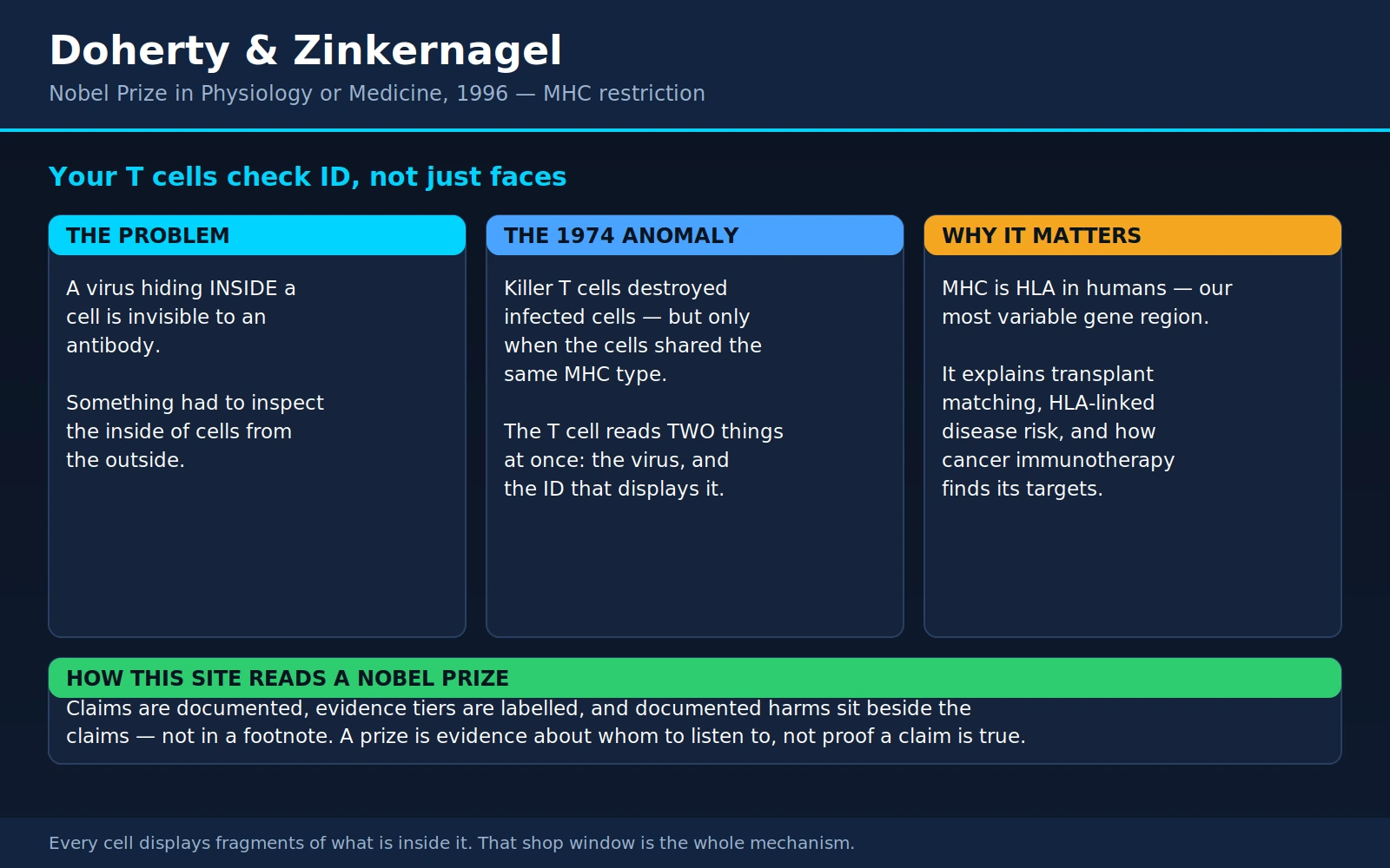
Table of Contents
- The Prize and the Two Men
- The Problem: A Virus Inside a Cell Is Invisible
- The 1974 Experiment, and the Anomaly That Mattered
- What It Means: The Shop Window
- Class I and Class II: Two Different Questions
- MHC Is HLA in Humans — the Most Variable Region We Have
- HLA and Disease: What a Risk Allele Actually Tells You
- Vaccines and Cancer Immunotherapy: Closing the Loop
- Autoimmunity and Molecular Mimicry
- Consumer HLA Testing and "Boost Your T Cells" Claims
- Doherty's Public Science
- Where Mainstream Medicine Agrees — and What Remains Debated
- Key Research Papers
- Connections
- Featured Videos
1. The Prize and the Two Men
The 1996 Nobel Prize in Physiology or Medicine was awarded jointly to Peter C. Doherty and Rolf M. Zinkernagel "for their discoveries concerning the specificity of the cell mediated immune defence." The work they were honoured for had been done twenty-three years earlier, over a few months in 1973, in a shared laboratory at the John Curtin School of Medical Research in Canberra, Australia. One of them was a postdoctoral fellow; the other was a graduate student. Neither had set out to answer the question they ended up answering.
What they found is one of those results that sounds modest in a sentence and turns out to underpin an entire field. A killer T cell cannot recognise a virus on its own. It recognises a fragment of the virus together with a molecule belonging to your own body — and it needs both, at the same time, on the same surface. That requirement, now called MHC restriction, is the reason your immune system can inspect the inside of every cell you own without opening a single one. It is also the reason transplants are rejected, the reason certain drugs cause catastrophic reactions in some people and not others, the reason a coeliac gene test is powerful in one direction and nearly worthless in the other, and the reason some tumours slip past immunotherapy.
Peter Doherty: the veterinarian
Peter Charles Doherty was born in Brisbane, Queensland, on 15 October 1940. He studied veterinary science at the University of Queensland and went to work with livestock — and that background is worth pausing on, because it shaped how he thought about disease for the rest of his career.
Veterinary training is not a lesser version of medical training. It is a differently shaped one. A veterinary student learns the anatomy, physiology and pathology of many species rather than one, which makes it very hard to mistake a quirk of a particular animal for a universal law of biology. Veterinarians deal in herds as well as individuals, so population thinking — how a disease moves through a group, why some animals in the group are spared — is built in from the first year. And a veterinarian who does post-mortems is looking at the whole diseased animal, its tissues, its inflammation, its damage, not a purified molecule in a tube.
Doherty then spent five years in Scotland as a neuropathologist at the Moredun Research Institute near Edinburgh, completing a part-time PhD through the University of Edinburgh Medical School in 1970 on the experimental pathology of louping-ill encephalitis — a tick-borne viral brain disease of sheep. So by the time he arrived in Canberra he had spent years looking down a microscope at infected brains and asking a specific, awkward question: how much of this damage is the virus, and how much of it is the immune response to the virus? That question is the whole of immunopathology, and almost nobody working in molecular immunology at the time was asking it.
In 1971 Doherty took a postdoctoral fellowship at the John Curtin School of Medical Research, part of the Australian National University in Canberra, and began working on lymphocytic choriomeningitis virus (LCMV) in mice — another virus that inflames the lining of the brain, and one where the damage is famously done by the animal's own T cells rather than by the virus itself. He is the first person with a veterinary qualification to win a Nobel Prize.
Rolf Zinkernagel: the physician
Rolf Martin Zinkernagel was born on 6 January 1944 in Riehen, a village just outside Basel, Switzerland. He took his MD at the University of Basel in 1970, trained in surgery and then in experimental medicine, and in 1973 travelled to Canberra to work on cell-mediated immunity to bacterial infection. He enrolled as a PhD student at the Australian National University, finishing in 1975.
By most accounts of the period the pairing was partly an accident of real estate: bench space at John Curtin was tight, and the new Swiss graduate student was put into the laboratory of the Australian postdoc who happened to have room. They were nominally working on different systems — Doherty on a virus, Zinkernagel on a bacterium — and they shared the mouse colony, the radioactivity counter and the cost of running experiments. Within months they were running experiments together.
Afterwards
Doherty went on to the Wistar Institute in Philadelphia and then to St Jude Children's Research Hospital in Memphis, where he chaired the immunology department, before returning to a position at the University of Melbourne. He was named Australian of the Year in 1997. The Peter Doherty Institute for Infection and Immunity in Melbourne — a joint venture of the University of Melbourne and the Royal Melbourne Hospital — carries his name.
Zinkernagel returned to Switzerland, becoming professor of experimental immunology at the University of Zurich and head of its Institute of Experimental Immunology, where he spent the rest of his research career working on the kinetics and localisation of antiviral immunity — the unglamorous question of where and when an immune response has to happen in order to work, rather than merely whether it happens at all.
2. The Problem: A Virus Inside a Cell Is Invisible
To see why the Canberra result mattered, you have to see the hole it filled.
By the early 1970s the antibody half of immunity was well understood in outline. B cells make antibodies; antibodies are Y-shaped proteins that circulate in blood and tissue fluid and bind to specific shapes on the outside of things. An antibody that binds a virus particle floating between cells can neutralise it — block it from docking onto a cell, tag it for a scavenging cell to eat, or trigger the complement cascade to punch holes in it. Emil von Behring's serum therapy in the 1890s had been built on exactly this, decades before anyone knew what an antibody was.
The problem is that antibodies work outdoors. They patrol the extracellular space. A virus spends only a few minutes of its life cycle there. Once a virus has bound a receptor and been drawn inside one of your cells, it is behind your own cell membrane, using your own ribosomes to manufacture more virus, and no antibody can reach it. From the outside, an infected cell looks like any other cell in your body: the same membrane, the same surface proteins, the same everything.
So there had to be a second system, and by the early 1970s its outline was known. Cytotoxic T lymphocytes — killer T cells — are white blood cells that do not make antibodies. They make contact with another cell and induce it to die, punching it with pore-forming perforin and injecting enzymes that trigger the cell's own suicide programme. Kill the factory and you stop the production line. This was clearly a real mechanism, and it was clearly essential: people and animals lacking functional T cells die of viral infections that barely trouble anyone else, a fact the site's severe combined immunodeficiency pages describe in detail.
What nobody knew was how a killer T cell picks the right cell.
The obvious guess was that it worked like an antibody. Perhaps viral proteins get inserted into the infected cell's surface membrane as the virus assembles itself, and the T cell simply binds those the way an antibody binds a free virus. It is a reasonable guess. Some viruses really do stud the cell surface with their own proteins. But it turns out to be wrong as a general explanation — and if it were the answer, there would have been no puzzle and no Nobel Prize.
The real question, stated as sharply as possible, is this: the immune system needs to read the contents of a cell from the outside, without opening the cell, and it needs to do this for every one of the roughly thirty trillion cells in a human body, continuously, for a lifetime. Whatever solves that problem has to be extraordinarily general — it cannot depend on any particular virus cooperating by putting its proteins on the surface — and it has to be cheap enough to run everywhere at once.
3. The 1974 Experiment, and the Anomaly That Mattered
The setup
Three things made the Canberra work possible.
The virus. Lymphocytic choriomeningitis virus is a natural pathogen of mice. Inject it into the brain of an adult mouse and the animal dies of meningitis about a week later — but it dies of its own immune response. Mice without functional T cells do not die; they simply carry the virus. LCMV is therefore the classic model of immunopathology, disease caused by the defence rather than the attacker, which is exactly what Doherty had been studying in sheep.
The mice. Laboratory mouse strains are inbred to the point of being genetically identical within a strain, and each strain has a defined type at the H-2 locus — the mouse version of the major histocompatibility complex, the gene cluster that had been identified decades earlier by George Snell precisely because it determines whether a tissue graft between two mice is accepted or rejected. At the time, H-2 was thought of as the transplant-rejection locus: an evolutionary oddity, a set of genes whose only known job was to cause a problem in an operation that does not occur in nature. Nobody knew what it was for.
The assay. Target cells are loaded with radioactive chromium-51, which stays inside a living cell and leaks out of a dying one. Mix target cells with T cells, wait a few hours, measure the radioactivity in the fluid. More counts in the fluid means more killing. It is a simple, quantitative readout of one cell destroying another.
The result
The experiment was straightforward. Take spleen cells from a mouse infected with LCMV — these contain the killer T cells raised against the virus. Put them onto target cells that are also infected with LCMV. Count the chromium released.
When the T cells and the target cells came from the same mouse strain, the targets were destroyed. That was expected.
When the T cells and the target cells came from different strains — but the targets were infected with the very same virus — nothing happened. The infected cells sat there unharmed.
Uninfected target cells of the matching strain were also left alone. So neither ingredient was sufficient on its own:
- Virus in the target, matching H-2 type → the target is killed.
- Virus in the target, mismatched H-2 type → the target survives.
- No virus, matching H-2 type → the target survives.
Doherty and Zinkernagel narrowed the requirement to the H-2 region itself by using semi-allogeneic mice — F1 hybrids carrying one parental H-2 type from each side — and by comparing strains that differ at H-2 but are otherwise identical, and strains that share H-2 but differ elsewhere. The rule held: what mattered was matching at H-2. Their two-page report appeared in Nature on 19 April 1974.
Why this was an anomaly, not a nuisance
This is the part of the story worth dwelling on, because it is the part that generalises.
Under the intuitive model — T cells see viral antigen the way antibodies do — the strain of the target cell should have been irrelevant. The virus is the same virus. A T cell raised against it should kill anything displaying it. The failure of cross-strain killing was, in that framework, simply a failed experiment: a bad batch of cells, an infection that had not taken, a technical fault of the kind that fills every immunologist's notebook.
Cross-strain experiments of this kind had been done before by other groups, and inconsistent results had been reported. The difference in Canberra was that Doherty and Zinkernagel decided the inconsistency was the finding. A veterinary pathologist's instinct is to take a strain difference seriously — strain and breed differences in susceptibility are ordinary, meaningful facts in animal medicine, not noise. And a well-controlled inbred mouse system made the anomaly reproducible enough to defend.
Six months later, in a second Nature paper, they set out what the result implied: a T cell recognises "altered self" — not the virus, and not the body's own marker, but something that exists only when the two are present together. In a longer 1975 paper in The Lancet they went further and gave the H-2 complex the job it had been missing. Its purpose was not to reject transplants, an event that never happens in the wild. Its purpose was to present the contents of a cell to the immune system. Transplant rejection is a side effect of a surveillance system, not a function.
They also laid out, honestly, two rival explanations of the mechanism and admitted they could not yet distinguish them:
- Altered self — the virus somehow modifies the H-2 molecule itself, so that the T cell sees a single changed shape.
- Dual recognition — the T cell carries two separate receptors, one for the H-2 molecule and one for the virus, and needs both engaged at once.
The answer, when the crystallographers finally supplied it thirteen years later, was a hybrid neither model quite predicted — and better than both. It is described in the next section. The naming stuck too: because the T cell's recognition is restricted to a particular MHC type, the phenomenon has been called MHC restriction ever since.
4. What It Means: The Shop Window
Here is the mechanism in plain language, as it is now understood.
Every nucleated cell in your body continuously advertises a sample of its own contents on its outer surface.
The machinery works like this. A cell is constantly making proteins and constantly destroying them — damaged ones, misfolded ones, ones it no longer needs, and a fraction of perfectly good freshly made ones. The destruction is done by the proteasome, a barrel-shaped protein shredder in the cytoplasm, which chops proteins into short fragments called peptides. A transporter called TAP pumps a selection of those peptides into the endoplasmic reticulum. There they are loaded, one peptide per molecule, into the groove of a freshly made MHC class I molecule. The loaded complex is then shipped to the cell surface and held there, facing outward, holding up its peptide.
A typical cell displays on the order of a hundred thousand MHC class I molecules on its surface, each holding a single short peptide — usually eight to ten amino acids long. Collectively, that display is a rotating sample of everything the cell is currently making inside.
Think of it as a shop window. The shop keeps its stock in the back, out of sight, but it puts a changing selection of that stock in the window facing the street. You do not have to go in and search the storeroom to know roughly what the shop sells. You look at the window.
A CD8 killer T cell walks the street looking in windows. Its T-cell receptor makes brief, low-affinity contact with MHC class I molecules on the cells it passes. If the peptide in the window is an ordinary self peptide — a fragment of actin, of a metabolic enzyme, of haemoglobin — the contact is uninformative and the T cell moves on. If the peptide is a fragment of a viral protein, the T cell binds, holds, and kills the cell.
Now the elegance of the design becomes visible:
- It works on any pathogen, without the pathogen's cooperation. A virus cannot avoid being made inside the cell — making itself is the entire point of a virus. And anything made inside the cell is available to the proteasome. The virus does not have to put proteins on the surface; the cell does that for it, and against it.
- It reads the interior without opening the cell. No immune cell has to breach a membrane to find out what is inside. The information comes out on its own.
- It runs everywhere, all the time, in every cell. The cost is a small tax on protein turnover, which the cell is doing anyway.
- It has a built-in identity check. The frame around the peptide is one of your MHC molecules. A T cell that has learned to read your frames is looking at your cells.
Why the T cell needs to see both — and why that was the surprise
The surprise in 1974 was not that T cells kill infected cells. It was that the identity of the frame is part of what is recognised.
Modern structural work shows why. The T-cell receptor does not have two binding sites, one for peptide and one for MHC. It has one, and it lands across the top of the MHC molecule diagonally, touching the two ridges of the MHC groove on either side and the peptide lying in the middle. What it binds is a composite surface — a shape that exists only when a particular peptide is sitting in a particular MHC molecule. Change the peptide and the shape changes. Change the MHC molecule and the shape changes too, and the ridges no longer sit where the receptor expects them.
That is why the Canberra cross-strain experiment failed. A T cell educated on one mouse's MHC molecules is reading a keyhole shape that a different mouse's MHC molecules do not make, whatever peptide is in it. The virus was there. The reader could not read the frame.
So the true answer is closer to "altered self" than to "dual recognition" — there is one receptor and one composite target, not two handshakes — but the alteration is not a chemical modification of the MHC protein. The alteration is a different peptide sitting in a groove.
The picture that settled it: 1987
In October 1987, Pamela Bjorkman, working with Don Wiley and Jack Strominger at Harvard, published the crystal structure of a human class I molecule, HLA-A2, in two companion papers in the same issue of Nature.
The first paper reported the structure itself. The molecule turned out to have a distinctive shape at its top: two long alpha-helices running roughly parallel, sitting on a floor of beta-pleated sheet, forming a long groove about 25 Ångströms end to end — and pointing away from the cell, straight up at whatever might come along to inspect it. Nothing about the amino-acid sequence had predicted it.
Then came the detail that made immunologists sit up. The groove was not empty. The electron-density maps showed extra material lying in it that could not be accounted for by the HLA protein itself, and it was too poorly resolved to be one defined molecule. That was the correct signature of a mixture — the natural set of many different peptides that the cell had loaded before the protein was purified. The groove had been photographed with its cargo still in it.
The second paper took the sequences of many different HLA alleles, mapped the positions where they differ onto the new structure, and found that the variable positions clustered in exactly two places: the floor and walls of the groove, and the upward-facing surfaces of the two helices. In other words, the parts of HLA that differ from person to person are precisely the parts that (a) determine which peptides can be held and (b) face the T-cell receptor.
Everything Doherty and Zinkernagel had inferred from mouse-strain mismatches thirteen years earlier was now visible as a shape. Because both papers are frequently cited interchangeably, it is worth keeping them straight: the paper at pages 506–512 is the structure determination; the paper at pages 512–518 is the interpretation of the groove and the mapping of T-cell recognition regions onto it.
5. Class I and Class II: Two Different Questions
There are two families of MHC molecule, and the cleanest way to keep them apart is to notice that they answer two different questions.
MHC class I — "What is happening inside this cell?"
- Where: on essentially every nucleated cell in the body.
- Loaded from: the cytosol — proteins the cell itself is making.
- Read by: CD8 T cells (killer T cells). The CD8 molecule physically grips a constant part of class I, which is what pairs the two.
- Consequence of a bad answer: the cell is destroyed.
Class I is surveillance of the self. The cell being inspected is the cell being reported on, and it reports on itself.
Two footnotes. Mature red blood cells have no nucleus and essentially no MHC class I, which is one reason a blood-stage malaria parasite is so hard for killer T cells to reach, and part of why red cells are matched by ABO and Rh rather than by HLA. And class I levels vary: interferon — the alarm signal a virus-infected cell releases — drives class I expression up, so an infected tissue turns up its own display volume.
MHC class II — "What did we find out there?"
- Where: only on professional antigen-presenting cells — dendritic cells, macrophages and B cells.
- Loaded from: endosomes — material the cell swallowed from outside itself and broke down internally.
- Read by: CD4 T cells (helper T cells), whose CD4 molecule grips class I's counterpart on class II.
- Consequence of a bad answer: instructions. Help B cells switch antibody class and mature their affinity; license killer T cells; summon and activate macrophages; shape the whole downstream response.
Class II is not surveillance of the presenting cell. It is a report on the neighbourhood. A macrophage that eats a bacterium is not saying "I am infected"; it is saying "I found this."
The specialists at that job are dendritic cells — the cell type Ralph Steinman discovered in 1973, the same year the Canberra experiments were running, and for which he shared the 2011 Nobel Prize. A dendritic cell samples a tissue, swallows what it finds, migrates to a lymph node, and there presents the results on class II to the CD4 T cells waiting in it. That journey is how a local infection becomes a system-wide response, and it is covered on the site's page for Beutler, Hoffmann and Steinman.
The exception that matters: cross-presentation
The tidy split above has one important leak, and it is deliberate. Dendritic cells can route material they swallowed from outside into the class I pathway — a manoeuvre called cross-presentation.
Without it there would be a gap in the defence. If a virus infects only liver cells, and no dendritic cell ever gets infected, then no dendritic cell would ever display a viral peptide on class I, and killer T cells specific for that virus would never be activated in the first place. Cross-presentation closes that gap: a dendritic cell that eats the debris of an infected liver cell can present the viral peptides on its own class I and prime the killer response. As section 8 explains, it is also the loophole that allows some vaccines to generate T-cell immunity they would not otherwise produce.
6. MHC Is HLA in Humans — the Most Variable Region We Have
In mice the locus is called H-2. In humans it is called HLA — human leukocyte antigen — because it was first detected on white blood cells, in the blood of people who had received multiple transfusions or been through several pregnancies and had made antibodies against other people's cells. Jean Dausset described the first such antigen in the late 1950s, work for which he shared the 1980 Nobel Prize with George Snell and Baruj Benacerraf. The name is a historical accident: HLA molecules are not confined to leukocytes and are not antigens in any interesting sense. They are peptide-display molecules that happened to be discovered by the immune reactions they provoke.
The genes sit close together on the short arm of chromosome 6, in a region of roughly four million base pairs. The main class I genes are HLA-A, HLA-B and HLA-C; the main class II genes are HLA-DR, HLA-DQ and HLA-DP. You inherit the whole block as a unit from each parent — a haplotype — because the genes are so close together that they are rarely separated by recombination.
The most polymorphic region in the human genome
Most human genes come in a handful of common versions. HLA genes do not. More than 40,000 distinct HLA alleles have been catalogued in the international IPD-IMGT/HLA database, the great majority of them in the six genes above. HLA-B alone accounts for thousands. This is, by a wide margin, the most variable region of the human genome.
That is not an accident, and the reason follows directly from what Doherty and Zinkernagel discovered.
Since an HLA molecule's groove determines which peptides it can hold, your HLA type determines which fragments of a pathogen your T cells can ever be shown. A virus that mutates the few peptides your particular HLA molecules present well becomes, to your immune system, considerably harder to see.
Now scale that up. Imagine a population in which everyone had identical HLA. A single pathogen mutation that escaped that one presentation repertoire would escape it in everybody at once. One lucky virus could sweep the entire population. That is not a hypothetical worry; it is what happens in genetically uniform livestock and in endangered species with collapsed genetic diversity.
Now imagine the opposite: a population in which almost everyone has a different HLA combination. A pathogen mutation that hides from one person's presentation repertoire is still visible to the next person's. There is no single escape mutation that works on everyone. The population as a whole cannot be swept, even though any given individual can be unlucky.
HLA diversity is therefore best understood as insurance held at the level of the population rather than the individual. It is maintained by a form of balancing selection: rare HLA types are favoured precisely because circulating pathogens have had less opportunity to adapt to them, so rare types are protected from being lost, and diversity accumulates over millions of years. Some HLA lineages are older than our species — humans and chimpanzees share allelic lineages inherited from a common ancestor.
Doherty and Zinkernagel themselves demonstrated the individual-level version of this in 1975, in a paper whose title states the finding: mice heterozygous at the H-2 complex — carrying a different allele from each parent, and therefore able to present a wider range of peptides — showed enhanced immunological surveillance compared with homozygotes. Two different grooves catch more peptides than two copies of the same groove.
What that variability costs: transplantation
The benefit of HLA diversity is paid for at the operating table.
Transplant rejection happens because HLA molecules are the single most conspicuous difference between two people's cells. A recipient's T cells, educated on the recipient's own HLA, encounter a donor organ covered in foreign HLA and react to it vigorously — an unusually large fraction of any person's T-cell repertoire will respond to any given foreign HLA molecule. That is why matching matters, and why the closer the match, the better the outcome and the less immunosuppression required.
Three consequences follow, and all of them are practical:
- Siblings have a one-in-four chance of being HLA-identical. Each parent has two haplotypes; a child gets one from each; there are four possible combinations; two siblings match when they drew the same pair. This is why sibling donors are sought first for bone marrow transplantation, and why the first successful kidney transplant was between identical twins.
- Unrelated matches are genuinely rare. With tens of thousands of alleles and six loci to match at high resolution, the number of possible types runs to astronomical figures. Finding an unrelated stranger who matches you closely at all six is a lottery, which is why donor registries need to be enormous.
- Matching is ancestry-linked, and that creates an equity problem. HLA haplotype frequencies differ substantially between populations, because the pathogen histories of those populations differed. Your best chance of a close match is among people with similar ancestry. When a donor registry is dominated by one ancestral group, patients from other groups — and particularly patients of mixed ancestry, whose haplotype combinations may be rare in every registry — wait longer and are matched less well. This is not a biological unfairness; it is a sampling one, and it is fixable by recruiting broadly.
The Murray & Thomas page covers the registry-diversity problem, and the workarounds — including haploidentical transplantation, which deliberately uses a half-matched family donor and manages the mismatch pharmacologically — in detail.
7. HLA and Disease: What a Risk Allele Actually Tells You
Read this paragraph before anything else in this section. Every association described below is a susceptibility marker. It is a statement about how a disease is distributed across a population, not a verdict on any individual. For almost every HLA-disease association known, the large majority of people carrying the risk allele never develop the disease. An HLA result changes a probability. It does not make a diagnosis. If you have seen one of these alleles on a consumer genetics report or a lab printout, that sentence is the most important thing on this page for you.
With that said, HLA turns up in association studies more often, and more strongly, than any other region of the human genome. The reason is the mechanism this whole page is about: HLA molecules choose which peptides get shown to T cells. A small change in the shape of a groove changes which self-peptides and which pathogen-peptides are displayed — and both of those can matter for disease.
HLA-B27 and ankylosing spondylitis
The first of these associations, and still the most famous, was published in 1973: David Brewerton's group in London found that the great majority of patients with ankylosing spondylitis — an inflammatory arthritis that fuses the spine and sacroiliac joints — carried a single HLA class I allele then written HL-A 27. It was a startling result at the time, and it was replicated within months.
The numbers, which the site covers in full on HLA-B27 Explained:
- 90–95% of people with ankylosing spondylitis carry HLA-B27.
- 6–8% of people of European ancestry carry it, with the figure varying enormously by ancestry — roughly 4% of African Americans, about 1% of Japanese, and up to 50% in some Indigenous populations of the Pacific Northwest.
- A carrier's lifetime risk of developing ankylosing spondylitis is only about 5–16%. Put the other way round: 85–95% of HLA-B27 carriers never develop spondyloarthritis at all.
So what is a B27 test good for? It is good for shifting a probability you were already entertaining. In a 30-year-old with months of inflammatory back pain — worse with rest, better with movement, more than half an hour of morning stiffness, waking in the second half of the night — a positive B27 substantially raises the probability of axial spondyloarthritis and a negative one lowers it, and that genuinely helps a rheumatologist decide about imaging and treatment. In a person with no symptoms, a positive B27 means almost nothing, and testing for it is not recommended. The clinical picture is doing the work; the allele is a modifier.
It is worth being honest that after fifty years nobody is certain why B27 does this. Three hypotheses remain live: that B27 presents a particular "arthritogenic" self-peptide resembling a bacterial one; that B27 has an unusual tendency to misfold in the endoplasmic reticulum and trigger a stress response; and that B27 forms unusual free heavy-chain dimers on the cell surface that engage other immune receptors. The association is beyond dispute; the mechanism is not. Sieper and Poddubnyy's Lancet review of axial spondyloarthritis (2017 Jul 1;390(10089):73–84) surveys the current state of play.
HLA-DQ2/DQ8 and coeliac disease — the negative result is the useful one
This is the association most worth understanding properly, because it is common, it is frequently tested, and it is almost universally misread.
Coeliac disease is an immune reaction to gluten peptides that damages the lining of the small intestine. The presentation step is unusually well worked out, and it is a textbook illustration of everything above. Gluten is rich in the amino acid glutamine. In the gut wall, an enzyme called tissue transglutaminase converts some of those glutamines into glutamate, which carries a negative charge. The grooves of HLA-DQ2 and HLA-DQ8 happen to have positively charged pockets that bind negatively charged residues especially well. So the deamidated gluten peptide slots into the DQ2 or DQ8 groove with high affinity, gets displayed to CD4 helper T cells, and the response follows. An HLA molecule with a differently shaped groove simply cannot present the peptide well enough to start the reaction.
Which produces this arithmetic:
- Essentially everyone with coeliac disease carries DQ2 or DQ8. The condition is close to impossible without one of them.
- But 30–40% of people of European ancestry carry DQ2 or DQ8 too — and only about 1% of the population has coeliac disease.
So a positive HLA-DQ result is nearly worthless on its own. It puts you in the same group as roughly a third of everyone around you, the overwhelming majority of whom will never develop coeliac disease. It is not a diagnosis, it is not a reason to go gluten-free, and it is not "the coeliac gene."
A negative result is the powerful one. If you carry neither DQ2 nor DQ8, coeliac disease is essentially off the table — permanently. A test with a very high negative predictive value is exactly the tool you want for ruling something out, and this is one of the best in clinical medicine.
The prospective evidence comes from a Dutch cohort published in Annals of Internal Medicine in 2007. Hadithi and colleagues followed 463 consecutive patients referred for small-bowel biopsy for suspected coeliac disease, of whom 16 turned out to have it (a prevalence of 3.46%). HLA-DQ typing had a sensitivity of 100% (95% CI 79–100%) and a negative predictive value of 100% (95% CI 98.6–100%); testing negative for both types gave a negative likelihood ratio of 0.00 and a post-test probability of coeliac disease of 0% (95% CI 0–1.4%). The authors also noted, correctly, that 16 cases is a small number and the confidence intervals are correspondingly wide — the upper bound on the residual probability is about 1.4%, not literally zero. It is a rule-out test, not a metaphysical certainty. But as rule-out tests go it is excellent, and it has been consistently supported since.
Where an HLA-DQ test genuinely earns its place:
- Someone already eating gluten-free whose antibody tests have normalised, and who does not want to endure a weeks-long gluten challenge to be tested properly. A negative HLA type ends the question without any challenge at all, because HLA does not change with diet.
- First-degree relatives of a coeliac patient deciding whether they need periodic antibody screening for the rest of their lives. A negative HLA type retires them from surveillance permanently.
- People with conditions carrying elevated coeliac risk — type 1 diabetes, autoimmune thyroid disease, Down syndrome, Turner syndrome — where lifelong repeated screening is otherwise recommended.
- Ambiguous biopsy or serology results where the question is whether coeliac disease is even biologically possible in this person.
Where it is misused: sold direct to consumers as a "coeliac gene test", with a positive result read as a diagnosis, or as licence to start a gluten-free diet. The correct step after a positive HLA-DQ result — or after any suspicion of coeliac disease — is coeliac serology (tTG-IgA) while still eating gluten, followed by biopsy if indicated. Starting the diet first destroys your ability to be tested and can leave the question unanswerable for years. The site's HLA-DQ2 and DQ8 genetic testing page and the main coeliac disease page walk through the sequence.
HLA-B*57:01 and abacavir — a test that is genuinely mandatory
Abacavir is an antiretroviral drug used in HIV treatment. In a minority of patients it causes a severe, sometimes fatal hypersensitivity reaction — fever, rash, gastrointestinal and respiratory symptoms — that gets worse, and can be lethal, on re-exposure. In the early 2000s the reaction was found to be strongly associated with a single class I allele, HLA-B*57:01. The mechanism is now well described: abacavir binds inside the B*57:01 groove itself and changes its shape, so that the molecule starts presenting a set of self-peptides it would never normally hold. The patient's T cells see a self cell displaying peptides they have never been tolerised to, and attack. It is, quite literally, "altered self" in the Doherty–Zinkernagel sense — with a drug doing the altering.
The definitive test of screening was PREDICT-1, published in the New England Journal of Medicine in 2008: a double-blind randomised trial of 1,956 patients across 19 countries, assigned either to prospective HLA-B*57:01 screening with carriers excluded from abacavir, or to standard care without screening. The results:
- Prevalence of HLA-B*57:01 in the cohort: 5.6%.
- Immunologically confirmed hypersensitivity reaction: 0% in the screened group versus 2.7% in the control group (P<0.001).
- Negative predictive value 100%; positive predictive value 47.9%.
- Clinically suspected hypersensitivity: 3.4% screened versus 7.8% unscreened (P<0.001).
Two honest readings of those numbers. First, screening did not merely reduce the reaction — it eliminated the immunologically confirmed form. Second, the positive predictive value of about 48% means that roughly half of carriers who took the drug would not have reacted. Medicine nonetheless withholds abacavir from all carriers, because the reaction is dangerous, alternative drugs exist, and there is no way to identify in advance which half of the carriers would be spared. That is a sensible asymmetry, not a contradiction, and it is worth understanding if you are ever told you "can't have" a drug because of a genotype.
Pre-treatment HLA-B*57:01 testing is now standard of care wherever abacavir is prescribed, and PREDICT-1 is routinely cited as the trial that proved a pharmacogenetic test could prevent a specific drug toxicity in practice rather than in principle.
HLA-B*15:02 and carbamazepine — and why ancestry is part of the rule
Carbamazepine is an anticonvulsant and mood stabiliser. It is also, in parts of Southeast Asia, the leading cause of Stevens–Johnson syndrome and toxic epidermal necrolysis — catastrophic reactions in which the skin and mucous membranes blister and detach, with substantial mortality. In 2004, Chung and colleagues reported in Nature an association so strong it barely needed statistics: in their Han Chinese cohort, essentially every patient with carbamazepine-induced Stevens–Johnson syndrome carried HLA-B*15:02 (2004 Apr 1;428(6982):486).
The prospective test came in 2011. Chen and colleagues recruited 4,877 carbamazepine-naive people across 23 hospitals in Taiwan and genotyped them all. The 7.7% who carried B*15:02 were advised not to take carbamazepine and given alternatives; the 92.3% who did not carry it took the drug. No case of Stevens–Johnson syndrome or toxic epidermal necrolysis occurred in any B*15:02-negative participant, against roughly ten cases expected from the historical incidence in that population (P<0.001).
The reason the recommendation is ancestry-targeted rather than universal is simply allele frequency. HLA-B*15:02 is common in Han Chinese, Thai, Malay and some South Asian populations, and rare in people of European or Japanese ancestry — so screening everyone in Europe would find almost nothing and prevent almost nothing, while screening in Taiwan or Thailand prevents a lot. Regulatory guidance therefore recommends B*15:02 testing before starting carbamazepine in people of Southeast Asian ancestry. (A separate allele, HLA-A*31:01, is associated with a broader range of carbamazepine hypersensitivity reactions in European and Japanese populations, and appears in some prescribing guidance as well.)
This is a case where ancestry-specific medicine is doing real, defensible work — not as a proxy for anything social, but because allele frequencies genuinely differ and the test's yield follows them. It is also a reminder that a drug reaction attributed to bad luck may have a readable genetic basis. The site's drug allergy page covers the broader picture.
Narcolepsy and HLA-DQB1*06:02 — the strongest association known
Narcolepsy type 1 — excessive daytime sleepiness with cataplexy, the sudden loss of muscle tone triggered by laughter or strong emotion — has the tightest HLA association in medicine. The allele HLA-DQB1*06:02 is present in 95–98% of people with narcolepsy type 1.
And it is present in about 25% of the general population. One person in four carries it. Narcolepsy type 1 affects roughly one person in two thousand. The allele is close to necessary and nowhere near sufficient — which is exactly why HLA typing is not used to diagnose narcolepsy.
Mignot's 2001 study of 420 people with narcolepsy-cataplexy and 1,087 controls across three ethnic groups found, as expected, that almost all patients carried DQB1*06:02, and went further: nine other class II alleles carried on the other chromosome modified the risk, some raising it and three lowering it. Risk is a property of the pair, not of one allele.
What makes narcolepsy type 1 so interesting is that the HLA association eventually explained itself. In 2000, Nishino and colleagues reported in The Lancet that hypocretin (orexin) was undetectable in the cerebrospinal fluid of most people with narcolepsy (2000 Jan 1;355(9197):39–40), and post-mortem work showed the loss of 85–95% of the small population of hypocretin-producing neurons in the hypothalamus — with the surrounding hypothalamus intact. A tightly targeted class II association plus the selective destruction of one small neuronal population is the signature of an autoimmune attack, and that is now the mainstream model. The site's narcolepsy page covers the diagnosis, including why a cerebrospinal fluid hypocretin measurement below 110 pg/mL is near-diagnostic where an HLA type is not.
Others you may encounter
- Type 1 diabetes — DR3-DQ2 and DR4-DQ8 haplotypes; the strongest genetic contribution to the disease, and overlapping with the coeliac alleles, which is why the two conditions cluster.
- Rheumatoid arthritis — the "shared epitope", a common amino-acid motif in the DRB1 groove found across several otherwise unrelated alleles.
- Multiple sclerosis — DRB1*15:01, the dominant genetic risk factor in populations of European ancestry.
- Psoriasis — HLA-C*06:02, strongest for early-onset disease.
- Behçet's disease — HLA-B*51, most strongly along the historical Silk Road populations.
Every one of these behaves the same way as the examples above. A meaningful shift in population-level risk; a very large majority of carriers who never develop anything; no diagnostic value on its own. Dendrou and colleagues' 2018 review in Nature Reviews Immunology is the best single survey of the whole landscape.
To repeat the point that section opened with: association is not diagnosis. A risk allele describes the distribution of a disease across a population. Your own status is settled by whether you have the disease.
8. Vaccines and Cancer Immunotherapy: Closing the Loop
Two practical fields run directly out of the 1974 result, and they turn out to be the same idea seen from opposite ends.
Why vaccine platforms behave differently
A vaccine's job is to teach the immune system in advance. But which part of the immune system it teaches depends on where the antigen ends up inside a cell — which is a direct consequence of the class I / class II split.
Protein subunit vaccines — a purified viral protein plus an adjuvant — deliver antigen to the outside of your cells. Antigen-presenting cells swallow it, break it down in endosomes, and load it onto class II. That drives CD4 helper responses and, through them, excellent antibody production. It is a strong platform when the goal is to neutralise a pathogen before it gets into a cell. What it does not naturally do is put antigen into the cytosol, which is where the class I pathway samples — so subunit vaccines tend to generate weaker CD8 killer T-cell responses.
Live-attenuated vaccines — a weakened but replicating virus — actually infect cells. The virus makes its proteins in the cytosol of the vaccinated person's own cells, which is exactly what the proteasome samples and the class I pathway displays. So a live-attenuated vaccine naturally generates both antibody and killer T-cell immunity, which is part of why it tends to produce long-lived protection from a small number of doses.
mRNA vaccines get to the same place by a different route. The mRNA is delivered into the cytosol, where the recipient's own ribosomes translate it. The resulting protein is newly made, inside the cell, in the cytosol — indistinguishable from a viral protein as far as the antigen-processing machinery is concerned. So it feeds the class I pathway and generates CD8 responses, while secreted and surface-displayed forms of the protein also drive antibody. The site's page on Karikó and Weissman covers how the platform was made to work; the point here is why where the protein is synthesised determines what kind of immunity you get.
Two honest qualifications. First, this is a tendency, not a law: adjuvants and cross-presentation let some subunit vaccines generate real T-cell responses, and viral-vector vaccines occupy a middle position. Second, more T-cell response is not automatically better — for many diseases neutralising antibody is what correlates with protection, and a vaccine that produces it reliably and safely is the right vaccine regardless of its T-cell profile.
Where killer T cells earn their keep is in durability and breadth. Antibody titres fall over months, and a variant that changes a few surface residues can escape antibody neutralisation across an entire population at once. T-cell recognition is harder to escape, for a reason that goes straight back to section 6: each person's HLA set presents a different subset of peptides drawn from the whole pathogen proteome. There is no single mutation that hides a virus from everyone's T cells simultaneously. Population-level HLA diversity is a defence that operates at the level of the species, and a virus cannot mutate its way around all of it.
Cancer: the tumour closes the shop window
Now run the same mechanism the other way.
A tumour cell is one of your own cells with a damaged genome. Mutated genes make mutated proteins; the proteasome shreds mutated proteins into mutated peptides; those peptides get loaded onto class I and displayed on the tumour cell's surface. These are neoantigens — peptide sequences that exist nowhere else in your body and that no T cell was ever tolerised to. A tumour advertising neoantigens on class I is, in principle, a legitimate target for exactly the killing machinery Doherty and Zinkernagel described.
That principle is the foundation of checkpoint inhibitor immunotherapy. Checkpoint inhibitors do not teach T cells anything new; they release the brakes on T cells that are already there and already recognising the tumour. That is why they work best in cancers with high mutation loads — melanoma, lung cancer, mismatch-repair-deficient tumours — because more mutations means more neoantigens means more for a T cell to see. The story of how those brakes were found is on the Allison & Honjo page.
Which sets up the elegant, unhappy closing of the loop. If you are a tumour under attack by T cells that recognise peptide-on-class-I, the way to survive is to stop displaying.
And that is exactly what tumours do. The routes include:
- Losing beta-2-microglobulin (B2M) — the small invariant light chain that every class I molecule needs in order to fold and reach the surface. Lose B2M and all class I disappears at once, whatever the HLA type.
- Losing the loading machinery — the TAP transporters, the peptide-loading complex, the immunoproteasome subunits.
- Loss of heterozygosity at the HLA locus — deleting one parental haplotype, which narrows the set of peptides the tumour can present without abolishing display entirely.
- Transcriptional silencing — simply turning class I expression down, often reversibly, sometimes by disabling the interferon signalling that would normally turn it up.
The clinical evidence is direct. Zaretsky and colleagues, in the New England Journal of Medicine in 2016, sequenced paired biopsies from four melanoma patients who had responded to pembrolizumab and then relapsed months to years later. Two had acquired loss-of-function mutations in JAK1 or JAK2, crippling their response to interferon-gamma; a third had a truncating mutation in B2M that caused loss of surface class I (2016 Sep 1;375(9):819–829). Sade-Feldman and colleagues then examined this at cohort scale in Nature Communications in 2017: B2M point mutations, deletions or loss of heterozygosity in 29.4% of patients with progressing disease, with B2M loss of heterozygosity enriched roughly threefold in non-responders (~30%) compared with responders (~10%) and associated with poorer overall survival — and loss of both copies of B2M found only in non-responders. Dhatchinamoorthy and colleagues' 2021 review in Frontiers in Immunology (2021 Mar 9;12:636568) collects the full range of escape routes.
Forty-three years after two researchers in Canberra worked out that a killer T cell must see a peptide inside a self-frame, the most consequential resistance mechanism in cancer immunotherapy turns out to be a tumour taking down the frame. A tumour hides by closing the shop window.
There is one more turn, and it is built into the system. Natural killer cells are triggered by the absence of class I — the "missing self" principle. A cell that has stopped displaying class I is, to an NK cell, suspicious by that very fact. That is presumably why the evolutionary arms race did not simply end with every virus and every tumour switching class I off. It is also why NK-directed approaches are of active interest for class-I-deficient tumours — though that remains a research programme, not established treatment, and tumours have counter-moves against NK cells too.
9. Autoimmunity and Molecular Mimicry
A system built on displaying your own peptides to cells licensed to kill has an obvious failure mode.
The safeguards are real and largely effective. Developing T cells are tested in the thymus against self-peptides displayed on thymic MHC, and those that bind self too strongly are deleted — a process that includes an ingenious trick in which the thymus, driven by the AIRE gene, displays proteins belonging to distant organs so that T cells can be screened against tissues they will not meet for years. This is the machinery Burnet and Medawar predicted decades before anyone could see it. Whatever escapes deletion is then kept in check in the periphery by regulatory T cells, which actively suppress self-reactive responses.
Autoimmune disease is what happens when both layers fail for a particular self-antigen.
Molecular mimicry: what it is, and how much it explains
Molecular mimicry is the hypothesis that a pathogen peptide can resemble a self peptide closely enough that T cells (or antibodies) raised against the infection cross-react with the body's own tissue. It is a natural prediction of everything on this page: since a T cell recognises a short peptide in a groove, and short peptides are only eight to fifteen amino acids long, coincidental resemblance between a microbial peptide and a human one is not merely possible but statistically inevitable.
Here is the honest status of the idea. Molecular mimicry is a well-supported mechanism in specific cases, and it is a plausible but unproven general explanation for autoimmunity as a whole.
The strongest cases:
- Rheumatic fever after group A streptococcal infection — antibodies against streptococcal M protein cross-react with cardiac myosin and heart valve tissue. This is the classical, best-established example.
- Guillain-Barré syndrome after Campylobacter jejuni infection — the bacterium's surface carbohydrates resemble gangliosides on peripheral nerves.
- Multiple sclerosis and Epstein-Barr virus — the epidemiological link is now very strong, and cross-reactivity between an EBV protein and a central nervous system protein has been described, though the full causal chain is still being worked out.
Against that, most autoimmune diseases have no single established trigger. The realistic picture is layered: a permissive HLA type, plus other genetic variants of small individual effect, plus something environmental — an infection, a drug, a change in the microbiome, a hormonal shift — which is often never identified, and may not be the same thing in two people with the same diagnosis. Claims that a particular infection or exposure "causes" a particular autoimmune disease should be read against that background. Cusick and colleagues' review in Clinical Reviews in Allergy & Immunology (2012 Feb;42(1):102–111) is a fair-minded survey of both the evidence and its limits.
Why HLA keeps showing up in autoimmunity
The mechanistic logic is simple, even where the details are not. A class II molecule that binds a particular self-peptide well is a class II molecule that can present that self-peptide to a CD4 T cell. If a self-reactive T cell survived thymic selection, the HLA type that displays its target antigen best is the HLA type that gives it the best chance of being activated.
Coeliac disease is the one case where this has been followed all the way through — a known trigger antigen (gluten), a known enzymatic modification (transglutaminase deamidation), a known structural reason why DQ2 and DQ8 present the modified peptide well, and a known T-cell response. That completeness is why coeliac disease is the model system for HLA-associated autoimmunity, and why the DQ2/DQ8 test behaves so cleanly. Nothing else is worked out to that depth. For ankylosing spondylitis, fifty years after the association was found, we still do not know the answer.
10. Consumer HLA Testing and "Boost Your T Cells" Claims
Direct-to-consumer genotyping reports increasingly include HLA-linked traits, and third-party services will re-analyse a raw data file and hand back a list of "immune risk variants." This section is about what to do with that.
First: know what you actually got
A consumer genotyping array does not usually type HLA directly. It reads a set of tag SNPs and imputes the likely HLA allele from patterns of linkage. Imputation is a genuine and useful technique, but its accuracy varies by allele and, importantly, by ancestry — it is generally most accurate for the populations that dominated the reference panels it was trained on, and less accurate elsewhere. High-resolution clinical HLA typing, of the sort used for transplant matching or before prescribing abacavir, is a different and more rigorous test.
So a consumer HLA result is a probabilistic inference from a research-grade array, reported outside any clinical context. Treat it as a prompt to ask a question, never as an answer.
Second: three questions that defuse almost any risk allele
When a report tells you that you carry a risk allele for something, ask:
- How common is this allele in people like me? If a third of the population carries it, it is not telling you much about you.
- How common is the disease? A tripled risk of something that affects one person in ten thousand is still one person in about three thousand.
- What fraction of carriers actually develop the disease? This is the number that matters most, and it is the one consumer reports are least likely to put in front of you. For essentially every HLA association in this article, the answer is "a small minority."
Two worked examples.
You learn you carry HLA-DQ2. So do roughly one in three people of European ancestry. About 1% of the population has coeliac disease. Your risk is genuinely higher than a non-carrier's — because non-carriers have essentially no risk — but you remain far more likely not to have coeliac disease than to have it. If you have symptoms, the right next step is tTG-IgA serology while still eating gluten. If you have no symptoms, the right next step is nothing. Starting a gluten-free diet on the strength of a genotype is the one clearly wrong move, because it makes the real test uninterpretable.
You learn you carry HLA-B27 and you feel fine. Between 85% and 95% of carriers never develop spondyloarthritis. There is nothing to monitor, no screening programme to enter, and no treatment to start. What the result buys you is a single piece of information to keep in your back pocket: if you ever develop persistent back pain that is worse with rest and better with movement, with more than half an hour of morning stiffness, beginning before age 45 — that is the moment to mention the B27 result to a doctor, because in that context it genuinely changes the picture.
Third: the genuine exceptions
Two HLA tests are standard of care before prescribing, and they are exceptions for a specific reason: a decision is attached to the result.
- HLA-B*57:01 before abacavir — universal, mandatory, validated in a randomised trial (PREDICT-1). Carriers get a different drug.
- HLA-B*15:02 before carbamazepine in people of Southeast Asian ancestry — validated prospectively in nearly 5,000 people in Taiwan. Carriers get a different drug. (HLA-A*31:01 appears in some guidance for European and Japanese populations.)
What makes these different from the rest is not that the associations are stronger in a statistical sense — the narcolepsy association is stronger than either. It is that there is a prospective, actionable choice: a drug that has not been given yet, an alternative that exists, and evidence that acting on the genotype prevents harm. Absent that structure, an HLA result changes nothing you would do.
High-resolution HLA typing for transplant or stem-cell donation is likewise a real clinical test with a real decision attached — and it is a different test from a consumer array.
Fourth: "boost your T cells"
Search engines will offer you supplements to strengthen T-cell immunity. Here is the position, tiered.
- Established. T-cell development and function genuinely depend on adequate nutrition. Protein-energy malnutrition causes thymic atrophy and impaired T-cell responses. Deficiencies of zinc, vitamin A, vitamin D, iron and selenium each impair aspects of cellular immunity. So do chronic sleep deprivation, uncontrolled diabetes, heavy alcohol use, and untreated chronic illness.
- Established. Correcting a real deficiency restores function toward normal. This is the whole of the honest case for nutritional support of immunity, and it is a good case — it is simply a case about deficiency, not about supplementation on top of sufficiency.
- Not supported. That any supplement improves T-cell surveillance in a well-nourished person. There is no evidence for it, and no plausible mechanism by which a nutrient already present in sufficient quantity would further improve antigen presentation or T-cell scanning.
- Not supported, and conceptually confused. The idea of "boosting" immunity as a general goal. The system Doherty and Zinkernagel described is a discrimination system. Its excellence lies in telling infected cells from healthy ones and self from non-self, not in intensity. An immune system turned up indiscriminately is not a stronger immune system — it is autoimmunity, allergy, and cytokine-driven tissue damage. Much of the harm in severe infection comes from the response rather than the pathogen, which is precisely what Doherty was studying in mice in 1973.
The useful framing is competence, not power. Feed it adequately, sleep, treat the chronic conditions that impair it, vaccinate so that it has been taught in advance, and let it discriminate.
11. Doherty's Public Science
Doherty has spent much of the past three decades writing and speaking for general readers, which is unusual among Nobel laureates and has been done consistently rather than occasionally.
He was named Australian of the Year in 1997, the year after the prize, and used the platform for science communication rather than ceremony. His books for non-specialists include The Beginner's Guide to Winning the Nobel Prize (2005), a deliberately unsentimental account of what a research career actually consists of; A Light History of Hot Air; Sentinel Chickens: What Birds Tell Us About Our Health and Our World, on birds as indicators of environmental and infectious threats; Pandemics: What Everyone Needs to Know (2013); The Knowledge Wars (2015), on evidence, expertise and public argument; The Incidental Tourist; and An Insider's Plague Year (2021), a month-by-month account of the first year of COVID-19 as he experienced it.
The consistent theme across all of it is pandemic preparedness — and specifically the argument that preparedness is mostly boring and mostly has to be paid for in advance: sustained surveillance of animal and human populations, funded basic science with no immediate application, vaccine platforms developed and tested before there is an emergency to use them on, manufacturing capacity that exists before it is needed, and a public with enough scientific literacy to follow an argument that changes as evidence arrives. Pandemics: What Everyone Needs to Know was published seven years before COVID-19.
The Peter Doherty Institute for Infection and Immunity in Melbourne, which bears his name, was the first laboratory outside China to grow SARS-CoV-2 from a patient sample, in late January 2020, and shared the isolate with reference laboratories and culture collections internationally within about 24 hours — an unusually direct illustration of the case he had been making for years about surveillance capacity and open sharing.
Zinkernagel's post-Nobel career was more conventionally academic. He led the Institute of Experimental Immunology at the University of Zurich until his retirement, continuing to work on the timing, location and kinetics of antiviral immune responses — and arguing, sometimes against the grain, that where and when an immune response occurs matters as much as its specificity.
12. Where Mainstream Medicine Agrees — and What Remains Debated
Settled
- MHC restriction is a fact. T cells recognise peptide presented by MHC, not free antigen. It is textbook material, structurally proven, and has been confirmed in every system examined for fifty years.
- The class I / class II division of labour. Class I samples the cytosol on essentially all nucleated cells and is read by CD8 T cells; class II samples endocytosed material on professional antigen-presenting cells and is read by CD4 T cells. Cross-presentation is a real and important exception.
- HLA is the most polymorphic region of the human genome, its diversity is maintained by pathogen-driven balancing selection, and HLA matching materially affects transplant outcome.
- Specific HLA-drug associations are strong enough to be mandatory pre-prescription tests — HLA-B*57:01 before abacavir universally, HLA-B*15:02 before carbamazepine in people of Southeast Asian ancestry.
- A negative HLA-DQ2/DQ8 result essentially excludes coeliac disease, and a positive one has little diagnostic value.
- Loss of MHC class I display is a genuine mechanism of resistance to checkpoint immunotherapy, demonstrated in patient tumours at both single-patient and cohort scale.
- An HLA association is a susceptibility marker, not a diagnosis. This is not a cautious hedge; it is the mainstream clinical position, reflected in guidelines that decline to recommend HLA testing outside specific decision points.
Unsettled or actively debated
- Why particular HLA alleles predispose to particular autoimmune diseases. Coeliac disease is worked out in structural detail. Almost nothing else is. For HLA-B27 and ankylosing spondylitis, three competing mechanistic hypotheses have coexisted for decades without resolution.
- How much of autoimmunity is molecular mimicry. Strong in specific cases; unproven as a general explanation. Bystander activation, epitope spreading and failure of regulatory control are competing or complementary accounts, and the relative contribution of each is unknown.
- Whether personalised neoantigen vaccines will deliver at scale. Predicting which mutated peptides a given patient's HLA molecules will present and which will actually provoke a useful T-cell response remains imperfect. Early trials are encouraging; the field is not yet settled.
- How to counter class-I-negative tumours. NK-cell approaches are conceptually well motivated by the "missing self" principle but are not established therapy, and tumours have evasion strategies against NK cells too.
- The individual predictive value of most non-drug HLA associations. Generally low, and the honest summary is that HLA typing outside transplantation and specific pharmacogenetic decisions rarely changes what anyone does.
- How best to close the ancestry gap in transplant matching. Broader registry recruitment, cord-blood banking, and haploidentical transplantation with post-transplant cyclophosphamide have all changed the picture substantially, but access and outcomes still differ by ancestry.
13. Key Research Papers
Every citation below was verified against the PubMed record — journal, year, volume and pages all confirmed — before being listed, and the described finding was checked against the paper's own abstract rather than inferred from its title.
- Zinkernagel RM, Doherty PC. Restriction of in vitro T cell-mediated cytotoxicity in lymphocytic choriomeningitis within a syngeneic or semiallogeneic system. Nature, 1974 Apr 19;248(5450):701–702. The founding experiment. Two pages establishing that virus-specific killing requires the effector and target cells to share H-2 type. (PMID 4133807)
- Zinkernagel RM, Doherty PC. Immunological surveillance against altered self components by sensitised T lymphocytes in lymphocytic choriomeningitis. Nature, 1974 Oct 11;251(5475):547–548. The follow-up six months later that names the concept: T cells recognise "altered self" rather than virus alone. (PMID 4547543)
- Doherty PC, Zinkernagel RM. A biological role for the major histocompatibility antigens. The Lancet, 1975 Jun 28;1(7922):1406–1409. The interpretive paper that reassigns the MHC from "the transplant rejection locus" to "the antigen presentation system", and sets out the two rival mechanistic models the authors could not yet distinguish. (PMID 49564)
- Doherty PC, Zinkernagel RM. Enhanced immunological surveillance in mice heterozygous at the H-2 gene complex. Nature, 1975 Jul 3;256(5512):50–52. Experimental support for heterozygote advantage at the MHC — the individual-level version of the argument for why HLA is so variable. (PMID 1079575)
- Bjorkman PJ, Saper MA, Samraoui B, Bennett WS, Strominger JL, Wiley DC. Structure of the human class I histocompatibility antigen, HLA-A2. Nature, 1987 Oct 8–14;329(6139):506–512. The structure determination. The first crystal structure of a class I molecule, revealing the peptide-binding groove — with unassigned electron density lying in it. (PMID 3309677)
- Bjorkman PJ, Saper MA, Samraoui B, Bennett WS, Strominger JL, Wiley DC. The foreign antigen binding site and T cell recognition regions of class I histocompatibility antigens. Nature, 1987 Oct 8–14;329(6139):512–518. The companion paper in the same issue — often confused with the one above. This is the interpretation: polymorphic residues map onto the groove and the T-cell-facing surface, explaining MHC restriction structurally. (PMID 2443855)
- Rock KL, Reits E, Neefjes J. Present Yourself! By MHC Class I and MHC Class II Molecules. Trends in Immunology, 2016 Nov;37(11):724–737. A readable modern review of both antigen-processing pathways, including cross-presentation. (PMID 27614798)
- Dendrou CA, Petersen J, Rossjohn J, Fugger L. HLA variation and disease. Nature Reviews Immunology, 2018 May;18(5):325–339. The best single survey of how HLA polymorphism translates into disease association, and of how far mechanism has and has not been established. (PMID 29292391)
- Brewerton DA, Hart FD, Nicholls A, Caffrey M, James DC, Sturrock RD. Ankylosing spondylitis and HL-A 27. The Lancet, 1973 Apr 28;1(7809):904–907. The first HLA-disease association, found the year the Canberra experiments were being run — and still the most-cited example. (PMID 4123836)
- Hadithi M, von Blomberg BM, Crusius JB, et al. Accuracy of serologic tests and HLA-DQ typing for diagnosing celiac disease. Annals of Internal Medicine, 2007 Sep 4;147(5):294–302. Prospective cohort of 463 patients referred for biopsy, 16 with coeliac disease. HLA-DQ typing: sensitivity 100% (95% CI 79–100%), negative predictive value 100% (95% CI 98.6–100%), post-test probability after a negative result 0% (95% CI 0–1.4%). The authors flag the small case number themselves. (PMID 17785484)
- Mallal S, Phillips E, Carosi G, et al. HLA-B*5701 screening for hypersensitivity to abacavir. New England Journal of Medicine, 2008 Feb 7;358(6):568–579. PREDICT-1. Randomised, double-blind, 1,956 patients, 19 countries. Screening eliminated immunologically confirmed hypersensitivity (0% vs 2.7%, P<0.001); negative predictive value 100%, positive predictive value 47.9%. (PMID 18256392)
- Chen P, Lin JJ, Lu CS, et al.; Taiwan SJS Consortium. Carbamazepine-induced toxic effects and HLA-B*1502 screening in Taiwan. New England Journal of Medicine, 2011 Mar 24;364(12):1126–1133. 4,877 carbamazepine-naive subjects genotyped; 7.7% carried HLA-B*1502 and were given alternatives. No Stevens–Johnson syndrome or toxic epidermal necrolysis occurred in any negative subject taking carbamazepine, against about ten cases expected historically (P<0.001). (PMID 21428768)
- Mignot E, Lin L, Rogers W, et al. Complex HLA-DR and -DQ interactions confer risk of narcolepsy-cataplexy in three ethnic groups. American Journal of Human Genetics, 2001 Mar;68(3):686–699. 420 patients and 1,087 controls: almost all patients carried DQA1*0102 and DQB1*0602, and nine further class II alleles carried in trans modified risk in either direction. (PMID 11179016)
- Sade-Feldman M, Jiao YJ, Chen JH, et al. Resistance to checkpoint blockade therapy through inactivation of antigen presentation. Nature Communications, 2017 Oct 26;8(1):1136. B2M point mutations, deletions or loss of heterozygosity in 29.4% of patients with progressing melanoma; B2M loss of heterozygosity enriched roughly threefold in non-responders (~30%) versus responders (~10%), with loss of both copies seen only in non-responders. (PMID 29070816)
Live PubMed Searches
- MHC restriction and T cell recognition
- HLA-B*57:01 abacavir screening
- HLA-DQ2 / DQ8 and coeliac disease
- MHC class I loss and immunotherapy resistance
- HLA disease association
14. Connections
- Notable Doctors — the full index of physicians and scientists profiled on this site.
- Nobel Prize in Physiology or Medicine — the complete roll of laureates from 1901 to the present, with the 1996 prize in context.
- Burnet & Medawar — clonal selection and acquired immune tolerance: why the system spares your own tissue in the first place, which is the assumption MHC restriction is built on.
- Murray & Thomas — transplantation, HLA matching, and the registry-diversity problem that follows directly from HLA polymorphism.
- Beutler, Hoffmann & Steinman — innate sensing and the dendritic cell, the professional class II presenter that starts every adaptive response.
- Allison & Honjo — checkpoint immunotherapy, which releases T cells that are already reading peptide-on-class-I — and which tumours evade by switching class I off.
- Brunkow, Ramsdell & Sakaguchi — regulatory T cells, the peripheral brake that catches self-reactive T cells escaping the thymus.
- Karikó & Weissman — mRNA vaccines, which work partly because protein made inside your own cytosol feeds straight into the class I pathway.
- Karl Landsteiner — the first typing of people by their cell-surface markers: ABO and Rh on red cells, decades before HLA typing did the same thing for tissues.
- Immunology — the site's immunology section, including the immunodeficiencies that show what happens when T-cell surveillance fails.
- HLA-B27 Explained — what a positive B27 result does and does not mean, in detail, with the carrier-risk numbers.
- Ankylosing Spondylitis — the disease itself: inflammatory back pain, diagnosis, and treatment.
- HLA-DQ2 and DQ8 Genetic Testing — the negative-predictive-value test explained in full, including when it is worth doing.
- Celiac Disease — the clearest worked example anywhere of an HLA type, a modified peptide, and a T-cell response fitting together.
- Celiac Disease Panel (tTG-IgA) — the serology that actually diagnoses coeliac disease, and why you must still be eating gluten when it is drawn.
- Narcolepsy — the strongest HLA association in medicine, and a case study in why a strong association is still not a diagnostic test.
- Drug Allergy — where HLA-linked severe reactions fit among the other mechanisms of drug hypersensitivity.