Kaelin, Ratcliffe & Semenza: How Your Cells Sense Oxygen
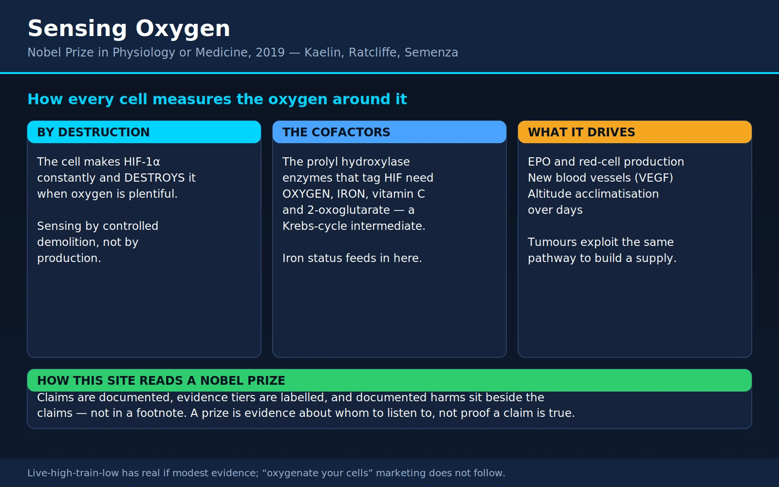
Table of Contents
- The Prize and the Three Men
- The Oldest Question in Physiology
- Semenza's HIF, 1992–1995
- Kaelin's Tumor Clue
- Ratcliffe Closes the Loop, 2001
- What Your Body Does with the Signal
- The Medicines This Built
- Altitude, Athletes, and the Honest Tiering
- Where This Touches Everyday Health
- Where Mainstream Medicine Agrees — and Where Claims Outrun Evidence
- Key Research Papers
- Connections
- Featured Videos
1. The Prize and the Three Men
In October 2019 the Nobel Assembly awarded the Prize in Physiology or Medicine jointly to William G. Kaelin Jr., Sir Peter J. Ratcliffe, and Gregg L. Semenza — "for their discoveries of how cells sense and adapt to oxygen availability." It is a citation that sounds almost too simple to be worth a Nobel until you sit with it. Every cell in your body burns oxygen, and oxygen supply changes constantly — when you climb a staircase, fly to Denver, form a clot, or stop breathing for fifteen seconds in the middle of the night. Cells respond within minutes, precisely, and differently in a kidney than in a muscle. Until the 1990s, nobody could say how a cell knows.
What makes this prize such a good story is that the three laureates reached the same molecular machine from three completely different directions, and none of them set out to discover a universal oxygen sensor.
Gregg Semenza, a pediatrician and molecular geneticist at Johns Hopkins, came at it through a gene. He wanted to know how the kidney turns on the gene for erythropoietin — EPO, the hormone that tells bone marrow to make red blood cells — when oxygen runs low. He was hunting a DNA switch. He found a protein sitting on that switch, and it turned out to be sitting on hundreds of others too.
Peter Ratcliffe was a kidney physician in Oxford, drawn in by a clinical fact he could not stop thinking about: people with failing kidneys become anemic, badly and predictably, and giving them iron does not fix it. The damaged kidney has stopped making EPO. Ratcliffe wanted to know what the kidney was measuring — and his laboratory then reframed the field by showing the response was not a kidney specialty at all. Liver, muscle, essentially any cell did it. Whatever the sensor was, it was ancient and everywhere.
William Kaelin, a cancer physician at the Dana-Farber Cancer Institute, came in through a rare inherited tumor syndrome most doctors never see: von Hippel–Lindau disease. Families with a broken copy of the VHL gene develop tumors that are strikingly, almost absurdly, full of blood vessels. Kaelin was not studying oxygen; he was studying a tumor suppressor gene. But VHL-defective cells were behaving as though they were suffocating — in a dish, in room air, with all the oxygen they could want.
2. The Oldest Question in Physiology
Oxygen is the terminal electron acceptor of aerobic metabolism — the substance at the end of the line that makes the energy chain in your mitochondria run. Without it, brain and heart muscle begin to die within minutes. Yet the oxygen actually available to a cell is not fixed: it varies with altitude, exercise, whether an artery is open, whether you are asleep and briefly not breathing, whether there is a wound, whether a tumor has outgrown its blood supply. Local tissue oxygen is a moving number.
Physiology has known this a long time. The French physiologist Paul Bert, working in the 1870s, established that it is the partial pressure of oxygen — not the percentage in the air — that matters to the body. His work founded barometric physiology and, incidentally, described oxygen toxicity at high pressure, a point that returns in section 10.
By mid-century the body's main long-term response was clear too: move to high altitude and you make more red blood cells, driven by erythropoietin from the kidney, isolated and cloned in the 1980s. So by 1990 the field knew the input (low oxygen) and one important output (more EPO, more red cells). What was missing was the middle. How does a cell measure oxygen? There was no known receptor for it, no channel, nothing like the lock-and-key arrangements that explain how cells detect hormones. Oxygen is a tiny, uncharged, freely diffusing molecule; it walks straight through membranes — you cannot put a doorman on a door that isn't there. And whatever the sensor was, it had to be graded (the EPO response is proportional to how low oxygen goes, not on/off) and fast.
3. Semenza's HIF, 1992–1995
Semenza took the classical molecular-genetics route: find the piece of DNA carrying the instruction, then find the protein that reads it. Working with the human EPO gene, his laboratory narrowed the low-oxygen response to a short stretch of DNA nearby — an enhancer, a control region that boosts transcription when the right protein latches on. Then they went looking for what latched on.
In 1992, Semenza and Guang Wang reported a nuclear protein that appeared within hours of low oxygen and bound exactly that sequence. They called it hypoxia-inducible factor — HIF. By 1995 they had purified and cloned it: a two-part protein pairing a subunit called HIF-1α with a partner known as ARNT (HIF-1β). Together they form a transcription factor, a molecular switch-thrower that sits on DNA and turns genes on.
The first surprise was scope. HIF did not just control EPO. Once the protein was in hand, the list of genes it bound ran to hundreds — blood vessel growth, glucose transporters, glycolytic enzymes, iron handling, cell survival and cell death. HIF was not a kidney gadget for making red cells. It was the master coordinator of a whole cellular program for living with less oxygen, present in essentially every cell type and evolutionarily ancient.
The second surprise is the hinge of the entire story. ARNT sat at a steady level regardless of oxygen. HIF-1α behaved completely differently: in low oxygen it accumulated rapidly; in normal oxygen it was almost undetectable. The obvious explanation would be that cells make HIF-1α only when needed. That is not what happens.
The cell manufactures HIF-1α continuously, all the time, and destroys it almost as fast as it is made whenever oxygen is plentiful. Its half-life in a well-oxygenated cell is a few minutes. Drop the oxygen and destruction stops; the protein already being produced simply survives, finds its partner, and switches on its gene program.
Read that again, because it explains everything that follows. The cell senses oxygen by controlled destruction, not by controlled production. It is the difference between a factory that ramps up when demand rises and a factory that runs flat out around the clock while a supervisor stands at the loading dock shredding everything that comes off the line — and stops shredding only when one particular supply runs out. It looks wasteful, and it buys something valuable: speed. Because the protein is already being made, the response does not wait for transcription and translation. It happens in minutes.
Which raised the obvious next question, and it took most of a decade to answer: what is doing the shredding, and how does the shredder know the oxygen level?
4. Kaelin's Tumor Clue
Von Hippel–Lindau disease is an inherited cancer-predisposition syndrome affecting roughly 1 in 36,000 people. Someone with VHL inherits one damaged copy of the VHL gene; when the remaining good copy is lost in some cell during life, tumors follow — hemangioblastomas in brain, spinal cord and retina; clear cell renal cell carcinoma, the commonest kidney cancer; pancreatic tumors; pheochromocytomas. Loss of the same gene also drives the great majority of sporadic clear cell kidney cancers, which is what made it interesting far beyond the families who carry it.
Kaelin's laboratory was studying VHL as a tumor suppressor, and the tumors handed over the clue. They are conspicuously soaked in blood vessels, and some produce so much EPO that the patient develops the opposite of anemia — exactly what you would expect from tissue that thinks it is suffocating. When Kaelin's group examined cells lacking functional VHL, they found high expression of hypoxia-response genes — VEGF, glucose transporters, the whole HIF catalogue — in full, normal oxygen. Put a working copy of VHL back in, and it switched off.
The interpretation was immediate: VHL is part of the machinery that shuts HIF off. In 1999, Ratcliffe's Oxford group showed the direct connection. The VHL protein is a component of a complex that attaches ubiquitin to HIF-1α — the cell's disposal tag, which delivers a protein to the proteasome to be destroyed. Maxwell and colleagues showed VHL binds HIF-α and targets it for oxygen-dependent destruction. Remove VHL and the tag is never applied: HIF survives indefinitely and the hypoxia program runs permanently. That is why VHL kidney tumors are so vascular, and in a very direct line why a drug discussed in section 7 exists.
But there was a gap in the middle, and it mattered most. VHL does not bind HIF-1α all the time — only when oxygen is present. Something had to be marking HIF-1α in an oxygen-dependent way. What was the mark?
5. Ratcliffe Closes the Loop, 2001
In April 2001 two papers appeared in the same issue of Science — one from Kaelin's laboratory with Michael Ivan as first author, one from Ratcliffe's with Panu Jaakkola as first author — and between them they closed the circle. Later that year, Ratcliffe's group and others identified the enzymes themselves.
The mark is a hydroxyl group — a single oxygen atom with a hydrogen, added to a specific proline amino acid in HIF-1α. Chemists call it prolyl hydroxylation. Once that hydroxyl is in place, VHL recognizes the modified protein and tags it for destruction. Without it, VHL slides past as though HIF were invisible.
The enzymes that do the stamping are a small family of prolyl hydroxylases — PHD1, PHD2 and PHD3, also called the EGLN proteins after the worm gene egl-9 in which the family's founding member was found. And here is the chemistry that makes the whole system work:
These enzymes use molecular oxygen as a substrate. Oxygen is not something they detect indirectly — it is a raw ingredient they physically consume. If oxygen is around, they stamp HIF and it dies. If oxygen is scarce, they slow or stop, HIF goes unstamped, VHL cannot find it, and it accumulates. The prolyl hydroxylases are the oxygen sensor; their reaction rate is the measurement. This is why the response is graded rather than on/off: enzyme activity falls smoothly as oxygen falls, so HIF rises smoothly in proportion. The cell counts oxygen by how fast one particular chemical reaction can run. A companion enzyme, FIH-1, hydroxylates a different site to mute HIF's activity rather than destroy it — so the system has both a coarse control and a fine one.
The cofactors: why this page lives on a nutrition site
The prolyl hydroxylases belong to the 2-oxoglutarate–dependent dioxygenases, and every member of that family needs the same short list of helpers. This is the part most popular accounts skip, and the part with the most everyday relevance:
- Molecular oxygen (O2) — the thing being measured, consumed in the reaction.
- Iron — each enzyme holds a single ferrous iron (Fe2+) atom at its catalytic center. No iron, no reaction. Iron is the atom that actually handles the oxygen chemistry.
- 2-oxoglutarate (α-ketoglutarate) — a co-substrate split during the reaction, and an intermediate of the citric acid cycle. The cell's oxygen sensor is chemically wired into its central metabolic pathway.
- Vitamin C (ascorbate) — not consumed in the main reaction, but required to keep the enzyme's iron in its usable reduced Fe2+ state. Catalytic iron occasionally gets oxidized to Fe3+ and the enzyme stalls; ascorbate reduces it back. Vitamin C plays the identical role for the collagen prolyl hydroxylases — the biochemical reason scurvy destroys connective tissue.
State that plainly, because it is the hinge of this page: your cells' oxygen sensor is an iron enzyme that needs vitamin C to keep working. Iron and ascorbate status are inputs to the machinery that decides how your body responds to low oxygen. That is well-established biochemistry — and section 9 is careful about what does and does not follow from it, because the honest answer is narrower than the supplement aisle would like.
6. What Your Body Does with the Signal
Once HIF is stabilized it goes to work on hundreds of genes, running a coherent strategy: get more oxygen in, build more pipes, and meanwhile burn fuel differently.
Make more red blood cells. HIF turns on the EPO gene in specialized kidney cells; EPO drives red cell production in the marrow, and more hemoglobin means more oxygen carried per liter of blood. A slow arm — days to weeks — because red cells have to be built.
Build more blood vessels. HIF turns on VEGF and related signals that recruit capillaries into oxygen-poor tissue. This is how a healing wound gets its blood supply, how trained muscle becomes more capillary-dense, and — unhelpfully — how a growing tumor arranges its own plumbing. It also directly explains those blood-drenched von Hippel–Lindau tumors.
Handle iron differently. HIF upregulates intestinal iron uptake machinery and transferrin and modulates hepcidin, the hormone gating how much iron enters the bloodstream. Expanding red cell production requires iron to build hemoglobin, so the system calling for more red cells also opens the supply lines. Our Iron pages cover that regulation in depth.
Switch fuel strategy. HIF upregulates glucose transporters and nearly every glycolytic enzyme while dialing down the flow of pyruvate into mitochondria. The cell shifts toward glycolysis — extracting energy from glucose without oxygen. Far less efficient per glucose molecule, but it works when oxygen is short, and cutting mitochondrial oxygen consumption also reduces damaging reactive oxygen species under hypoxic stress.
That last arm connects to another laureate on this site. Otto Warburg observed in the 1920s that tumor cells consume glucose voraciously and produce lactate even with plenty of oxygen — the "Warburg effect," which he blamed on broken mitochondria. HIF supplies much of the modern explanation: tumors often have constitutively active HIF, because the interior is genuinely hypoxic or because mutations (VHL loss classically) stabilize it regardless of oxygen. A glycolytic tumor is often one running the hypoxia program with the sensor jammed. Warburg had the observation right and the mechanism wrong.
Altitude acclimatization is this system doing its job
Fly from sea level to 3,500 metres and you can watch the program run on a schedule. Within minutes, breathing deepens and quickens. Within a day or two, HIF has stabilized in kidney cells and blood EPO rises sharply, often several-fold. Over the following days and weeks, red cell mass climbs, muscle capillary density increases, and cellular metabolism shifts. The unpleasant part — acute mountain sickness, with headache, nausea and poor sleep — reflects the lag between arriving and adapting.
7. The Medicines This Built
Within two decades the mechanism had produced two genuinely new drug classes, running the pathway in opposite directions. Both deserve honest reporting rather than a triumphal one.
HIF prolyl-hydroxylase inhibitors (HIF-PHIs) for anemia of kidney disease
The logic is direct. If prolyl hydroxylase destroys HIF, a drug that inhibits it leaves HIF standing and the cell behaves as though oxygen were low: it makes EPO. A pill, in other words, that persuades the kidney to make its own erythropoietin rather than an injection of the manufactured hormone. For people with chronic kidney disease who have spent years on injected erythropoiesis-stimulating agents plus intravenous iron, that is appealing — and HIF-PHIs improve iron mobilization, which injected EPO does not. Several exist: roxadustat, daprodustat, vadadustat, plus molidustat and enarodustat in some markets.
The regulatory picture differs by country, and the differences are informative rather than arbitrary:
- Roxadustat is approved in China, Japan and the European Union for anemia of chronic kidney disease. In August 2021 the U.S. FDA declined to approve it, following an advisory committee that voted overwhelmingly against it over cardiovascular safety signals — thrombosis and thromboembolic events in particular.
- Daprodustat was approved by the FDA in February 2023 for adults on dialysis at least four months. Its pivotal ASCEND trials met a non-inferiority standard against conventional erythropoiesis-stimulating agents for major adverse cardiovascular events in the dialysis population. The approval did not extend to patients not on dialysis, where results were less reassuring.
- Vadadustat was approved in Japan and later in the U.S. (2024) for dialysis patients, after an earlier rejection for the non-dialysis population.
The pattern should be stated plainly: the cardiovascular safety debate around this class is real and not fully resolved. Meta-analyses generally find HIF-PHIs comparable to erythropoiesis-stimulating agents for correcting hemoglobin, with thrombotic signals that vary by agent and population. Notice what the concern is, biologically: these drugs do not narrowly switch on EPO — they stabilize HIF, which controls hundreds of genes including VEGF and whole metabolic and vascular programs. The worries about vascular effects and tumor biology are the direct consequence of the same mechanism that makes the drugs work. That does not make them bad drugs; it makes them drugs whose risk-benefit calculation genuinely differs between patients.
Belzutifan: blocking HIF-2α directly
The second drug runs the pathway the other way, tracing back with unusual directness to Kaelin's VHL tumors: if those cancers grow because HIF-2α is permanently active, blocking it should shrink them. That long looked impossible — transcription factors were considered "undruggable" — until structural work revealed an unexpected internal cavity in HIF-2α. Nobody set out to build a kidney cancer drug by studying oxygen, and three decades later a syndrome once managed almost entirely by repeated surgery has a pill that shrinks its tumors.
Belzutifan (Welireg) is the result: a first-in-class small-molecule HIF-2α inhibitor. The FDA approved it in August 2021 for adults with von Hippel–Lindau disease needing treatment for VHL-associated renal cell carcinoma, CNS hemangioblastomas or pancreatic neuroendocrine tumors who do not require immediate surgery. In the pivotal trial reported by Jonasch and colleagues, roughly half of patients had an objective response in their kidney tumors, with responses also seen in hemangioblastomas and pancreatic lesions. In December 2023 the approval was extended to advanced clear cell renal cell carcinoma after prior PD-1/PD-L1 and antiangiogenic therapy — connecting this pathway to the immunotherapy story on our Allison and Honjo page.
Its most characteristic side effect is exactly what the biology predicts: anemia. Block HIF-2α and you turn down EPO — the same lever the HIF-PHIs push the other way. There is a satisfying symmetry in a drug class whose main adverse effect is the condition the other class was invented to treat.
8. Altitude, Athletes, and the Honest Tiering
Because this pathway produces red blood cells, it has a long and complicated relationship with sport — and a less respectable one with wellness marketing.
🟢 Live high, train low — real, modest, variable
The best-supported altitude method is "live high, train low": sleep and live at moderate altitude (roughly 2,000–2,500 m) to trigger the HIF/EPO response, but descend to do hard sessions where the air is thick enough to sustain full intensity. Altitude gives the adaptation; sea level preserves training quality.
The evidence supports a real effect, smaller and less consistent than the marketing suggests. Studies typically find red cell and hemoglobin mass rising a few percent after 3–4 weeks of adequate daily hypoxic exposure (usually cited as 12–16+ hours a day), with performance gains that matter at elite level and vary enormously between individuals. Some athletes are strong responders, some essentially non-responders — though iron status is one of the few reliably identified requirements, since you cannot build hemoglobin without iron, which is why elite camps monitor ferritin. Short trips or arriving iron-deficient reliably produce nothing.
🟡 Hypoxic tents and altitude simulation — plausible, dose-dependent, over-marketed
Nitrogen-enriched sleeping tents simulate the "live high" half at home, and the physiology is legitimate — the same signal reaches the same sensor. The problem is dose: a genuine response demands many hours per night for weeks, and compliance is poor because sleeping in a sealed tent is unpleasant. Intermittent hypoxic training — brief exposures during a workout — has much weaker evidence and is best regarded as unproven. Anyone with significant cardiovascular or pulmonary disease should not be self-administering hypoxia.
🔴 EPO doping — the dark mirror
Injected recombinant EPO thickens the blood far beyond what altitude achieves, and it did enormous damage to endurance sport from the late 1980s onward: raised hematocrit increases viscosity and thrombotic risk, and a cluster of unexplained deaths among young cyclists is widely attributed to it. Detection difficulty drove the biological passport, which watches an athlete's own blood parameters for implausible movement.
HIF-PHI drugs are on the World Anti-Doping Agency's Prohibited List — under S2, hypoxia-inducible factor activators, alongside cobalt and argon — and roxadustat has already produced sanctions. The point is not that athletes may cheat, but that this is a serious drug class with serious cardiovascular questions. Any product marketed as a "natural HIF activator" is playing with a pathway whose deliberate pharmaceutical manipulation took years of trials and still produced a split regulatory verdict.
🔴 "Hypoxia training" gadgets and elevation masks
Training masks that restrict airflow do not simulate altitude. Altitude reduces the partial pressure of oxygen; a restrictive mask makes breathing harder against resistance while leaving the oxygen fraction unchanged. They train respiratory muscles somewhat and make a workout feel harder, but they do not produce the hypoxic stimulus that drives HIF and EPO, and controlled studies show no hematological adaptation. Calling them altitude simulators is simply wrong about the physiology.
9. Where This Touches Everyday Health
This is not an exotic pathway. It is running in your cells right now, and several very common clinical situations are best understood through it.
Anemia of chronic kidney disease
The condition that pulled Ratcliffe into the field remains its most direct application. Damaged kidneys lose the cells that make EPO, so the marrow never gets the order. The resulting anemia is not an iron problem — though iron deficiency very often coexists and must be corrected too — which is why iron tablets alone do not fix it, and why the options are erythropoiesis-stimulating agents or, now, HIF-PHI tablets. Anyone with kidney disease and persistent fatigue should have hemoglobin, ferritin and transferrin saturation checked. See Nephrology and Anemia.
Sleep apnea and intermittent hypoxia
Obstructive sleep apnea subjects the body to something this pathway did not evolve for: not sustained low oxygen but intermittent hypoxia — dozens or hundreds of cycles per night of desaturation followed by rapid reoxygenation. That pattern activates HIF-1α while generating bursts of reactive oxygen species and driving sympathetic activation, and is now regarded as a major reason untreated apnea raises risks of hypertension, arrhythmia, cardiovascular events and metabolic dysfunction — a plausible answer to why apnea harms the cardiovascular system out of proportion to the oxygen debt. Some people with untreated apnea also develop a mildly raised red cell count, the EPO arm of the same response. See Obstructive Sleep Apnea.
Iron deficiency
Iron sits at two places in this story at once. First, it is the raw material for hemoglobin, so no amount of EPO signal produces red cells without it. Second, and more subtly, iron is the catalytic metal in the oxygen-sensing enzymes themselves. Severe iron deficiency and iron chelation demonstrably alter this pathway in laboratory and human physiology studies, and iron infusion measurably blunts the hypoxic pulmonary pressure response.
What follows is modest and specific: correcting a genuine iron deficiency matters, and matters more than usual if you are anemic, pregnant, at altitude, or on an agent that stimulates red cell production. What does not follow is that extra iron beyond repletion improves oxygen sensing in a healthy person. Iron overload is genuinely harmful — oxidative damage, and organ injury in hemochromatosis — so supplementation should follow a blood test, not a theory. Check ferritin with transferrin saturation; see Iron Deficiency and Iron-Deficiency Anemia.
Vitamin C, without overclaiming
Here is the careful version of a claim that is easy to inflate. Ascorbate is a required cofactor for the prolyl hydroxylases — settled biochemistry, not speculation. It keeps the enzymes' iron reduced so catalysis continues, and cells depleted of ascorbate show altered HIF activity in the laboratory.
What this justifies is not being deficient. The amount needed to saturate these enzymes is within reach of ordinary dietary intake, and plasma ascorbate plateaus at modest intakes because absorption is regulated and the kidney excretes the surplus. What it emphatically does not justify is the claim that gram doses "optimize oxygen sensing," improve endurance, or treat cancer through this pathway — the work on ascorbate and HIF in tumors remains laboratory work. The honest statement: enough vitamin C is required for the sensor to function; more than enough has not been shown to make it function better. See Vitamin C, and our Linus Pauling page for how a real cofactor role became a much larger claim.
Tumors exploiting the pathway
A solid tumor outgrows its blood supply, its interior turns hypoxic, HIF stabilizes, and the tumor recruits vessels, shifts to glycolysis, and becomes more invasive and more radiation-resistant (radiation needs oxygen to work well). Hypoxic tumors have worse outcomes across many cancer types, and in VHL-mutant kidney cancer the sensor is broken outright — which is why Oncology and Warburg's century-old observation now sit in the same conversation.
10. Where Mainstream Medicine Agrees — and Where Claims Outrun Evidence
🟢 Where the evidence is solid
- The mechanism itself. HIF, VHL and the prolyl hydroxylases are among the best-characterized systems in cell biology, reproduced in thousands of laboratories. Not a contested area.
- Iron, 2-oxoglutarate, oxygen and ascorbate as required cofactors — straightforward enzymology.
- Correcting anemia of kidney disease with erythropoiesis-stimulating agents or approved HIF-PHIs improves hemoglobin, with the drug-specific caveats above.
- Belzutifan shrinks VHL-associated tumors and is approved on trial evidence.
- Treating obstructive sleep apnea reduces the intermittent hypoxia driving cardiovascular strain — one of the highest-value interventions on this page, requiring no supplement at all.
- Supplemental oxygen for people who are genuinely hypoxemic (advanced COPD, severe pulmonary disease) extends life. Measured need, measured benefit.
🟡 Real but limited, or still open
- Live-high-train-low works, modestly, with high individual variability and demanding dose requirements.
- Long-term safety of systemic HIF stabilizers is not settled, and the FDA/EMA divergence on roxadustat reflects genuine expert disagreement rather than bureaucratic caprice.
- Hyperbaric oxygen therapy for its approved indications — decompression sickness, carbon monoxide poisoning, gas gangrene, certain non-healing diabetic foot ulcers, late radiation tissue injury, compromised skin grafts — has an evidence base, and the mechanism is interesting: hyperoxic-then-normoxic cycling can transiently raise growth-factor signaling in poorly perfused tissue. See Hyperbaric Oxygen Therapy for the indication-by-indication review.
🔴 Where claims outrun the evidence
- "Oxygenate your cells." The commonest misreading of everything above, and it inverts the biology. In a healthy person breathing room air, arterial hemoglobin is already 95–99% saturated — there is essentially no headroom, and delivery is limited by blood flow and hemoglobin, both near ceiling. Oxygenated water, "stabilized oxygen" drops, oxygen bars and ozone products do not do what their labels imply, and HIF biology supports none of them.
- Hyperbaric oxygen for general wellness, anti-aging, longevity or unproven neurological indications. There are real indications and a large market well beyond them, often at considerable out-of-pocket cost; the approved indication list is precisely what distinguishes the two. It is not risk-free: ear and sinus barotrauma is common, and oxygen toxicity — the phenomenon Paul Bert described in the 1870s — is why protocols strictly limit pressure and duration. More oxygen is not linearly better; at high partial pressures it becomes a poison.
- "HIF-activating" or "hypoxia-mimetic" supplements. Compounds that genuinely activate HIF are prohibited in sport and, in pharmaceutical form, carry cardiovascular warnings. Anything sold over the counter claiming this effect is either inert or, if truly active, unregulated exposure to a pathway controlling vessel growth and metabolism. Neither is attractive.
- Extrapolating from cofactor to megadose. Iron and vitamin C are required for the sensor. Requirement is not dose-response. Beyond repletion, neither has been shown to improve oxygen sensing, and iron in excess is directly harmful.
- Elevation training masks as altitude simulators. Wrong physiology, as described above.
The through-line is worth stating once, clearly. This pathway is a regulator, not a fuel tank. It does not make more oxygen available; it decides what a cell does about the oxygen it has. Understanding that distinction dissolves most of the marketing built on top of it.
11. Key Research Papers
- Semenza GL, Wang GL. A nuclear factor induced by hypoxia via de novo protein synthesis binds to the human erythropoietin gene enhancer at a site required for transcriptional activation. Mol Cell Biol 1992;12(12):5447-54 — the paper that named HIF.
- Wang GL, Jiang BH, Rue EA, Semenza GL. Hypoxia-inducible factor 1 is a basic-helix-loop-helix-PAS heterodimer regulated by cellular O2 tension. Proc Natl Acad Sci U S A 1995;92(12):5510-4 — purification and cloning; HIF-1α plus ARNT.
- Maxwell PH, Wiesener MS, Chang GW, et al. The tumour suppressor protein VHL targets hypoxia-inducible factors for oxygen-dependent proteolysis. Nature 1999;399(6733):271-5 — Ratcliffe's group links VHL to HIF destruction.
- Ivan M, Kondo K, Yang H, et al. HIFα targeted for VHL-mediated destruction by proline hydroxylation: implications for O2 sensing. Science 2001;292(5516):464-8 — Kaelin's half of the simultaneous solution.
- Jaakkola P, Mole DR, Tian YM, et al. Targeting of HIF-α to the von Hippel-Lindau ubiquitylation complex by O2-regulated prolyl hydroxylation. Science 2001;292(5516):468-72 — Ratcliffe's half, in the same issue.
- Epstein AC, Gleadle JM, McNeill LA, et al. C. elegans EGL-9 and mammalian homologs define a family of dioxygenases that regulate HIF by prolyl hydroxylation. Cell 2001;107(1):43-54 — identifies the prolyl hydroxylase enzymes themselves.
- Semenza GL. Oxygen sensing, hypoxia-inducible factors, and disease pathophysiology. Annu Rev Pathol 2014;9:47-71 — a readable synthesis by one of the field's founders.
- Singh AK, Carroll K, Perkovic V, et al. Daprodustat for the treatment of anemia in patients undergoing dialysis. N Engl J Med 2021;385(25):2325-2335 — the ASCEND-D trial behind the U.S. approval.
- Jonasch E, Donskov F, Iliopoulos O, et al. Belzutifan for renal cell carcinoma in von Hippel-Lindau disease. N Engl J Med 2021;385(22):2036-2046 — the trial that turned Kaelin's tumor clue into a drug.
- Ha JT, Hiremath S, Jun M, et al. Hypoxia-inducible factor prolyl hydroxylase inhibitors in kidney disease. NEJM Evid 2024;3(9):EVIDoa2300189 — systematic review and meta-analysis of efficacy and cardiovascular safety.
- Millet GP, Roels B, Schmitt L, et al. Combining hypoxic methods for peak performance. Sports Med 2010;40(1):1-25 — review of live-high-train-low and related altitude protocols.
Live PubMed Searches
- Hypoxia-inducible factor discovery
- HIF prolyl hydroxylase inhibitor anemia
- Belzutifan von Hippel-Lindau
- Altitude training hemoglobin
- Intermittent hypoxia sleep apnea
12. Connections
- All Notable Doctors
- Nobel Prize in Physiology or Medicine — the full roll of laureates, 1901 to the present
- Otto Warburg — the glycolytic shift in tumors he described in the 1920s, which HIF largely explains
- Hans Krebs — 2-oxoglutarate, the co-substrate every prolyl hydroxylase consumes, is an intermediate of his citric acid cycle
- James Allison & Tasuku Honjo — checkpoint immunotherapy; belzutifan's advanced-kidney-cancer approval follows PD-1 blockade
- Linus Pauling — a cautionary case study in how a genuine vitamin C cofactor role became a much larger claim
- Iron — the catalytic metal at the heart of every oxygen-sensing enzyme, and the raw material for hemoglobin
- Iron Deficiency — symptoms, testing and correction, including why it blunts any altitude or EPO response
- Vitamin C — ascorbate keeps the prolyl hydroxylases' iron reduced; requirement, not megadose
- Hematology — blood disorders, red cell production and the conditions this pathway governs
- Anemia — the many causes, and why kidney anemia does not respond to iron alone
- Iron-Deficiency Anemia — the most common anemia worldwide
- Polycythemia Vera — too many red cells from the other direction, via a marrow mutation rather than the oxygen sensor
- Nephrology & Hepatology — kidney disease, the clinical problem that pulled Ratcliffe into oxygen sensing
- Obstructive Sleep Apnea — intermittent hypoxia and why untreated apnea strains the cardiovascular system
- Ferritin Test — the iron-stores blood test to check before supplementing anything
- Hyperbaric Oxygen Therapy — the approved indications, and the wellness claims that do not follow from HIF biology
- Oncology — tumor hypoxia, vessel recruitment and radiation resistance