Hodgkin, Huxley & Eccles: The Electrical Nerve Impulse, and Why Electrolytes Matter
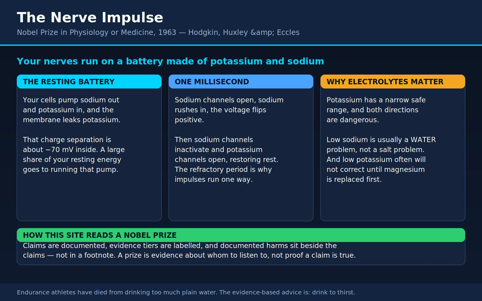
Table of Contents
- The Prize and the Three Men
- The Squid Giant Axon
- The Resting Membrane: Your Cells Run on a Battery
- The Action Potential: One Millisecond, in Detail
- The Voltage Clamp and the Equations
- Eccles and the Synapse: A Man Who Changed His Mind
- Why Your Electrolytes Matter
- Electrolyte Supplements and Sports Drinks, Tiered Honestly
- Channelopathies: The Diseases That Prove the Mechanism
- What This Means for You Today
- Where Mainstream Medicine Agrees — and What Remains Debated
- Key Research Papers
- Connections
- Featured Videos
1. The Prize and the Three Men
In 1963 the Nobel Prize in Physiology or Medicine went to three men — Sir John Carew Eccles, Alan Lloyd Hodgkin and Andrew Fielding Huxley — for their discoveries concerning the ionic mechanisms involved in excitation and inhibition in the peripheral and central portions of the nerve cell membrane. That is a mouthful of a citation, and it hides one of the most satisfying answers in all of biology. Between them, these three worked out what a nerve impulse actually is.
The answer turned out to be startlingly simple in outline and beautiful in detail. A nerve impulse is not a current flowing along a wire. It is a wave of dissolved minerals moving briefly across a membrane — sodium rushing in, potassium rushing out, through gates in the cell's outer skin that open and close in response to voltage. Every thought you have ever had, every heartbeat, every twitch of a finger, is that. It runs on salt.
That is why this page does double duty on this site. It is the story of a discovery, and it is also our explainer for the question readers ask most often about minerals: why do electrolytes matter so much? The answer is not a slogan from a sports-drink advertisement. It is that the nervous system and the heart are electrical machines whose working fluid is dissolved potassium, sodium, calcium, magnesium and chloride — and the machine's tolerances are tight.
Alan Hodgkin (1914–1998)
Hodgkin was born in Banbury, Oxfordshire, into a Quaker family, and came to Trinity College, Cambridge, where he began working on nerve conduction as an undergraduate. He had a physicist's instinct for a biological problem: rather than describe the impulse, he wanted to measure it, model it, and predict it. He was also, by his own later account, extraordinarily lucky in his collaborators and in his timing — a point he made with some care in his 1976 memoir of the work, which remains the best first-person account of how the discovery actually unfolded, complete with the false starts.
Andrew Huxley (1917–2012)
Huxley arrived in Hodgkin's laboratory as a Cambridge undergraduate in 1939 and stayed a collaborator for more than a decade. He carried one of the most remarkable surnames in British intellectual life: he was a grandson of Thomas Henry Huxley, the comparative anatomist and combative defender of Darwin known as "Darwin's bulldog," and a half-brother of the novelist Aldous Huxley and of the biologist Julian Huxley. Andrew's own gift was mathematical and mechanical. He built apparatus, and he could carry a differential equation through to a number — a skill that turned out to be the hinge on which the whole discovery swung. After the nerve work he went on to a second career-defining contribution in muscle physiology, where the sliding-filament theory of contraction bears his name.
John Eccles (1903–1997)
Eccles was Australian, born in Melbourne, and came to Oxford as a Rhodes Scholar to work under Charles Sherrington, the great analyst of the reflex. He spent much of his career back in the southern hemisphere — Sydney, then Otago in New Zealand, then the Australian National University in Canberra — and he is the reason this page has a genuinely admirable story in it rather than merely an impressive one.
For years, Eccles was the leading champion of the losing side of a famous argument. The question was how one nerve cell passes a signal to the next: by a direct electrical connection, or by releasing a chemical messenger. The debate was nicknamed "sparks versus soup." Eccles argued forcefully for sparks. Then, in the early 1950s, he built the apparatus that could settle it — and his own experiments demolished his own position. He accepted the result publicly, rebuilt his thinking around chemical transmission, and went on to do the work that won him a share of the Nobel Prize.
Eccles credited the philosopher Karl Popper, whom he had come to know in New Zealand, with making that reversal not just bearable but welcome. Popper's argument is that a scientific theory earns its keep by being falsifiable, and that a decisive refutation is a success of the method rather than a personal defeat. Eccles took that seriously and later wrote about the episode as one of the best things that had happened to him in science: he had stated a hypothesis clearly enough that it could be killed, and then he had gone and killed it himself. We tell that story properly in section 6, because it is rarer and more valuable than any single experimental result.
2. The Squid Giant Axon
Here is a fact worth sitting with: the electrical basis of human thought was worked out on a squid.
The problem in the 1930s was one of scale. Nerve fibres in a mammal are microscopically thin — typically a few thousandths of a millimetre across. To find out what the inside of a nerve fibre is doing electrically, you have to get inside it, and there was no way to put an electrode inside something that small without destroying it. Everything known about nerve impulses had been inferred from electrodes placed on the outside, which is rather like trying to work out how an engine functions by listening to the bonnet.
The way through came from an unrelated corner of biology. In the 1930s the British zoologist J. Z. Young, studying the anatomy of squid, established that a large pale tube running through the animal's mantle — which earlier anatomists had taken for a blood vessel — was in fact a single, enormous nerve fibre. Squid escape by contracting the mantle and firing a jet of water, and that escape reflex has to fire fast and all at once. Since conduction speed rises with fibre diameter, evolution's solution was brute size: the squid giant axon can reach roughly a millimetre across — the figure varies with species and specimen, but it is hundreds of times the diameter of an ordinary nerve fibre, and thick enough to see with the naked eye.
That changed everything. An axon a millimetre wide is an axon you can thread a fine wire electrode down the inside of. You can measure the voltage across its membrane directly. You can even squeeze out the contents like toothpaste from a tube and replace them with a solution of your own choosing, then ask what happens.
It is worth being explicit about how this discovery came about, because the lesson is easy to lose. Nobody funded J. Z. Young to cure a disease. He was studying squid anatomy for its own sake, because squid are interesting animals and their nervous systems had not been properly described. The single most consequential tool in twentieth-century neurophysiology fell out of curiosity-driven zoology. Hodgkin, who benefited from it more than anyone, said as much repeatedly. When people argue about whether basic research pays for itself, the squid giant axon is the example to reach for.
Hodgkin and Huxley did their squid work at the Marine Biological Association laboratory in Plymouth, on England's south coast, where the animals could be caught fresh and used within hours — the axon does not travel well. In 1939 they succeeded for the first time in pushing a capillary electrode down the axis of a living axon and recording the potential difference across its membrane from the inside. What they saw was not what the textbooks predicted. The prevailing theory said the impulse should be a transient collapse of the resting voltage to about zero. Instead, the voltage did not merely collapse: it overshot, swinging past zero and making the inside of the fibre briefly positive with respect to the outside. They published a short note in Nature that autumn, and then the war stopped them.
The interruption was total and it lasted the better part of a decade. Hodgkin spent the war on airborne radar; Huxley worked on gunnery problems. The overshoot — a result that nobody could yet explain — sat unexamined until they returned to Plymouth in the late 1940s and picked the problem up where they had dropped it.
3. The Resting Membrane: Your Cells Run on a Battery
Before you can understand an impulse, you have to understand the state the cell is in when nothing is happening. That state is not "off." It is loaded, like a drawn bow.
The gradients
Every cell in your body maintains a deliberate, expensive imbalance of two minerals across its outer membrane:
- Potassium is concentrated inside the cell. Roughly speaking, the fluid inside a cell holds something like thirty to forty times more potassium than the fluid outside it. Around 98 per cent of the potassium in your body is inside cells, not in your blood — a fact with important consequences we return to in section 7.
- Sodium is concentrated outside the cell. The reverse gradient, roughly ten- to fifteen-fold, in the other direction.
Neither of these gradients is a natural resting state. Left alone, salts diffuse until they are evenly spread. Maintaining the imbalance takes continuous work, and the machine that does the work is one of the most important proteins in biology.
The sodium-potassium pump
The sodium-potassium pump — formally the Na⁺/K⁺-ATPase — is a protein embedded in the membrane of essentially every animal cell. It burns ATP, the cell's energy currency, and with each cycle it throws three sodium ions out of the cell and drags two potassium ions in. It is running in you right now, in every cell, without pause, from before you were born until after you die.
It was identified in 1957 by the Danish physiologist Jens Christian Skou, working on crab nerve, who found an enzyme that split ATP only when both sodium and potassium were present — the signature of a pump handling both ions in one cycle. Skou received the Nobel Prize in Chemistry in 1997 for it, a full forty years later. Hodgkin and Huxley's account of the impulse required such a pump to exist; Skou found it.
Here is the fact most readers find genuinely surprising. Running that pump is one of the largest single items on your body's energy budget. Estimates vary by tissue and by measurement method, but the sodium-potassium pump is routinely reckoned to consume something on the order of a fifth to a third of a resting cell's ATP — and in nervous tissue, considerably more. Your brain is about two per cent of your body weight and takes something like twenty per cent of your resting energy, and detailed accounting of what grey matter spends that energy on puts the great majority of it into restoring the ion gradients that signalling has just spent. When you are lying still in a dark room thinking about nothing, a large fraction of the calories you are burning is going into pushing sodium out of cells and potassium in.
You are, in a real and unromantic sense, mostly paying to keep your salt sorted.
How a mineral gradient becomes a voltage
Now the physics, in plain language. The membrane is not a perfect wall. It is studded with channels — protein pores — and at rest, the ones that are mostly open are potassium channels. So potassium, which is crowded inside, leaks out down its concentration gradient.
But potassium ions carry a positive charge. Every one that leaves takes a positive charge with it and leaves behind an unbalanced negative charge inside the cell. Within a very short time the growing negative charge inside starts pulling potassium back, and an equilibrium is reached where the outward push of concentration exactly balances the inward pull of charge.
That equilibrium is a voltage. The inside of the cell settles at roughly −70 millivolts relative to the outside — the exact figure varies by cell type, from about −60 mV in some neurons to about −90 mV in cardiac muscle at rest. The membrane is a capacitor with a charge separation across it. It is, quite literally, a battery, and every cell in your body carries one.
Two refinements matter. First, the resting voltage does not sit exactly where potassium alone would put it, because the membrane also leaks a little sodium inward, dragging the voltage a few tens of millivolts less negative. Second, the pump is itself slightly electrogenic — three positives out for two in is a net export of positive charge — so it contributes a small amount to the negativity directly, on top of the much larger contribution it makes by maintaining the gradients in the first place.
The number to hold onto is this: −70 millivolts across a membrane about five nanometres thick works out to an electric field strength of roughly 100,000 volts per centimetre. It is a tiny voltage across an unimaginably thin barrier, and the field is ferocious. That is the field that voltage-gated channels sense.
4. The Action Potential: One Millisecond, in Detail
An action potential — a nerve impulse — takes about a millisecond. Here is what happens in it.
The sequence
- Something depolarises the patch of membrane. A signal arrives from another neuron, or a sensory receptor is stimulated, and the voltage inside the cell becomes a little less negative — say from −70 mV toward −55 mV.
- Voltage-gated sodium channels open. These are the crucial devices. They are shut at rest, and they open when the membrane depolarises past a threshold. Sodium, which has been held outside at high concentration and is electrically attracted to the negative interior, pours in. Both forces — concentration and charge — push the same way, so the influx is fast and forceful.
- The voltage flips positive. Incoming sodium makes the inside less negative, which opens more sodium channels, which lets in more sodium. It is a runaway loop, and it is why an action potential is all-or-none: once threshold is crossed, the thing goes off completely. The interior overshoots zero and peaks at something in the region of +30 to +40 mV — the overshoot Hodgkin and Huxley first saw in 1939 and could not then explain.
- Sodium channels inactivate. This is the subtle part, and it is the piece Hodgkin and Huxley pinned down. The sodium channel does not simply close again when the voltage falls; it has a separate, slower inactivation mechanism — think of a second gate that swings shut behind the first — that shuts it a fraction of a millisecond after it opened, while the membrane is still depolarised. The sodium flood stops itself.
- Voltage-gated potassium channels open. These respond to the same depolarisation but more slowly. By the time they are properly open, sodium entry is already shutting down. Potassium now streams out, carrying positive charge with it, and the interior plunges back toward negative.
- The membrane overshoots downward, then settles. Potassium channels are slow to close as well as slow to open, so for a few milliseconds the membrane is briefly more negative than its resting value — the after-hyperpolarisation. Then the channels close, the resting leak reasserts itself, and the membrane is back at −70 mV, ready to fire again.
The sodium and potassium that moved are gradually put back where they belong by the pump, but — and this is a point that surprises people — not urgently. A single action potential exchanges such a minute fraction of the cell's total ion content that a large axon whose pump has been poisoned can still fire many thousands of impulses before its gradients noticeably run down. The impulse spends the battery in almost imperceptible sips. The pump's job is to keep up over hours, not to refill between spikes.
How it travels
An action potential at one point on the membrane depolarises the membrane just next to it, which pushes that patch past threshold, which fires it, which depolarises the patch beyond. The impulse regenerates itself as it goes, at full amplitude, all the way along the fibre. This is why a nerve signal does not fade with distance the way a signal in a wire does — it is not being transmitted, it is being re-created at every step.
The refractory period, and why signals do not reverberate
Immediately after firing, a patch of membrane is absolutely refractory: its sodium channels are inactivated, and no stimulus of any strength will fire it again until they have reset. This is followed by a relatively refractory period in which a stronger-than-usual stimulus is required.
The refractory period is not an inconvenience; it is essential engineering. It is why an impulse travelling down an axon does not turn round and come back. The membrane behind the wave front is refractory, so the wave can only advance into fresh membrane ahead of it. Without that, a single stimulus anywhere would set up an impulse ricocheting endlessly up and down the fibre, and the nervous system would be a permanent screaming feedback loop. The refractory period also caps how fast a neuron can fire, which matters because neurons encode intensity as frequency, not amplitude. Every action potential is the same size. A bright light or a hard pinch does not produce a bigger spike; it produces more spikes per second.
Saltatory conduction and myelin
The squid's answer to "make it fast" was to make the axon enormous, which works but does not scale — a human arm built on squid principles would need to be the width of a tree trunk. Vertebrates found a better solution: myelin.
Myelin is a fatty insulating sheath wrapped around the axon by supporting cells — Schwann cells in the peripheral nerves, oligodendrocytes in the brain and spinal cord. The wrapping is not continuous. It is interrupted every millimetre or so by bare gaps called nodes of Ranvier, and the voltage-gated sodium channels are concentrated at those nodes. The impulse therefore does not creep along every micron of membrane; it jumps from node to node, regenerating only at the gaps. This is saltatory conduction, from the Latin saltare, to leap.
The speed difference is dramatic. The squid giant axon, unmyelinated and a millimetre wide, conducts at roughly 20–25 metres per second. A myelinated human motor nerve fibre a fraction of that diameter conducts at up to about 120 metres per second — well over 250 miles an hour. Myelin is also, therefore, the thing whose loss is catastrophic: in multiple sclerosis, immune attack strips myelin from central nervous system axons, and conduction slows, becomes unreliable, or fails outright. The symptoms of demyelination are the symptoms of a nervous system whose cables have lost their insulation.
The domino analogy, and exactly where it breaks
Textbooks reach for a row of dominoes or a burning fuse, and both analogies get two things right. The first is self-propagation: each unit triggers the next, so the signal travels at constant strength without needing a push from behind. The second is all-or-none behaviour: a domino either falls or it does not; a fuse either catches or it does not. Nudge a domino gently and nothing happens. Nudge it past its tipping point and it falls the whole way, at full size, every time.
Now the ways the analogy misleads, which are more instructive than the ways it works:
- A fuse is consumed; an axon is not. Burn a fuse and it is gone. Fire an axon and it resets itself within a couple of milliseconds and is ready to go again. A neuron can fire hundreds of times a second, indefinitely.
- A fuse burns its own fuel; an axon barely touches its reserves. As above, one impulse moves a negligible fraction of the cell's ions. The fuse analogy makes the impulse sound expensive. In terms of ions moved per spike, it is astonishingly cheap; the expensive part is the standing pump that keeps the gradients topped up.
- The axon has a built-in stopping mechanism the analogy has no equivalent for. Sodium-channel inactivation shuts the influx down after a fraction of a millisecond regardless of what the voltage is doing. Nothing in a row of dominoes corresponds to that, and it is the single most important detail for understanding both the refractory period and how a whole class of drugs works.
- Direction is not intrinsic. A fuse lit at one end burns one way. But an axon stimulated in the middle in a laboratory will propagate impulses in both directions from the stimulus point. One-way traffic in the body is a consequence of where signals start — at the axon hillock, one end — combined with the refractory zone behind the wave, not of any inherent polarity in the membrane itself.
5. The Voltage Clamp and the Equations
Knowing that sodium comes in and potassium goes out is a qualitative story. Hodgkin and Huxley wanted a quantitative one, and there was a maddening obstacle in the way.
The problem, and the trick that solved it
The membrane's channels respond to voltage, and the currents they carry change the voltage. Everything is coupled to everything. If you depolarise the membrane and watch, you cannot tell which part of what you are seeing is the channels' response and which part is the voltage running away from you. You cannot measure the input-output relationship of a system whose output feeds straight back into its input.
The solution was the voltage clamp, developed in the late 1940s by Kenneth Cole and George Marmont in the United States. It is an elegant piece of electronics: a feedback amplifier continuously measures the membrane voltage, compares it with a value the experimenter has chosen, and injects exactly enough current to hold the membrane at that value no matter what the channels do. The voltage is clamped. The runaway loop is broken.
And the current the amplifier has to inject to hold the line is a direct, moment-by-moment readout of the current the membrane's own channels are carrying. You are no longer watching the system's output; you are reading it off a meter.
Hodgkin and Huxley adopted the technique and refined it substantially, adding a long axial wire inside the axon so the whole length of the measured region was held at one voltage at once — a "space clamp" — and improving the feedback circuitry. They then did the decisive experiment for identifying the ions: they changed the sea water. Replace much of the sodium in the bathing fluid with an impermeant substitute, and the inward current shrinks in exact proportion. Do the same with potassium, and the outward current changes as predicted. There was no longer any doubt about which ion carried which phase of the impulse.
The 1952 papers
The results were published in The Journal of Physiology in 1952, as a series of five closely spaced papers. They are worth listing separately, because they are frequently confused with one another — same two authors, same journal, same year:
- Measurement of current-voltage relations in the membrane of the giant axon of Loligo (with Bernard Katz) — the method paper, describing the voltage clamp as they used it.
- Currents carried by sodium and potassium ions through the membrane of the giant axon of Loligo — the ion-substitution experiments that separated the total current into a sodium component and a potassium component.
- The components of membrane conductance in the giant axon of Loligo — how each conductance depends on voltage and time.
- The dual effect of membrane potential on sodium conductance in the giant axon of Loligo — the paper that establishes inactivation as a distinct process from activation.
- A quantitative description of membrane current and its application to conduction and excitation in nerve — the famous one, the synthesis, the paper people mean when they say "Hodgkin and Huxley 1952."
A small pleasing detail: the fifth paper appeared in The Journal of Physiology volume 117, issue 4 — and the paper by Brock, Coombs and Eccles announcing intracellular recording from motor neurons is in the same issue, seventy pages earlier. Both halves of the 1963 Nobel Prize are bound together in one volume of one journal.
The equations, and three weeks of turning a handle
The synthesis paper does something no previous account of nerve conduction had attempted. It reduces the membrane to a small set of differential equations. The membrane is treated as a capacitor in parallel with three conductances — one for sodium, one for potassium, one small "leak" — and the sodium and potassium conductances are made to depend on voltage and time through three gating variables, conventionally written m, h and n: m for sodium activation, h for sodium inactivation, n for potassium activation. Each obeys its own first-order equation whose rate constants were fitted to the voltage-clamp data.
Four coupled equations, then. And then the real test: solve them, without putting any information about the shape of an action potential into the solution, and see whether an action potential falls out.
The Cambridge computer, EDSAC, was unavailable — it was being worked on. So Huxley solved the equations by hand. He used a Brunsviga mechanical calculator, a hand-cranked machine of brass gears and sliding registers, and integrated the system numerically step by tiny step. It took him about three weeks of steady work. He could not know while he was turning the handle whether the answer would be a beautiful curve or garbage, and there was no shortcut to finding out.
What came out was an action potential. Not a rough approximation of one: the computed curve reproduced the shape, the amplitude, the overshoot, the duration and the after-hyperpolarisation of the real thing. The model also predicted the speed at which an impulse should travel down the axon — a number that had never been part of the fitting — and the predicted velocity came out close to the velocity measured in a real squid axon. It reproduced the refractory period, and the threshold behaviour, and the response to sub-threshold stimuli, all of them consequences of the equations rather than inputs to them.
That is what a quantitative theory in biology looks like, and in 1952 there was essentially nothing else like it. The Hodgkin-Huxley model is generally reckoned one of the founding moments of computational biology and computational neuroscience: the first time a complex biological behaviour had been reduced to equations that predicted new measurements correctly. More than seventy years on, it is still taught to every neuroscience student, and it is still used — extended, modified, and equipped with new channel types, but recognisably the same framework — in simulations of neurons, cardiac muscle and neural circuits. Very little theoretical work in biology has aged that well.
6. Eccles and the Synapse: A Man Who Changed His Mind
Hodgkin and Huxley explained how a signal travels along a neuron. Eccles explained what happens when it reaches the end and has to get to the next one.
Sparks versus soup
By the 1930s the existence of the synapse — the junction between two neurons, established by Cajal's work — was accepted. How it worked was not. One camp held that the electrical impulse jumped the gap directly. The other held that the arriving impulse released a chemical that diffused across and acted on the next cell — a position supported by Otto Loewi's and Henry Dale's work on acetylcholine at the nerve-heart and nerve-muscle junctions.
Eccles led the electrical camp, and he was not a gentle opponent. His argument had real substance: chemical transmission looked far too slow to account for the speed of reflexes, and diffusion across a gap is a sluggish business compared with a spark. It was a serious hypothesis, defended by a serious scientist, and it was wrong.
The experiment that settled it
What settled it was a technique. In the early 1950s, Eccles — with Lawrence Brock and John Coombs — succeeded in pushing a glass microelectrode with a tip well under a micrometre across into the body of a living motor neuron in a cat's spinal cord, without killing the cell, and recording its membrane potential directly while its inputs were stimulated. Nobody had ever listened in on a central neuron from the inside before.
What they recorded were two kinds of small, graded voltage changes:
- Excitatory postsynaptic potentials (EPSPs) — brief depolarisations that nudge the cell toward threshold. A single one is usually far too small to fire the cell.
- Inhibitory postsynaptic potentials (IPSPs) — brief hyperpolarisations that push the cell further away from threshold, making it harder to fire.
The IPSP was the killing blow to the electrical hypothesis, and it is the finding worth understanding properly. Before this, inhibition in the nervous system was widely imagined as a subtraction — a withholding, an absence of excitation, a matter of not sending a signal. Eccles showed it is nothing of the kind. Inhibition is an active process with its own machinery: an inhibitory synapse opens its own set of channels, letting chloride ions in and potassium out, which drives the membrane potential down, away from firing. Inhibition is a signal in its own right, with its own transmitters and its own receptors, and it costs energy to produce.
The timing and behaviour of these potentials, and the drugs that blocked them, fitted chemical transmission and not the electrical model. Eccles accepted it. Within a few years he and his colleagues had worked out the ionic basis of the inhibitory potential in detail — which conductances change, and by how much — and Eccles became one of the leading investigators of chemical synaptic transmission in the central nervous system.
Why this is the good story
Scientists change their minds less often than the popular account of science suggests, and rarely with grace. Eccles did it in public, at the height of his reputation, on the basis of data he had gone to great trouble to collect precisely because it might refute him. He credited Popper for the framing — that a hypothesis is valuable in proportion to how decisively it can be killed, and that killing your own is a contribution rather than an embarrassment — and he wrote about the experience afterward as something close to a liberation. He had been wrong, he had found out he was wrong by his own hand, and the finding out was the whole point.
It is also worth adding a coda that historians of the field enjoy: Eccles was not entirely wrong, merely wrong about the general case. Electrical synapses do exist — direct pore-to-pore connections called gap junctions, found in the mammalian brain, the retina, and in cardiac and smooth muscle, where they are essential for synchronising large sheets of cells. They are the minority arrangement, not the rule, and Eccles was defending them as the rule. But the sparks did not vanish; they took their proper, smaller place.
Integration: what a neuron actually does
Eccles's recordings also revealed the computational job a neuron performs. A single motor neuron in your spinal cord receives synapses from thousands of other neurons — excitatory and inhibitory, arriving at different places on the cell body and dendrites, at different times, with different strengths.
None of them individually decides anything. Each contributes a small EPSP or IPSP, and these sum — across space, because inputs at different points on the membrane add together, and across time, because a potential that has not yet decayed adds to the next one arriving. If the running total at the axon hillock crosses threshold, the neuron fires an action potential. If it does not, nothing happens.
That is the whole computation, and it is the reason the nervous system can do more than relay: a neuron is a device that continuously weighs thousands of votes and produces one all-or-none output. Every model of neural computation from the 1950s to today's artificial networks is a descendant of that picture, which came out of a glass pipette in a cat's spinal cord.
7. Why Your Electrolytes Matter
Everything above has a direct, practical consequence, and it is the reason this page exists on a health site rather than only in a history of science.
Your nervous system and your heart are electrical machines built out of dissolved minerals. The voltage that makes a nerve impulse possible is made of potassium. The impulse itself is made of sodium. The release of neurotransmitter at every synapse is triggered by calcium. Magnesium gates and modulates a great deal of the rest. "Electrolytes" is not a marketing category invented to sell coloured drinks; it is the name of the working fluid your nerves and heart run on, and the tolerances are narrow because the physics does not negotiate.
This is also why electrolyte disturbance is dangerous in a way that most nutritional problems are not. A vitamin deficiency generally takes weeks to months to cause harm, and correcting it takes weeks. A severe potassium disturbance can stop a heart in minutes. Both directions — too much and too little — are dangerous, which is the crucial point that "more electrolytes are better" gets exactly backwards.
Read the rest of this section as an explanation of why these things matter and when to seek care — not as a self-treatment guide. Electrolyte abnormalities are diagnosed with a blood test and corrected under supervision, and several of them are made worse by well-intentioned attempts to fix them at home.
Potassium: the narrowest margin in your bloodstream
Blood potassium is normally held between roughly 3.5 and 5.0 mmol/L. That is the entire safe range: a window about one and a half units wide, defended tightly by your kidneys and by the sodium-potassium pump.
Why so tight? Because the resting potential of every excitable cell — and therefore how easily it fires — is set primarily by the potassium gradient across its membrane. Change the potassium outside the cell and you change the resting potential of the heart, which changes how its cells depolarise, repolarise and conduct. The heart is the organ with the least tolerance for getting this wrong.
An essential caveat about the blood test: since around 98 per cent of your body's potassium is inside cells, the number on your blood result is a reading of the small extracellular fraction. It is what matters for your heart's electrical stability — but it is a poor measure of your total body potassium, and it can shift substantially without any change in stores at all, because things like insulin, adrenaline and blood pH drive potassium into or out of cells within minutes.
Hypokalemia (low potassium) commonly comes from diuretics, prolonged vomiting or diarrhoea, some kidney disorders, and — importantly — from magnesium depletion, discussed below. Symptoms are vague until they are not: fatigue, muscle weakness and cramps, constipation, and palpitations. On an electrocardiogram, low potassium characteristically produces flattened T waves, ST-segment depression, and prominent U waves — an extra bump after the T wave — with lengthening of the interval over which the ventricles repolarise. That lengthening is what makes severe hypokalemia dangerous: it predisposes to torsades de pointes, a chaotic ventricular rhythm that can degenerate into cardiac arrest. Low potassium also markedly increases the toxicity of digoxin, which is worth knowing if you or a relative takes it.
Hyperkalemia (high potassium) is the more feared direction, because it is frequently silent right up to the point of collapse. It arises most often from kidney impairment, from drugs (ACE inhibitors, ARBs, spironolactone and other potassium-sparing diuretics, trimethoprim, NSAIDs), from tissue breakdown, and from potassium supplements or salt substitutes taken by someone whose kidneys cannot clear the load. On the ECG it produces a characteristic progression: tall, narrow, peaked or "tented" T waves first; then flattening and loss of the P wave with a lengthening PR interval; then widening of the QRS complex, until the trace degenerates into a slow sine wave shortly before the heart stops.
Severe potassium disturbance in either direction is a medical emergency. Not a supplement question, not a diet question — an emergency-department question. If a blood test shows a markedly abnormal potassium, or if there are palpitations, profound weakness or fainting alongside it, that is a same-day medical problem.
One useful piece of context: a high potassium result is sometimes not real. If red cells rupture in the tube on the way to the laboratory, or if the patient clenched a fist repeatedly during the blood draw, or if the platelet or white cell count is very high, potassium leaks out of cells into the sample and the analyser faithfully reports it. This is called pseudohyperkalemia, and it is the reason a laboratory will often ask for a repeat sample rather than acting on a surprising result in a well-looking person. That is good practice, not incompetence.
Sodium: almost always a water problem, not a salt problem
This is the single most widely misunderstood item in clinical chemistry, and getting it straight is genuinely useful.
Blood sodium is normally 135–145 mmol/L. It is a concentration — sodium divided by water. So when the number is low, the usual reason is not that you are short of sodium. It is that you have too much water relative to the sodium you have. Hyponatremia is, in the overwhelming majority of cases, a disorder of water balance.
The common causes make the point:
- SIADH — the syndrome of inappropriate antidiuretic hormone. The hormone that tells your kidneys to retain water is secreted when it should not be, so you hold on to water and dilute yourself. Triggers include many cancers, pneumonia and other lung disease, brain injury, and a long list of medicines — SSRIs and carbamazepine among the most common.
- Thiazide diuretics. A leading cause in older people, and a reason a repeat blood test is standard a week or two after starting one.
- Heart failure, cirrhosis and kidney disease, where the body holds water even while total body sodium may be high.
- Adrenal insufficiency and hypothyroidism.
- Drinking very large volumes of plain water — in endurance events, in some psychiatric conditions, and in the pattern sometimes called beer potomania, where fluid intake is high and solute intake very low.
Symptoms track how low the sodium is and, crucially, how fast it fell. A slow drift to 128 mmol/L over months may cause little more than mild unsteadiness and poor concentration — though even that is now recognised to raise the risk of falls and fractures in older people. The same value reached in a few hours can cause nausea, headache, confusion, seizures and brain swelling, because the brain has had no time to adapt.
Why the treatment is not a do-it-yourself matter, and this part is important. When blood sodium has been low for more than a day or two, brain cells adapt by shedding internal solutes so they do not swell. That adaptation is protective — and it means that if the blood sodium is then raised too quickly, the adapted brain cells find themselves in a suddenly concentrated environment and lose water rapidly. The result is osmotic demyelination syndrome (historically called central pontine myelinolysis): destruction of the myelin sheaths in the brainstem and elsewhere, typically appearing a few days after the correction, and capable of causing severe, sometimes permanent disability — difficulty swallowing and speaking, paralysis, and in the worst cases a "locked-in" state.
This is why European guidance limits correction of chronic hyponatremia to roughly 10 mmol/L in the first 24 hours and about 8 mmol/L in each 24 hours after that, with several expert groups recommending tighter limits still in high-risk patients. It is a rate limit, not a target — the danger is in the speed, not the destination. Correcting hyponatremia is a hospital procedure with repeat blood tests every few hours, and salt tablets bought online are not a version of it. In SIADH in particular, taking more salt without restricting water can simply be excreted, leaving the sodium concentration where it was.
Hypernatremia — sodium too high — is also usually a water problem, in the other direction: not enough water, most often in older or frail people who cannot get to a drink or do not feel thirst reliably. It carries its own mirror-image rule about correcting slowly.
Calcium: the dial that sets how twitchy your nerves are
Calcium does two very different jobs, and confusing them is a common source of muddle. It is the mineral of bone, and it is also the trigger for neurotransmitter release, muscle contraction and a great deal of intracellular signalling. It is the second job that matters here.
Low calcium makes nerves and muscles more excitable, not less — which strikes most people as backwards, so it is worth explaining. Calcium ions sit on the outer face of the cell membrane and partly shield its negative surface charge. Remove some of that shielding and the membrane's voltage-gated sodium channels experience a stronger local electrical pull toward opening: they now open with less depolarisation than usual. The cell has, in effect, had its threshold lowered. Nerves start firing spontaneously.
Clinically that produces tetany: tingling around the mouth and in the fingertips, muscle cramps and twitching, and in more severe cases carpopedal spasm — an involuntary cramped posture of the hand and wrist. Two classic bedside signs come from this:
- Chvostek's sign — tapping over the facial nerve in front of the ear produces a twitch of the facial muscles. Worth knowing that this sign is not reliable on its own: a noticeable proportion of people with completely normal calcium show it.
- Trousseau's sign — inflating a blood-pressure cuff above the systolic pressure for a few minutes brings on carpopedal spasm in the hand below it. This one is considerably more specific.
Severe hypocalcemia can cause laryngospasm, seizures and dangerous heart rhythm disturbance. The commonest cause in practice is damage to or removal of the parathyroid glands during thyroid surgery; others include severe vitamin D deficiency, kidney disease, pancreatitis, and magnesium depletion.
High calcium does the opposite: it makes membranes less excitable, producing fatigue, muscle weakness, constipation, excessive thirst and urination, and in more severe cases confusion — the old teaching mnemonic being "stones, bones, groans and psychiatric overtones." It is most often caused by an overactive parathyroid gland or by cancer.
One technical point that saves confusion when reading your own results: about half the calcium in blood is bound to albumin and is not biologically active. If albumin is low, the total calcium reported will look low even when the active, ionised calcium is perfectly normal. Laboratories therefore report a corrected value or measure ionised calcium directly. A low total calcium in someone with low albumin is often not a calcium problem at all.
Magnesium: the one that gates the others
Magnesium is a cofactor for hundreds of enzymes, including the ones that make and use ATP — which means the sodium-potassium pump itself depends on it. It also blocks certain channels physically, and modulates calcium's entry into cells.
Here is the clinical fact that deserves to be far better known than it is:
Hypokalemia that will not correct, no matter how much potassium is given, is frequently caused by unrecognised magnesium depletion — and it will not resolve until the magnesium is replaced.
The mechanism is now well described, and it is a satisfying piece of channel physiology. Certain potassium channels in the kidney tubule (the ROMK channels) are the route by which potassium is excreted into the urine. Normally, magnesium inside the cell physically plugs those channels from within, restraining potassium loss. When intracellular magnesium falls, the plug is removed, the channels open more freely, and the kidney dumps potassium into the urine faster than it can be replaced. You can pour potassium in the top; it goes straight out the bottom. Replace the magnesium and the block is restored, and the potassium finally holds.
Low magnesium also causes hypocalcemia that resists calcium supplementation, by impairing both the secretion of parathyroid hormone and the response of tissues to it. And intravenous magnesium is the standard treatment for torsades de pointes, the rhythm that low potassium and long QT intervals predispose to — even when magnesium levels look normal.
Common causes of magnesium depletion include loop and thiazide diuretics, chronic alcohol use, prolonged diarrhoea, poorly controlled diabetes, and long-term proton pump inhibitor therapy — a link recognised by regulators, and a reason for periodic checking in people who have taken a PPI for years.
As with potassium, the blood test is a limited instrument: well under one per cent of body magnesium is in the blood, so a normal serum magnesium does not exclude depletion of the stores inside cells. Clinicians who suspect it will often treat on the clinical picture rather than wait for the number to fall.
8. Electrolyte Supplements and Sports Drinks, Tiered Honestly
Electrolyte products are among the fastest-growing categories in the supplement aisle, and the marketing rests on a genuine physiological truth stretched well past where the evidence supports it. Here is the honest tiering.
🟢 Well supported
- Food and water are sufficient for ordinary activity and ordinary sweating. A normal diet delivers far more sodium than anyone needs, along with potassium, magnesium and calcium. If you have working kidneys and you are not exercising for hours in heat, going for a walk, a gym session or a hot afternoon does not create an electrolyte deficit that needs a product to fix. Your kidneys adjust sodium excretion across an enormous range without you noticing.
- Sodium replacement during prolonged endurance exercise in heat has real evidence behind it. Once you are several hours into sustained effort with heavy sweat losses — a marathon, a long ride, a hot-weather hike, occupational work in heat — replacing sodium along with fluid is sensible, and it is a genuine part of preventing the low-sodium problem described below. This is the one everyday scenario where sports drinks are doing the job they advertise.
- Oral rehydration solution for significant fluid losses from illness. For diarrhoeal illness, properly formulated oral rehydration salts are one of the highest-value interventions in all of medicine. This is not the same product as a sports drink, and it is worth knowing the difference when someone in the house is ill.
- Correcting a documented deficiency, under supervision. If a blood test shows low magnesium, low potassium or low calcium, replacing it is straightforward medicine. The evidence supports the treatment; what it does not support is guessing at the diagnosis.
- Getting potassium from food. Potassium-rich whole foods — potatoes, beans, avocado, leafy greens, fish, meat, fruit — deliver potassium in amounts and at rates the body handles easily. Most people in Western countries get less potassium than is recommended, and food is the right way to close that gap. See our potassium page for the detail.
🟡 Plausible, but oversold or conditional
- Daily electrolyte powders for people who are not endurance athletes. They are not usually harmful in someone with healthy kidneys, but the premise — that ordinary life depletes you — is not well founded. If one helps you drink enough water because you like the taste, that is a real if modest benefit. It is not replacing something you were losing.
- Magnesium for muscle cramps. Widely recommended, and the evidence is genuinely mixed — trials in older adults with nocturnal leg cramps have been largely disappointing, while people who are actually magnesium-depleted do improve. Reasonable to try; not reliable enough to promise.
- Coconut water as a rehydration drink. Respectable potassium content, but low in sodium — and sodium is the electrolyte that actually matters for replacing sweat losses. For long, hot efforts it is the wrong tool.
- "Electrolyte" claims for fatigue, brain fog or headaches in people with normal blood work. Possible in individual cases; not established, and easily used to delay looking for the real cause.
🔴 Not supported, and some of it is dangerous
- Drinking to a schedule rather than to thirst during endurance events. This is the important corrective, because the popular advice has run the wrong way for decades. The historical guidance to "drink as much as you can tolerate" and "stay ahead of your thirst" caused real deaths.
The problem is exercise-associated hyponatremia, and it is caused by drinking too much plain water, not too little. Sweat is more dilute than blood, so a long effort loses proportionally more water than sodium. If you then replace the losses with a large volume of plain water — and especially if antidiuretic hormone is elevated, which exercise makes likely — the sodium in your blood is progressively diluted. Symptoms are nausea, headache, bloating and confusion, all of which are easily mistaken for dehydration, which leads the sufferer or a well-meaning bystander to give them more water. In the worst cases the brain swells, with seizures and death. Marathon runners have died this way.
A study of runners in the 2002 Boston Marathon found that a substantial minority of those sampled — on the order of one in eight — finished with hyponatremia, and a small number were critically low. The strongest risk factor was gaining weight during the race, which is only possible if you drank more than you sweated out. International consensus guidance since then has settled on the simple, evidence-based instruction: drink to thirst. Thirst is a well-calibrated instrument. It does not need to be overridden by a schedule.
- "Adrenal fatigue" salt-loading protocols. "Adrenal fatigue" is not a recognised diagnosis, and reviews of the evidence have not found a consistent condition behind the label. The protocols built on it — sea salt in water first thing in the morning, escalating salt intake for fatigue — treat a diagnosis that has not been established with an intervention that has no evidence for it. Salt loading is broadly tolerated by healthy kidneys, which is why it seems harmless, but it is not harmless in heart failure, kidney disease or poorly controlled hypertension, and it delays investigation of fatigue that may have a real and treatable cause. Genuine adrenal insufficiency (Addison's disease) is an entirely different, diagnosable and dangerous condition requiring hormone replacement — not a salt regimen.
- Unmeasured high-dose potassium supplementation. This is the one to take most seriously. Potassium is the electrolyte the heart is least forgiving about, and the kidney is the only meaningful route for getting rid of an excess. Anyone with reduced kidney function, or taking an ACE inhibitor, ARB, spironolactone or amiloride, can accumulate potassium far faster than they clear it.
It is not an accident that oral potassium supplements are dose-capped in many countries — in the United States, over-the-counter solid oral potassium products are restricted to 99 mg per unit, a small fraction of a day's dietary intake. That cap exists because concentrated oral potassium has caused both gastrointestinal injury and dangerous hyperkalemia. Potassium-based salt substitutes deserve the same caution and are frequently overlooked, because they look like ordinary seasoning: they are a genuinely useful blood-pressure measure for many people, and a real hazard for someone with kidney impairment or on a potassium-sparing drug.
- Using electrolyte products to self-treat an abnormal blood result. If a panel comes back abnormal, the correct response is to find out why, not to buy the corresponding mineral. An abnormal sodium is usually a water-handling problem that salt will not fix. An abnormal potassium may be a kidney or drug problem that supplements will worsen. A stubborn low potassium may actually be a magnesium problem. The number is a clue to a cause, not a shopping instruction.
9. Channelopathies: The Diseases That Prove the Mechanism
There is a particular kind of confidence you get in a scientific theory when the theory predicts a whole family of diseases that nobody had connected before. Hodgkin and Huxley's channels were, at the time, inferred entities — conductances in a set of equations. Decades later, molecular biology found the actual proteins, and then found what happens when their genes are faulty. The diseases are called channelopathies, and they map onto the mechanism with uncomfortable precision.
Heart rhythm
- Long QT syndrome is the clearest example. The QT interval on an ECG measures how long the ventricles take to repolarise — that is, how long the potassium currents take to reset the membrane after each beat. Faults in the genes for those potassium channels (KCNQ1, KCNH2) prolong it; a fault in the cardiac sodium channel gene (SCN5A) that lets a small sodium current persist when it should have inactivated prolongs it too. A prolonged QT predisposes to torsades de pointes and sudden death, sometimes in young, apparently healthy people. Strikingly, the subtypes have different triggers that follow directly from which current is affected: exercise and swimming for the commonest potassium-channel form, sudden loud noises and emotional shock for another, and sleep or rest for the sodium-channel form. Many ordinary medicines — certain antibiotics, antipsychotics, antiemetics — also prolong the QT interval by blocking the same potassium channel, which is why drug interaction warnings about QT prolongation matter, and why low potassium and low magnesium make all of it worse.
- Brugada syndrome arises most often from loss-of-function faults in the cardiac sodium channel SCN5A. It produces a distinctive ST-segment pattern in the right-sided chest leads and carries a risk of ventricular fibrillation, characteristically at rest or during sleep. Fever unmasks and worsens it, because the channel's behaviour is temperature-sensitive — a direct, visible consequence of channel biophysics.
Muscle
- Hyperkalemic periodic paralysis is caused by gain-of-function mutations in the skeletal muscle sodium channel SCN4A. The channels fail to inactivate properly; muscle fibres depolarise persistently and then cannot fire at all, producing attacks of weakness or paralysis, often triggered by rest after exercise or by potassium-rich food.
- Hypokalemic periodic paralysis produces similar attacks from the opposite direction, most often from faults in a calcium channel gene (CACNA1S), with attacks precipitated by carbohydrate-heavy meals or rest after exertion — both circumstances that drive potassium into cells.
- Myotonia congenita comes from faults in a muscle chloride channel: the membrane becomes over-excitable and muscles are slow to relax after contracting.
Brain
- Epilepsies. Several genetic epilepsy syndromes are channelopathies. Dravet syndrome is caused by loss-of-function mutations in the sodium channel gene SCN1A; faults in the potassium channel genes KCNQ2 and KCNQ3 cause neonatal seizure syndromes. There is a genuinely important clinical consequence here that shows the mechanism paying its way: in Dravet syndrome, drugs that block sodium channels — carbamazepine, phenytoin, lamotrigine — can make seizures worse, because the underlying defect impairs sodium channels chiefly in inhibitory neurons, and blocking them further removes the brakes rather than the accelerator.
- Migraine. Familial hemiplegic migraine, a rare inherited form, is caused by mutations in a calcium channel gene (CACNA1A), a sodium channel gene (SCN1A) — and, in a pleasing closing of the circle, in ATP1A2, which encodes a subunit of the sodium-potassium pump itself.
The pharmacology that came out of it
Understanding channels did not only explain diseases; it explained and then guided a large slice of the drug cabinet.
- Local anaesthetics — lidocaine, bupivacaine and relatives — are voltage-gated sodium channel blockers. That is the entire mechanism. Block the sodium channels in a nerve and it cannot generate an action potential, so pain signals never leave the site. Every dental injection is applied Hodgkin-Huxley.
- Antiarrhythmic drugs are classified by which current they act on: the class I agents block sodium channels; the class III agents block potassium channels and lengthen repolarisation. The classification scheme is the ionic mechanism.
- Anticonvulsants such as carbamazepine, phenytoin and lamotrigine work largely by binding sodium channels in a use-dependent way — that is, they preferentially block channels that are firing rapidly and repeatedly, which is precisely what distinguishes a seizure focus from normal brain activity. The selectivity comes straight from the inactivation kinetics Hodgkin and Huxley characterised.
- Digoxin, one of the oldest cardiac drugs, works by inhibiting the sodium-potassium pump — and its notorious narrow safety margin is inseparable from potassium, since low potassium increases its binding and can turn a therapeutic dose into a toxic one.
- Tetrodotoxin, the poison of the pufferfish served as fugu, is the classic experimental sodium channel blocker: it plugs the channel from the outside with extraordinary potency and selectivity. It is what pufferfish poisoning is — progressive paralysis with a clear mind, because sodium channels are silenced while consciousness is not — and it became the tool that let researchers isolate, count and eventually purify the channels themselves.
Modern work on the channels of sensation continues in the same line: the 2021 Nobel Prize to David Julius and Ardem Patapoutian was for identifying the specific ion channels that open in response to heat, capsaicin and mechanical pressure — the receptors that turn touch and temperature into the electrical language Hodgkin and Huxley described.
10. What This Means for You Today
Three practical questions follow from all of this: when is an electrolyte test worth having, what commonly disturbs electrolytes, and what symptoms should prompt checking rather than supplementing.
When an electrolyte panel is worth having
Electrolytes are measured on a basic metabolic panel or the fuller comprehensive metabolic panel — both cheap, routine, and available from any clinician. Note that magnesium is not included on either panel by default and usually has to be requested specifically, which is one reason magnesium depletion is under-diagnosed.
Situations where checking is genuinely worthwhile:
- Starting or changing a diuretic, particularly a thiazide. A check a week or two afterward is standard practice for good reason.
- Taking an ACE inhibitor, ARB, spironolactone or amiloride, especially in combination or alongside NSAIDs.
- Any degree of chronic kidney disease, which changes the entire risk calculation for potassium.
- Heart failure, where both the condition and its treatments disturb sodium and potassium.
- Prolonged vomiting, diarrhoea or poor intake — more than a day or two, particularly in the very young or very old.
- Long-term proton pump inhibitor use, where magnesium is the item to ask about specifically.
- Chronic heavy alcohol use, which depletes magnesium, potassium and phosphate together.
- Before starting potassium supplements or a potassium-based salt substitute, if you have any kidney concern or take any of the drugs above.
- New confusion or unsteadiness in an older person, where low sodium is a common and eminently correctable contributor that is easily attributed to age instead.
Drugs that commonly disturb electrolytes
- Thiazide diuretics — lower sodium, potassium and magnesium; raise calcium.
- Loop diuretics (furosemide and relatives) — lower potassium, magnesium and calcium.
- ACE inhibitors, ARBs, spironolactone, amiloride, trimethoprim, NSAIDs, heparin — raise potassium, and the risk compounds when several are combined.
- Proton pump inhibitors — lower magnesium with long-term use.
- SSRIs, carbamazepine, some chemotherapy agents — lower sodium through SIADH.
- Corticosteroids and some laxatives — lower potassium.
- Digoxin — does not itself disturb electrolytes much, but its toxicity is powerfully amplified by low potassium and low magnesium.
- Insulin and salbutamol-type inhalers — drive potassium into cells and lower the blood level temporarily, which is why they are used deliberately to treat hyperkalemia.
None of this is a reason to stop a prescribed medicine. Diuretics and ACE inhibitors save a great many lives; the point is that they warrant periodic blood monitoring, and that anyone taking them should think twice before adding an electrolyte product on their own initiative.
Symptoms that warrant checking rather than supplementing
The symptoms of electrolyte disturbance are frustratingly non-specific, which is exactly why they should send you toward a blood test rather than a bottle:
- Muscle weakness or cramps that are new, persistent, or accompanied by other symptoms
- Palpitations, or a sense that the heartbeat is irregular
- Unusual fatigue in someone taking a diuretic or with kidney disease
- Confusion, unsteadiness or a fall in an older person — check the sodium
- Tingling around the mouth or in the fingers, or hand cramping — check the calcium
- Cramps or an abnormal heart rhythm that do not settle when potassium is replaced — check the magnesium
Seek urgent care for severe muscle weakness or paralysis, fainting, a seizure, confusion coming on over hours, or palpitations with light-headedness. And if you already have a blood result showing a markedly high or low potassium or a markedly low sodium, that is a same-day medical conversation, not something to manage at home.
The single most useful habit is a modest one: know your own numbers if you are on a drug that moves them. Ask what your last potassium, sodium and kidney function were. It is a cheap test, and the physiology on this page is the reason it is worth asking about.
11. Where Mainstream Medicine Agrees — and What Remains Debated
Settled, and not seriously disputed
- The ionic theory of the nerve impulse is established fact, not a leading hypothesis. It has been confirmed by every subsequent technique — patch clamping of single channels, molecular cloning of the channel proteins, and X-ray structures showing the actual pores. The Hodgkin-Huxley description has been refined and extended, never overturned.
- The sodium-potassium pump, chemical synaptic transmission, and active inhibition via inhibitory postsynaptic potentials are all firmly established.
- The clinical consequences described in section 7 — the ECG signatures of potassium disturbance, the dangers of over-rapid sodium correction, magnesium-dependent hypokalemia — are standard, guideline-level medicine, not fringe positions.
- Exercise-associated hyponatremia is caused by over-drinking, and drink-to-thirst is the consensus preventive advice. This has been the position of international consensus panels since the mid-2000s.
Genuinely debated
- The optimal rate of correcting hyponatremia. Everyone agrees rapid correction causes osmotic demyelination; expert groups differ on exactly how conservative the limits should be, and there is active debate about whether the standard limits are too cautious in some patients and not cautious enough in others.
- How much potassium people should eat. Recommended intakes vary between authorities, and most populations fall well short of all of them. There is good trial evidence for potassium-based salt substitution reducing stroke and cardiovascular events at a population level — alongside real, individual-level risk in people with kidney impairment. Reconciling those two facts in public health advice is unresolved.
- What magnesium status actually means. Serum magnesium is a poor proxy for body stores, no better test is in routine use, and consequently the true prevalence of magnesium depletion — and who benefits from replacing it — is not well established.
- How far the Hodgkin-Huxley formalism should be pushed. The model was built on a squid axon with two main channel types. Real mammalian neurons carry dozens, with additional behaviour it does not capture. Computational neuroscientists disagree about when the classical framework is the right level of description and when a different one is needed — a healthy argument about the scope of a good model, not about whether it is right.
Not supported
- That ordinary daily life depletes electrolytes in a way that requires supplementation.
- That "adrenal fatigue" is a diagnosable condition treatable with salt loading.
- That more electrolytes are safer than fewer. For potassium in particular, the risk curve has a bottom in the middle and rises steeply at both ends.
- That thirst is an unreliable guide during endurance exercise. The evidence points the other way, and overriding thirst is what has caused the documented deaths.
12. Key Research Papers
- Hodgkin AL, Huxley AF. Action potentials recorded from inside a nerve fibre. Nature 1939;144(3651):710-711 — the first intracellular recording, and the unexplained overshoot. Predates PubMed indexing; linked by DOI.
- Hodgkin AL, Katz B. The effect of sodium ions on the electrical activity of giant axon of the squid. J Physiol 1949;108(1):37-77
- Hodgkin AL, Huxley AF, Katz B. Measurement of current-voltage relations in the membrane of the giant axon of Loligo. J Physiol 1952;116(4):424-48 — first of the 1952 series: the voltage-clamp method.
- Hodgkin AL, Huxley AF. Currents carried by sodium and potassium ions through the membrane of the giant axon of Loligo. J Physiol 1952;116(4):449-72 — the ion-substitution experiments.
- Hodgkin AL, Huxley AF. A quantitative description of membrane current and its application to conduction and excitation in nerve. J Physiol 1952;117(4):500-44 — the synthesis paper containing the model and the hand-computed action potential.
- Hodgkin AL. Chance and design in electrophysiology: an informal account of certain experiments on nerve carried out between 1934 and 1952. J Physiol 1976;263(1):1-21 — Hodgkin's own account of how the work actually proceeded.
- Brock LG, Coombs JS, Eccles JC. The recording of potentials from motoneurones with an intracellular electrode. J Physiol 1952;117(4):431-60 — in the same issue as the Hodgkin-Huxley synthesis paper.
- Coombs JS, Eccles JC, Fatt P. The specific ionic conductances and the ionic movements across the motoneuronal membrane that produce the inhibitory post-synaptic potential. J Physiol 1955;130(2):326-74 — inhibition as an active ionic process.
- Eccles JC. The synapse: from electrical to chemical transmission. Annu Rev Neurosci 1982;5:325-39 — Eccles's own retrospective on changing his mind.
- Skou JC. The influence of some cations on an adenosine triphosphatase from peripheral nerves. Biochim Biophys Acta 1957;23(2):394-401 — the discovery of the sodium-potassium pump. Note: this original is also indexed as later reprints under different PMIDs; this is the 1957 primary record.
- Narahashi T, Moore JW, Scott WR. Tetrodotoxin blockage of sodium conductance increase in lobster giant axons. J Gen Physiol 1964;47(5):965-74
- Catterall WA, Raman IM, Robinson HP, Sejnowski TJ, Paulsen O. The Hodgkin-Huxley heritage: from channels to circuits. J Neurosci 2012;32(41):14064-73 — modern assessment of the model's continuing use.
- Attwell D, Laughlin SB. An energy budget for signaling in the grey matter of the brain. J Cereb Blood Flow Metab 2001;21(10):1133-45 — where the brain's energy actually goes.
- Palmer BF, Clegg DJ. Physiology and Pathophysiology of Potassium Homeostasis: Core Curriculum 2019. Am J Kidney Dis 2019;74(5):682-695
- Huang CL, Kuo E. Mechanism of hypokalemia in magnesium deficiency. J Am Soc Nephrol 2007;18(10):2649-52 — why low potassium will not correct until magnesium is replaced.
- Almond CS, Shin AY, Fortescue EB, et al. Hyponatremia among runners in the Boston Marathon. N Engl J Med 2005;352(15):1550-6
- Hew-Butler T, Rosner MH, Fowkes-Godek S, et al. Statement of the Third International Exercise-Associated Hyponatremia Consensus Development Conference, Carlsbad, California, 2015. Clin J Sport Med 2015;25(4):303-20 — the drink-to-thirst consensus. Co-published in Br J Sports Med the same year.
- Spasovski G, Vanholder R, Allolio B, et al. Clinical practice guideline on diagnosis and treatment of hyponatraemia. Eur J Endocrinol 2014;170(3):G1-47 — including the correction-rate limits. Co-published in Nephrol Dial Transplant and Intensive Care Med.
- Schwartz PJ, Ackerman MJ. The long QT syndrome: a transatlantic clinical approach to diagnosis and therapy. Eur Heart J 2013;34(40):3109-16
- Cannon SC. Channelopathies of skeletal muscle excitability. Compr Physiol 2015;5(2):761-90 — the periodic paralyses and related disorders.
Live PubMed Searches
- Hodgkin Huxley action potential
- Exercise-associated hyponatremia
- Hypokalemia magnesium repletion
- Long QT syndrome channelopathy
- Sodium-potassium ATPase energy
13. Connections
- All Notable Doctors
- The Nobel Prize in Physiology or Medicine — the full roll of laureates, 1901 to the present
- Cajal & Golgi — the neuron doctrine: proof that the nervous system is made of separate cells with gaps between them
- Loewi & Dale — chemical neurotransmission, the "soup" side of the argument Eccles eventually joined
- Carlsson, Greengard & Kandel — signal transduction and the cellular basis of memory, the next chapter after the impulse
- Willem Einthoven — the electrocardiogram, which is how the ion currents of this page are read at the bedside
- Julius & Patapoutian — the ion channels for touch, pressure and heat: how sensation becomes an electrical signal
- Potassium — the mineral that sets the resting voltage of every cell you have
- Hypokalemia (Low Potassium) — causes, symptoms and why it may not correct without magnesium
- Hyperkalemia (High Potassium) — the silent one, and its ECG progression
- Sodium — and hyponatremia, which is nearly always a water problem
- Magnesium — and hypomagnesemia, the deficiency that hides behind other deficiencies
- Calcium — and hypocalcemia, tetany, and the Chvostek and Trousseau signs
- All Minerals — the complete mineral index
- Basic Metabolic Panel — the routine test that reports your electrolytes (magnesium must be requested separately)
- Magnesium Test — what it measures, and why a normal result does not exclude depletion
- Neurology — conditions of the nerves and brain
- Epilepsy — including the channelopathy epilepsies and why sodium-channel drugs can worsen some of them
- Multiple Sclerosis — what happens to conduction when myelin is lost
- Cardiology — the heart, the organ least forgiving of electrolyte error
- Long QT Syndrome — a potassium- and sodium-channel disorder read directly off the ECG
- Arrhythmia — when the heart's electrical timing goes wrong