Cajal & Golgi: The Neuron Doctrine, and the Prize They Shared While Disagreeing
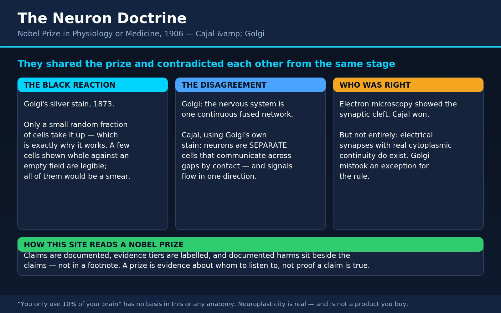
Table of Contents
- The Prize and the Two Men
- The Problem: A Grey Felt You Cannot See Into
- Golgi's Black Reaction, 1873
- Cajal Picks It Up, 1887
- The Disagreement: Reticulum or Neuron
- Stockholm, December 1906
- Who Was Right — and the Honest Nuance
- What the Neuron Doctrine Bought
- Cajal's Other Findings: Growth Cones and the Harsh Decree
- Golgi's Other Legacy: An Apparatus and a Fever Clock
- Reading Brain Claims Today
- Where Mainstream Science Agrees / What Remains Debated
- Key Research Papers
- Connections
- Featured Videos
1. The Prize and the Two Men
On 11 December 1906, two men stood up in Stockholm on the same afternoon, accepted the same Nobel Prize in Physiology or Medicine, and told the audience two incompatible things about the same tissue. The citation said they were honored "in recognition of their work on the structure of the nervous system." What it did not say was that they could not agree on what that structure was — and that one of them was about to use his Nobel Lecture to attack the other's central idea while the other sat listening.
Camillo Golgi (1843–1926) was an Italian physician and pathologist. He was born in Corteno, a mountain village in the province of Brescia that has since been renamed Corteno Golgi in his honor, and he trained in medicine at the University of Pavia, graduating in 1865. He spent most of his working life at Pavia, eventually as professor of general pathology and, for a period, rector of the university. He was methodical, institutional, well connected, and by the end of his life a senator of the Kingdom of Italy. He also invented, almost single-handedly and in a converted hospital kitchen, the staining method that made modern neuroanatomy possible.
Santiago Ramón y Cajal (1852–1934) was Spanish, from the village of Petilla de Aragón in Navarre, and by his own cheerful account he was an appalling child. He was rebellious, obsessed with drawing, and repeatedly in trouble — the most famous incident being a homemade cannon he built as a boy and fired at a neighbor's wooden gate, destroying it and landing him briefly in the village jail. His father, Justo Ramón, a barber-surgeon who had clawed his way up to a medical degree and a professorship of applied anatomy, responded to this behavior by pulling him out of school and apprenticing him — first to a barber, then to a cobbler. Cajal wanted to be an artist. His father regarded that as a catastrophe and refused.
What eventually converted the boy was, of all things, bones. His father took him to a local ossuary to collect human skeletal remains and taught him osteology from the specimens, and Cajal — who could draw anything he could see — discovered that anatomy was a subject where looking hard and drawing well were the entire job. He took a medical degree at Zaragoza in 1873, served as an army medical officer in Cuba during the Ten Years' War, came home with malaria and tuberculosis, recovered, and by 1877 had a doctorate from Madrid. He held anatomy chairs at Valencia, Barcelona, and finally Madrid.
He never stopped drawing. Over roughly fifty years he produced somewhere near three thousand ink drawings of nervous tissue, made at the bench with the microscope in front of him, and a great many of them are still reproduced in neuroscience textbooks today — not as historical curiosities but because they remain the clearest available depiction of what they show. The boy who was forbidden to be an artist ended up drawing the nervous system better than anyone before or since, and it is not clear that anyone has beaten him yet.
These two shared the prize. They disliked each other's conclusions, and by most accounts they barely spoke in Stockholm. The story of how they got there — and of how the argument was eventually settled, and of the small but real sense in which the loser was not wholly wrong — is one of the most instructive episodes in the history of medicine, and it is worth telling carefully.
2. The Problem: A Grey Felt You Cannot See Into
To understand why this was hard, put yourself at a microscope in 1870 with a thin slice of brain on the stage.
By that date, cell theory was already settled for most of the body. Everyone accepted that liver, skin, muscle, and blood were built from discrete cells — the proposition Schleiden and Schwann had advanced in the late 1830s and that had held up everywhere it was tested. Nervous tissue was the glaring exception, and the reason was purely technical: nobody could see into it.
Stains available at the time — carmine, hematoxylin, and their relatives — color everything. In liver, that is fine, because liver cells are compact, roughly polygonal, and packed like paving stones; a stain that colors all of them still leaves the boundaries legible. Nerve tissue is not like that. A single neuron is a cell body a few thousandths of an inch across with a spray of branching processes that may run for millimeters or, in the case of a motor axon, for a meter. Millions of them are interwoven in a volume the size of a sugar cube. Stain the lot, and what you get down the eyepiece is a dense, uniform grey felt — the nineteenth-century anatomists called it the Punktsubstanz or "dotted substance" — in which you can make out cell bodies and you can make out a matted thicket of fibers, but you cannot follow any single fiber from where it starts to where it ends, and you cannot say whether the thicket is made of separate threads touching or of one continuous fused mesh.
That is not a small gap in the picture. It is the question, and it decides how you are allowed to think about everything the nervous system does.
If the brain is a continuous network — a single fused reticulum in which all the processes run into one another and the "cells" are just thickenings along a common web — then nervous activity is a diffuse, spreading, field-like phenomenon. Excitation propagates through the mesh. There are no units. There is no direction of flow imposed by anatomy. Localization of function becomes suspect, because there is nothing to localize in. Learning cannot mean changing a connection, because there are no connections in the ordinary sense — there is one thing.
If instead the brain is built of separate cells that contact one another without merging, then everything changes. There are units. There are junctions between units. Signals must be handed across those junctions somehow, which immediately raises the question of how. Connections can be selectively strong or weak, which gives you a physical substrate for learning. Circuits can be traced. Pathways can have a direction. Damage can be localized. Essentially every concept modern neurology and neuroscience runs on — the reflex arc, the sensory pathway, the synapse, the circuit, the neurotransmitter, the receptor, plasticity — presupposes that the second answer is the correct one.
In 1870 nobody could tell which it was, because the tissue would not show itself. What was needed was not a better argument. It was a better stain.
3. Golgi's Black Reaction, 1873
In 1872, Golgi — then not yet thirty, and short of money — took a post as chief resident physician at the Pio Luogo degli Incurabili, a hospice for the chronically ill in the small town of Abbiategrasso, outside Milan. It was a step away from the research world, not toward it. He set up a laboratory in the hospital kitchen and worked there in the evenings, reportedly by candlelight, on the problem of staining nervous tissue.
What he found in 1873 he called la reazione nera — the black reaction. The method is startlingly simple. Harden a block of nervous tissue in potassium dichromate (bichromate of potash) for a period of days to weeks. Then transfer it to a solution of silver nitrate. Inside the tissue, silver chromate precipitates as a fine, dense, opaque deposit — and it does so inside a small number of cells, filling them completely: cell body, every branch of the dendritic tree, the axon and its collaterals, out to the finest twigs, all of it rendered in solid black against a clear amber background.
Golgi announced it in a short communication, "Sulla struttura della sostanza grigia del cervello" ("On the structure of the grey matter of the brain"), in the Gazzetta Medica Italiana. Lombardia, 1873, volume 33, pages 244–246. That is a regional Italian medical weekly, in Italian, with essentially no international circulation. The most important methodological advance in the history of neuroanatomy was published in a venue where almost nobody who needed to read it would ever see it, and for roughly a decade almost nobody did.
Why the method's biggest weakness is what makes it work
Here is the part that still surprises people. The Golgi stain does not stain all the cells. It stains a small, apparently scattered subset — the usual estimate is on the order of one to five percent — and it leaves the rest completely untouched. Why it selects the cells it selects has never been fully explained. Investigators have looked hard at the question: a 1987 study in the Journal of Comparative Neurology examined whether the impregnation is genuinely random or biased toward particular cell types, and the question of what governs the selection remains open more than 150 years after Golgi first mixed the solutions. The most honest summary is that the mechanism is understood in outline — something about local tissue chemistry determines where the silver chromate crystal nucleates and then grows to fill the cell — and not in detail.
And that unexplained capriciousness is precisely why the method solved the problem. Consider the alternative. A stain that colored every neuron completely would produce a solid black block: perfect information, zero legibility. What the reader needs is not more signal, it is contrast — a handful of cells shown in their entirety, in isolation, with the surrounding thicket rendered invisible. That is exactly what the black reaction delivers. For the first time, an anatomist could look at a single neuron the way you look at a bare tree in winter: whole, in outline, against an empty sky.
It is worth sitting with that, because it is a general lesson about scientific instruments and it recurs constantly. The most useful technique is very often not the one that shows you the most. It is the one that throws away the right ninety-nine percent. Modern neuroscience has rediscovered the same principle repeatedly — sparse genetic labeling, single-cell sequencing, single-molecule imaging — and every one of them is Golgi's insight wearing new clothes.
Golgi's original preparations survive. A 2019 study in Frontiers in Neuroanatomy examined the actual histological slides held in Pavia, and confirmed both what he saw and the quality of what he made. He was, by any measure, an outstanding technician.
4. Cajal Picks It Up, 1887
In 1887 Cajal, then thirty-five and holding a chair at Valencia, traveled to Madrid on academic business and called on Luis Simarro Lacabra, a psychiatrist and neurologist who had trained in Paris and kept up with continental technique. Simarro showed him preparations made with Golgi's method.
Cajal's reaction, which he described later in vivid terms, was the reaction of a man who has been trying to read a book in the dark and is handed a lamp. He went home and began making Golgi preparations by the hundred, and then by the thousand. Within about two years he had overturned the field.
He did it by being better at the method than its inventor, in three specific ways.
He repeated the impregnation. Cajal found that running the dichromate–silver sequence twice on the same tissue — the "double impregnation" — produced far more consistent and complete filling. Golgi's version was notoriously capricious; a batch could simply fail. Cajal's version was still capricious, but it worked often enough to support industrial-scale production of slides.
He used young animals. This is the change that mattered most, and it is a beautiful piece of reasoning. Adult nervous tissue is a nightmare for this method because mature axons are wrapped in thick myelin sheaths, which both obstruct the impregnation and swell the fibers so that the tissue is even more crowded. Cajal reasoned that embryonic and newborn animals — chick embryos, newborn mice, young birds — have neurons that are largely unmyelinated, thinner, more widely spaced, and simpler in their branching, while still being recognizably the same cells that the adult will have. He was, in effect, reading the finished manuscript by looking at the draft. He also gravitated toward tissues with a naturally orderly architecture — cerebellum, retina, olfactory bulb, spinal cord — where the cells line up in layers and a break in the pattern is obvious.
He out-worked everybody. This is not a rhetorical flourish. Cajal made and examined tens of thousands of preparations across his career, frequently working twelve to fifteen hours a day, and he drew what he saw immediately, at the bench, in ink. He worked largely alone — without a large school, without significant institutional funding, in a country then at the periphery of European science, and often at his own expense.
The journal he published himself
In May 1888 — the year he called his año cumbre, his peak year — Cajal did something that tells you a great deal about his situation. Unable to get his findings in front of the people who mattered, he founded his own journal, the Revista Trimestral de Histología Normal y Patológica ("Quarterly Review of Normal and Pathological Histology"), wrote most of it himself, paid for the printing out of his own pocket, and mailed copies to the leading histologists of Europe. It was in Spanish, which almost none of them read. A good many copies presumably went straight into the wastebasket.
So in October 1889 he took a different approach: he packed his own microscope and a case of his own slides, traveled to the congress of the German Anatomical Society in Berlin, set the microscope up on a table, and stood beside it asking people to look. Among those who looked was Albert von Kölliker, then the most eminent anatomist in Europe. Kölliker looked, and was convinced, and thereafter did two things that changed Cajal's life: he championed the work publicly, and he learned enough Spanish to read the Revista himself. Within a few years Cajal was internationally known.
The lesson embedded in that anecdote is one Cajal himself drew in his Advice for a Young Investigator (1897): a scientific finding that nobody can see is not yet a finding. He did not win the argument by writing a better polemic. He won it by putting the preparation under a microscope and letting the most skeptical man in the room look through the eyepiece himself.
The discipline of drawing what is there
One more thing about Cajal is worth stating plainly, because it is the methodological heart of why he was right and Golgi was wrong when they were looking at the same slides.
The Golgi method has a specific, dangerous failure mode. Because it impregnates only a scattered few percent of cells, a fiber will frequently vanish in the middle of its course — the impregnation simply stops. Two fibers from two different cells will cross in the plane of section and appear, at the resolution of a light microscope, to be one continuous fiber. A dendrite from one cell will run alongside an axon from another and look fused. The method that finally let you see individual neurons also produced a steady stream of convincing artifacts suggesting they were joined.
What protected Cajal from those artifacts was, of all things, his failed career as an artist. When you draw a structure — slowly, in ink, following each process by hand — you are forced to make a decision about every ambiguity, and you notice the ambiguity as you make it. You cannot skim. Cajal's practice was to look, draw, then change focus and look again, then change the section, then find the same configuration in a different animal and a different age. He built his conclusion out of thousands of independently observed instances, in which the fibers always ended, always in free terminals, always without fusing. Golgi, working with the same stain, saw the same ambiguous crossings and read them as continuity, because continuity is what he already believed.
That is the difference between drawing what you see and drawing what your theory expects, and it is not a nineteenth-century problem. It is exactly the problem that image analysis, region-of-interest selection, and post-hoc statistics present today.
5. The Disagreement: Reticulum or Neuron
Golgi's view, held from the 1870s to his death, was the reticular theory. In its mature form he argued that the axons of nerve cells branch and then fuse with one another, forming a single continuous rete nervosa diffusa — a diffuse nervous network extending throughout the grey matter. The cell bodies and dendrites, in his account, were largely nutritive rather than conductive; the real business of the nervous system was conducted by the fused axonal web. There were no independent units. The nervous system was one thing.
Cajal, from 1888 onward, argued the opposite. Every observation told him that nerve cells are independent, discrete units that contact one another but never merge — the doctrine of contiguity, not continuity. Axons end in free terminal arborizations. Those terminals apply themselves closely to the dendrites and cell bodies of other neurons, like a hand laid on a shoulder, and the signal is passed at that point of contact across some kind of interruption.
His four strongest lines of evidence, all of them things he had drawn hundreds of times:
- Free endings, everywhere. Follow any well-impregnated axon far enough and it terminates in a spray of fine branches that simply stop. It does not run into anything.
- Orderly layered tissues. In the cerebellum and the retina, cell types are stacked in defined strata with defined partners. A continuous fused mesh has no reason to be organized that way. A wiring diagram does.
- Development. In embryos, Cajal could watch nerve fibers grow outward from individual cell bodies into empty territory, with a motile tip leading the way. They arrived as extensions of a single cell. Nothing fused to make them.
- Degeneration. Cut a nerve, and the damage propagates only through the injured cell's own territory and stops. It does not spread indefinitely through a shared network, which is what continuity would predict.
Cajal was not the first person to suspect cell independence. The Swiss embryologist Wilhelm His had argued for it in 1886–1889 on developmental grounds, and the Swiss psychiatrist August Forel had argued for it from degeneration studies at almost the same moment. Their reasoning was sound but indirect. Cajal's contribution was that he could put the picture in front of you. In 1891 the German anatomist Heinrich Wilhelm Waldeyer gathered the evidence into a review article and gave the unit a name: the neuron. Waldeyer coined the word and the phrase "neuron doctrine"; he did comparatively little of the observing. It is a fair summary that His and Forel reasoned it, Cajal proved it, and Waldeyer named it.
The law of dynamic polarization
Cajal then took a second step, and this one was arguably more original than the first. If neurons are separate, they are not merely separate — they have a direction.
In 1891, working from the same preparations and from a close reading of which cell part faces which, Cajal proposed what he called the law of dynamic polarization: within a neuron, the signal flows in one direction only — received by the dendrites and cell body, transmitted away along the axon. (The Belgian anatomist Arthur van Gehuchten arrived at a similar formulation independently at almost the same time.) Cajal derived it from pure anatomy, before any of the electrical measurements that would eventually confirm it existed.
This is the sentence that turns a pile of cells into a nervous system. It means that a chain of neurons is a pathway, with an input end and an output end. It means information moves through the brain along traceable routes. It means the concepts of "afferent" and "efferent," "upstream" and "downstream," "sensory" and "motor" have real anatomical referents. Every circuit diagram in every neuroscience textbook is drawn under Cajal's law, and it is still, in its main lines, correct — with genuine exceptions that were discovered much later, including dendrites that release transmitter and axons that carry signals backward into the cell body.
One further thing was still missing, and neither man supplied it: a name for the point of contact. That came in 1897, when the English physiologist Charles Sherrington — needing a word for the junction while writing the nervous-system chapters of Michael Foster's Textbook of Physiology — coined synapse, from the Greek for "to clasp together."
6. Stockholm, December 1906
The Nobel Committee's decision to divide the 1906 prize between Golgi and Cajal was, on its face, elegant. One man had invented the method; the other had used it to see further than anyone. The tissue they both worked on was the same tissue. The citation covered them both without adjudicating between them.
What happened at the ceremony is one of the strangest episodes in the prize's history.
Golgi delivered his Nobel Lecture on 11 December 1906 under the title "The neuron doctrine — theory and facts." He used it to attack the neuron doctrine. Not to note reservations, not to argue that the evidence was incomplete: to reject it, in front of the Karolinska faculty, the Swedish court, and his co-laureate, who was sitting in the audience and would be speaking next.
He opened by asserting that the neuron doctrine was generally going out of favor — which was, by 1906, simply not true; the doctrine had been the mainstream position for well over a decade. He restated the diffuse nervous network. He questioned whether nerve cells were functionally independent at all. He was, as far as one can tell from the text, entirely sincere: this was not showmanship, it was a man of sixty-three defending in public the position he had held since his thirties, on the largest stage he would ever have, in the belief that the field had gone wrong.
Cajal then gave his own lecture, "The structure and connexions of neurons," and presented his evidence. He did not use it to counter-attack.
Privately he was another matter, and this is where the episode becomes genuinely illuminating rather than merely astonishing. In his autobiography, Recuerdos de mi vida (translated as Recollections of My Life), Cajal describes the pairing as a cruel irony of fate — joining, like Siamese twins united at the shoulders, scientific adversaries of such contrasting character — and he does not spare himself in the telling. He records his own indignation, his effort to hold his tongue, and his awareness that his pride was involved. The account is unflattering to Golgi's obstinacy and unflattering to Cajal's vanity, in roughly equal measure, and it is the better for it. By most accounts the two men had almost nothing to say to one another during the visit.
What the episode actually shows
It is tempting to file this under "amusing anecdote," and it is one. But it repays a closer look, because it is a clean, well-documented illustration of how scientific disputes really end — which is almost never the way the tidy version claims.
The tidy version says: evidence accumulates, the wrong party is persuaded, consensus updates. That is not what happened here. By 1906 the evidence was already lopsided and the community had already moved. Golgi was not persuaded, and he never was; he died in 1926 still defending the reticulum, twenty years after the prize and nearly forty years after Cajal's first decisive papers. He was not a crank — he was a superb experimentalist, a Nobel laureate, and the man who had built the tool. He was simply committed, and commitment of that depth is not dislodged by data.
What settled the question was not Golgi changing his mind. It was that the field's younger workers found Cajal's account more useful, built on it, and eventually constituted the field. The physicist Max Planck's famously bleak formulation — that science advances one funeral at a time — is usually quoted as cynicism. Here it is closer to plain description, and it is not really a criticism of Golgi. Deep priors are what make an expert good at anything; they are also what makes an expert the last person to abandon a position.
The practical corollary matters for anyone reading medical claims: an eminent, credentialed, prize-winning expert holding a position is not, on its own, evidence that the position is correct. Golgi held the highest possible credential in exactly the relevant field and was wrong about its central question, and the way you could have known that in 1906 was not by weighing his eminence against Cajal's but by looking at the preparations. Look at the evidence; the résumé does not substitute.
7. Who Was Right — and the Honest Nuance
Cajal was right, and this is not a close call or a matter of interpretation.
The light microscope could not settle it, because the gap between two neurons is about 20 nanometers wide and the best light microscope of 1906 could not resolve anything finer than roughly 200 nanometers. The two men were arguing about a structure ten times smaller than the smallest thing either of them could possibly have seen. That is why fifty years of skilled, honest observation could not close the question: the answer was physically below the instrument's floor.
The electron microscope changed the floor. In the mid-1950s, Sanford Palay working with George Palade, and independently Eduardo De Robertis with H. Stanley Bennett, published electron micrographs of central nervous system synapses. What the images showed was exactly what Cajal had inferred and could never have seen: two distinct cell membranes, presynaptic and postsynaptic, running parallel and separated by a narrow, continuous cleft; a cluster of small round vesicles on the presynaptic side; a thickening of the membrane on the postsynaptic side. Two cells, touching, not fused. Palay's 1956 paper in the Journal of Biophysical and Biochemical Cytology is the standard citation. The reticular theory was finished.
The part that gets flattened, and shouldn't
What usually follows in popular accounts is a morality tale: Cajal the visionary artist versus Golgi the stubborn reactionary, virtue rewarded, obstinacy punished. That version is satisfying and it is not accurate, and the inaccuracy matters because it teaches a bad habit of mind — the habit of treating a scientific dispute as a contest between a good person and a bad one.
Golgi was wrong about the main question. He was not wrong about everything, and the residue of truth in his position is real and specific. Three points, stated precisely:
First, direct cytoplasmic coupling between neurons exists. In 1957, Edwin Furshpan and David Potter reported in Nature that transmission at the crayfish giant motor synapse was electrical rather than chemical — current passing directly from one cell into the next. The structures responsible are gap junctions: arrays of channels, built in mammals from proteins called connexins, that span both membranes and create a continuous aqueous pore from the cytoplasm of one cell into the cytoplasm of the other. Ions and small molecules up to roughly 1 kilodalton pass straight through. These are electrical synapses, they are found throughout the mammalian brain — in the retina, the inferior olive, the thalamic reticular nucleus, and in networks of cortical inhibitory interneurons — and they are especially abundant in the developing brain. In a literal anatomical sense, at these junctions the cytoplasm of two neurons is continuous.
Second, genuine functional syncytia exist in other tissues. Cardiac muscle is coupled by gap junctions at the intercalated discs, which is precisely why the heart contracts as a coordinated unit rather than as a bag of independent twitching cells; smooth muscle in the gut and uterus behaves similarly. And in the brain itself, astrocytes — which vastly outnumber neurons in some regions — are extensively gap-junction-coupled into networks through which small molecules and ions move freely. Tissue that behaves as a continuous network is not a fantasy. Golgi was not imagining an impossible kind of biology; he was applying a real kind of biology to the wrong tissue.
Third — and this is where precision matters most — none of that rescues the reticular theory. Golgi's claim was not "some neurons are electrically coupled." His claim was that nerve cells have no individuality: that their axons fuse into a single continuous web which is the true functional unit, and that the cell bodies are essentially nutritive appendages hanging off it. That claim is false. A gap junction connects two cells that remain unambiguously two cells, each with its own complete plasma membrane, its own nucleus, its own protein synthesis, its own identity and lineage. The channels are discrete, regulated pores — they open and close in response to voltage, pH, and calcium — not a merger. And the overwhelming majority of signaling in the adult mammalian brain is chemical, across a cleft, exactly as Cajal described.
The accurate summary is therefore: the reticular theory was wrong, and the phenomenon it wrongly generalized is real. Golgi mistook an exception for the rule. That is a very common way to be wrong, it is a much more interesting way to be wrong than simple stubbornness, and it is worth more than a morality tale. There is also a fourth point that is sometimes made in Golgi's favor and should be handled carefully: he insisted that the nervous system functions as an integrated whole rather than as a collection of isolated units, and modern network neuroscience does take large-scale integration extremely seriously. That instinct was good. But it is a claim about function, and he defended it with a claim about anatomy that turned out to be false. Being right about the destination does not make you right about the road.
8. What the Neuron Doctrine Bought
The neuron doctrine is not an interesting historical detail. It is the load-bearing assumption underneath essentially all of modern neurology, psychiatry, and neuroscience, and the fastest way to see that is to follow what it made thinkable.
If there is a gap, something must cross it. This is the question the neuron doctrine forces and the reticular theory forbids, and it was answered within a generation. In 1921 Otto Loewi showed that a chemical released from a stimulated vagus nerve could slow a second, separately perfused frog heart — the first direct demonstration that nerves signal by releasing a substance. Henry Dale identified the substance and mapped the pharmacology. Loewi and Dale shared the 1936 Nobel Prize for chemical neurotransmission — a discovery that only makes sense if Cajal was right, because in a fused reticulum there is nothing for a transmitter to cross.
If there are transmitters, there are specific ones, and they can go wrong. That line runs directly to Arvid Carlsson, Paul Greengard, and Eric Kandel, who shared the 2000 Nobel Prize for signal transduction in the nervous system: Carlsson establishing dopamine as a transmitter in its own right and thereby making levodopa treatment for Parkinson's disease possible, Greengard working out the cascades of protein phosphorylation that a transmitter sets off inside the receiving cell, Kandel demonstrating in Aplysia that learning physically changes the strength of specific synapses. Every psychiatric and neurological drug in the pharmacopoeia — every antidepressant, antipsychotic, anticonvulsant, anesthetic, opioid, and stimulant — acts somewhere in that scheme.
If connections can change, learning has a physical substrate. Cajal saw this coming, and said so in print. In his 1894 Croonian Lecture to the Royal Society he proposed that mental exercise produces greater branching and growth of neuronal processes, strengthening existing connections and creating new ones — a remarkably direct anticipation of what is now called synaptic plasticity. The modern version began in 1973 with Bliss and Lømo's description of long-term potentiation in the rabbit hippocampus, and has grown into the central mechanistic account of memory. Richard Nicoll's 2017 history of the field in Neuron is a good entry point.
If the brain is a network of units with directed connections, it can be modeled. The entire tradition of computational and network neuroscience — and, at a considerable remove, artificial neural networks — takes Cajal's picture as its starting premise: discrete units, directed connections, weights on those connections that change with experience. Whatever one thinks of how far that abstraction can be pushed, it is Cajal's abstraction.
And it made clinical neurology coherent. Localization — the idea that a specific deficit implicates a specific structure or pathway — requires discrete traceable circuits. So does the concept of a demyelinating disease that slows conduction along particular tracts, or a stroke whose deficits map onto an arterial territory, or an epilepsy that begins in a focus and spreads along known routes.
9. Cajal's Other Findings: Growth Cones and the Harsh Decree
The growth cone, and how a nerve finds its target
In 1890 Cajal, working on chick embryo spinal cord, described the structure at the leading tip of a growing nerve fiber: a motile, club-shaped, constantly changing expansion, throwing out and retracting fine processes as it advanced. He called it the cono de crecimiento — the growth cone — and he was looking at the mechanism by which the nervous system wires itself. A 2009 study in the Journal of the History of the Neurosciences went back to Cajal's own surviving preparations and publications and confirmed what he had seen and how he had seen it.
He then made an inference that was, for 1890, close to reckless. A growth cone travels a long distance through developing tissue and arrives at a specific target. Cajal proposed that it is guided there by diffusible chemical signals released by the target itself — a chemotactic or "neurotropic" hypothesis. He had no candidate molecule, no biochemistry, and no way to test it. He simply reasoned that a tip which behaves as though it is searching is probably following something.
He was right, and the confirmation is one of the great stories in twentieth-century biology. Working in Italy in the 1940s — partly in a bedroom laboratory after being barred from academic work under the Fascist racial laws — Rita Levi-Montalcini found that a mouse tumor transplanted into a chick embryo caused nerve fibers to grow toward it even when the two tissues were not in contact, which means the signal was diffusible. With Stanley Cohen she isolated the molecule responsible: nerve growth factor. They shared the 1986 Nobel Prize. Cajal's guess about diffusible target-derived signals, made from a drawing of a moving tip, was correct, and the modern field of axon guidance — netrins, semaphorins, ephrins, slits — is its direct descendant.
"Everything may die, nothing may be regenerated"
Cajal's most famous single sentence is also his most frequently amputated one. In Degeneration and Regeneration of the Nervous System (published in Spanish in 1913–14, and in Raoul May's English translation in 1928), he wrote of the adult central nervous system that its nerve paths are something fixed, ended, immutable — that everything may die, nothing may be regenerated.
Quoted there, it is a counsel of despair, and it is quoted there constantly. But Cajal's next sentence is the one that shows what kind of scientist he was: it is for the science of the future, he wrote, to change, if possible, this harsh decree. He was reporting an observation and explicitly declining to convert it into a law of nature. He left the door open on purpose.
The modern position, stated honestly, is that he was largely right about the observation and right to leave the door open.
Peripheral nerves regenerate; central ones largely do not. Cut a nerve in the arm and, given intact connective-tissue sheaths and a reasonable gap, axons regrow at roughly a millimeter a day — slowly, imperfectly, but genuinely. Schwann cells in the peripheral nerve clear the debris and form guidance tracks. Cut a tract in the spinal cord and essentially nothing regrows across the lesion. The failure is not because central neurons are inherently incapable: it is a combination of an actively hostile environment (myelin-associated inhibitors such as Nogo-A, and the chondroitin sulfate proteoglycans of the glial scar) and a reduced intrinsic growth program in mature central neurons. The decisive demonstration came in 1981, when Sam David and Albert Aguayo showed in Science that adult rat central neurons will extend axons over long distances if you give them a graft of peripheral nerve to grow through. The neurons could do it. The neighborhood was the problem. That is exactly the kind of finding Cajal's open door was written for, and it is why central nervous system injury and spinal cord injury remain so much harder to treat than a severed nerve in a limb.
Adult neurogenesis: real, limited, and genuinely contested in extent. Cajal's "nothing may be regenerated" also implied that no new neurons are added to the adult brain, and this is where the honest answer requires more care than most sources give it.
Joseph Altman reported new neurons in the adult rat brain in the 1960s and was largely ignored for two decades. In 1998 Peter Eriksson and colleagues published evidence of new neurons in the adult human hippocampus in Nature Medicine, using tissue from cancer patients who had received the DNA label BrdU during life. In 2013, Kirsty Spalding and colleagues used a beautifully indirect method — carbon-14 from Cold War atmospheric nuclear testing, incorporated into DNA at the moment a cell is born — to date human hippocampal neurons, and estimated a modest but real ongoing turnover in the dentate gyrus continuing into old age.
Then, in 2018, two large studies were published within a month of each other and reached opposite conclusions from human post-mortem tissue. Sorrells and colleagues, in Nature, found that markers of newly generated neurons in the human hippocampus declined steeply through childhood and were essentially undetectable in adults. Boldrini and colleagues, in Cell Stem Cell, examined subjects aged 14 to 79 and found comparable numbers of immature neurons and neural progenitors across the whole age range. In 2019 Moreno-Jiménez and colleagues reported in Nature Medicine that immature neurons remained detectable into the ninth decade of life — and, importantly, argued that the discrepancy was substantially methodological, driven by how long tissue sat before fixation and how long it was fixed, both of which degrade the very markers the counting depends on. Single-cell and single-nucleus transcriptomic studies since then have not closed the question; a 2023 analysis in Neuron reviewing those datasets is candidly subtitled with the question of whether they reconcile the controversy or fuel it.
The honest state of the field, stated without spin:
- Adult neurogenesis in the human hippocampus is probably real, and it is disputed in magnitude, with credible investigators on both sides and a substantial part of the disagreement traceable to tissue handling and marker specificity rather than to biology.
- It is confined, in humans, to at most one or two small regions — principally the dentate gyrus of the hippocampus. It is not brain-wide.
- Even the most generous published estimates are small relative to the whole brain. Nobody claims the adult brain replaces damaged cortex, or regrows a stroke.
- It therefore does not support any product, supplement, program, or protocol claiming to "regrow your brain," and any marketing that cites this literature as if the question were settled in its favor is misrepresenting it.
Cajal, who declined to turn his observation into a law, would probably have recognized this as exactly the state of affairs he had in mind.
10. Golgi's Other Legacy: An Apparatus and a Fever Clock
Golgi lost the argument he cared most about. He is nonetheless one of the more consequential figures in the history of cell biology, for reasons largely unrelated to it.
The Golgi apparatus
In 1898, using a silver-and-osmium variant of his staining chemistry on Purkinje cells from the cerebellum of a barn owl and a cat, Golgi described a delicate ribbon-like structure lying inside the cell body, near the nucleus. He called it the apparato reticolare interno — the internal reticular apparatus — and published it in the bulletin of the Pavia medical society.
Then a familiar thing happened: for decades, a large fraction of cell biologists insisted it was not real. The objection was reasonable on its face. It was visible only with a capricious heavy-metal impregnation of exactly the kind known to produce artifacts; silver methods precipitate metal along all sorts of things; and no other technique showed it. The suspicion that it was a fixation or staining artifact — a smear of silver, not an organelle — ran from roughly 1910 into the 1950s, which is to say it outlived Golgi himself by a quarter of a century.
It was settled the same way the neuron doctrine was settled, by the same instrument, in the same decade. Electron microscopy in the 1950s showed the structure directly: a stack of flattened membrane-bound cisternae, present in essentially every eukaryotic cell, with a defined entry face and exit face. It is now understood as the cell's central sorting and finishing department — proteins arrive from the endoplasmic reticulum, are modified (glycosylation happens here), and are packaged and dispatched to their destinations. George Palade's Nobel Prize in 1974, shared with Albert Claude and Christian de Duve, was for the work that established this. Anna Dröscher's 1998 centenary review in Brain Research Bulletin traces the whole arc from the 1898 description to the electron-microscope vindication.
The symmetry is worth noticing and not overplaying. Golgi's method persuaded him of something false about the nervous system and something true about the cell, and in both cases the light microscope was inadequate to settle it. He was not a man with bad judgment. He was a man working at the resolution limit of his instrument, which is a different thing, and which is a hazard every generation of researchers faces with whatever their current instrument happens to be.
Malaria: reading the fever as a clock
Golgi's other major contribution was clinical, and it is the one a patient would have felt first.
Between about 1885 and 1893, Golgi studied the blood of malaria patients and worked out the relationship between the parasite's life cycle and the pattern of the fever. He showed that the fever paroxysm coincides with the moment the infected red blood cells rupture and release a new generation of parasites — the observation still known as Golgi's law. He then used that clock to do something clinically decisive: he demonstrated that tertian malaria, with a fever spike every 48 hours, and quartan malaria, with a spike every 72 hours, are caused by different parasites with different developmental cycles, not by different intensities of one infection. Those parasites are known today as Plasmodium vivax and Plasmodium malariae.
This was the beginning of malaria as a set of distinguishable diseases rather than one fever, and it made the periodicity of a patient's fever chart a diagnostic instrument. It also fits alongside Ronald Ross's demonstration, in 1897–98, that the parasite is transmitted by mosquitoes — work that won the 1902 prize. Golgi described what the parasite does inside the patient; Ross described how it gets there.
Golgi also has his name on the Golgi tendon organ, the stretch receptor in tendon that reports muscle tension to the spinal cord, and on the Golgi–Mazzoni corpuscles. A man who is remembered by an organelle, a stain, a sensory receptor, and a law of fever periodicity has not had a small career, whatever happened in Stockholm.
11. Reading Brain Claims Today
This section is short and blunt, because it is the practical part. The history above is directly relevant to a set of claims you will meet constantly in health marketing, and it settles several of them.
🟢 "You only use 10% of your brain" is false. Not exaggerated — false, with no basis in this anatomy or any other. Cajal and Golgi between them mapped the cellular architecture of the nervous system and found no reserve, no dormant sector, no unused ninety percent. Everything since agrees. The brain is about two percent of body weight and consumes around twenty percent of the body's resting energy; evolution does not maintain that bill for idle tissue. There is no region of the brain you can destroy without a deficit — the whole discipline of clinical neurology is the catalogue of what goes wrong when small areas are damaged. Functional imaging shows activity distributed across the entire brain over the course of a day. Despite all of this, the myth is remarkably durable: surveys of teachers and of the general public repeatedly find something in the region of half of respondents endorsing it, which is why it appears in every published list of "neuromyths." When a product's pitch opens with the 10% claim, you have learned something useful about the product before you have heard a word about what it does.
🟡 Neuroplasticity is real, and it is bounded. Synapses do strengthen and weaken with use; dendritic spines appear and disappear; cortical maps reorganize after injury or intensive training; skill practice produces measurable structural change. That is genuine science, it descends directly from Cajal's 1894 speculation, and it is the mechanism behind why rehabilitation after a stroke works at all. What it does not mean is unlimited change, change in any desired direction, rapid change, or change you can purchase. Plasticity is a property of tissue, not a service. Nobody sells it to you.
🟡 "Rewiring your brain" is usually marketing outrunning evidence. The phrase is not meaningless — connection strengths do change with experience — but in commercial use it almost always implies far more than the underlying study supports: that a short program produces large, durable, general change in how you think or feel. Ask three questions of any such claim. What was actually measured — a brain image, or an outcome you would care about? Compared with what — a real control group, or the same people before and after? And how long did the effect last after the program ended? Most "rewiring" claims fail at least two of the three.
🟡 Brain-training games improve the trained task, with weak transfer. This is the honest finding and it has been examined exhaustively. The pattern in the literature is consistent: practice a task and you get better at that task; you get somewhat better at very similar tasks; and you get little or no reliable improvement on general cognition, intelligence measures, or everyday functioning. The comprehensive 2016 review in Psychological Science in the Public Interest found the evidence for far transfer to be weak, and meta-analyses of working-memory training reach the same conclusion. This is not merely an academic dispute: in January 2016 the U.S. Federal Trade Commission settled deceptive-advertising charges against Lumos Labs, the maker of Lumosity, over claims that its games reduced cognitive decline and improved performance at work and school. If you enjoy the games, play them. Do not buy them as medicine.
🔴 "Regrow your brain cells" supplements and protocols are not supported. As set out in section 9, adult human neurogenesis is confined to at most a very small region, is small in magnitude, and is actively disputed in extent by serious investigators. No supplement, device, or program has been shown to increase it in humans in a way that produces a measurable clinical benefit. Marketing that cites Boldrini or Moreno-Jiménez while omitting Sorrells is quoting one side of a live scientific disagreement as though it were the verdict.
The unifying rule, and the one this whole page argues for: the claim is only as good as what was actually observed, and the credential of the person making it does not upgrade the observation. A Nobel laureate defended the reticular theory from the podium in Stockholm in 1906 and he was wrong, and the way to know that was to look at the preparations.
12. Where Mainstream Science Agrees / What Remains Debated
Where mainstream science agrees
- Neurons are discrete cells. The neuron doctrine is correct and was directly confirmed by electron microscopy in the 1950s, which resolved the synaptic cleft, the presynaptic vesicles, and the two separate membranes.
- The Golgi stain works because it impregnates only a small subset of cells, giving contrast rather than completeness. Why it selects those particular cells is still not fully explained.
- Cajal's law of dynamic polarization — dendrites and soma receive, axon transmits — is correct in its main lines and remains the organizing principle of circuit anatomy.
- Most signaling in the adult mammalian brain is chemical, across a synaptic cleft, using released transmitters and postsynaptic receptors.
- Electrical synapses formed by gap junctions are real, are widespread, and do involve genuine cytoplasmic continuity between two otherwise distinct cells. Astrocytes are extensively gap-junction coupled.
- The Golgi apparatus is a real organelle, not a staining artifact, present in essentially all eukaryotic cells, and central to protein processing and trafficking.
- Peripheral nerves regenerate; central axons largely do not, for environmental as well as intrinsic reasons.
- Synaptic plasticity is the leading mechanistic account of learning and memory.
- The "10% of your brain" claim is false.
- Brain-training programs reliably improve the trained task, with little evidence of far transfer to general cognition.
What remains debated
- The extent of adult human hippocampal neurogenesis. Genuinely unsettled. Sorrells (2018) versus Boldrini (2018) and Moreno-Jiménez (2019) is a live disagreement, with much of it turning on post-mortem interval, fixation protocol, and antibody specificity rather than on biology. Anyone who tells you it is settled, in either direction, is overstating.
- The functional weight of electrical synapses in the adult human brain. Their existence is not in question; how much of adult cortical computation depends on them is an active research area.
- The mechanism of Golgi impregnation. Still not fully explained, more than 150 years on, including whether the selection is truly random or systematically biased toward certain cell types.
- How far synaptic plasticity accounts for memory. Long-term potentiation is the leading candidate mechanism, and the mapping between specific synaptic changes and specific remembered content is far from complete.
- Whether any intervention meaningfully promotes central nervous system regeneration in humans. Multiple approaches — growth factors, inhibitor blockade, scar modification, cell grafts — look promising in animals; clinical translation for spinal cord injury remains limited.
- How much structural brain change a given amount of training produces, and whether it matters functionally. Plasticity is real; the effect sizes reported in training studies are frequently small, and replication has been uneven.
13. Key Research Papers
Golgi's and Cajal's own foundational works date from 1873 to 1906, were published in Italian and Spanish journals, and are not indexed in PubMed. They are given here with full bibliographic detail, followed by topic searches for the modern literature that discusses them.
- Golgi C. Sulla struttura della sostanza grigia del cervello [On the structure of the grey matter of the brain]. Gazzetta Medica Italiana. Lombardia 1873;33:244–246. — the announcement of the black reaction. Related literature on PubMed
- Golgi C. Sulla infezione malarica. Archivio per le Scienze Mediche 1886;10:109–135. — the malarial fever cycle and the separation of tertian from quartan fevers. Related literature on PubMed
- Golgi C. Intorno alla struttura delle cellule nervose. Bollettino della Società Medico-Chirurgica di Pavia 1898;13:3–16. — the description of the internal reticular apparatus, now the Golgi apparatus. Related literature on PubMed
- Ramón y Cajal S. Estructura de los centros nerviosos de las aves. Revista Trimestral de Histología Normal y Patológica 1888;1:1–10. — the first of the decisive papers, in the journal he founded and paid for himself. Related literature on PubMed
- Ramón y Cajal S. Textura del sistema nervioso del hombre y de los vertebrados. Madrid: Nicolás Moya; 1899–1904. — the three-volume synthesis, later expanded as the French Histologie du système nerveux de l'homme et des vertébrés (1909–1911). Related literature on PubMed
- Ramón y Cajal S. Estudios sobre la degeneración y regeneración del sistema nervioso. Madrid: Moya; 1913–1914. English translation by R.M. May, Degeneration and Regeneration of the Nervous System, Oxford University Press, 1928. — the source of "everything may die, nothing may be regenerated," and of the sentence about the science of the future that usually gets dropped. Related literature on PubMed
- Golgi C. The neuron doctrine — theory and facts. Nobel Lecture, Stockholm, 11 December 1906. — Ramón y Cajal S. The structure and connexions of neurons. Nobel Lecture, Stockholm, 11 December 1906. Both published in Nobel Lectures, Physiology or Medicine 1901–1921, Elsevier, 1967. Related literature on PubMed
Modern peer-reviewed literature:
- Glickstein M. Golgi and Cajal: the neuron doctrine and the 100th anniversary of the 1906 Nobel Prize. Curr Biol 2006;16(5):R147-51
- De Carlos JA, Borrell J. A historical reflection of the contributions of Cajal and Golgi to the foundations of neuroscience. Brain Res Rev 2007;55(1):8-16
- Bentivoglio M, Cotrufo T, Ferrari S, et al. The original histological slides of Camillo Golgi and his discoveries on neuronal structure. Front Neuroanat 2019;13:3
- Shimono M, Tsuji N. Study of the selectivity of the impregnation of neurons by the Golgi method. J Comp Neurol 1987;259(1):122-30
- Dröscher A. The history of the Golgi apparatus in neurones from its discovery in 1898 to electron microscopy. Brain Res Bull 1998;47(3):199-203
- Palay SL. Synapses in the central nervous system. J Biophys Biochem Cytol 1956;2(4 Suppl):193-202
- Garcia-Marin V, Garcia-Lopez P, Freire M. The growth cone as seen through Cajal's original histological preparations and publications. J Hist Neurosci 2009;18(2):197-210
- Pereda AE. Electrical synapses and their functional interactions with chemical synapses. Nat Rev Neurosci 2014;15(4):250-63
- Nicoll RA. A brief history of long-term potentiation. Neuron 2017;93(2):281-290
- David S, Aguayo AJ. Axonal elongation into peripheral nervous system "bridges" after central nervous system injury in adult rats. Science 1981;214(4523):931-3
- Sorrells SF, Paredes MF, Cebrian-Silla A, et al. Human hippocampal neurogenesis drops sharply in children to undetectable levels in adults. Nature 2018;555(7696):377-381
- Boldrini M, Fulmore CA, Tartt AN, et al. Human hippocampal neurogenesis persists throughout aging. Cell Stem Cell 2018;22(4):589-599.e5
- Tosoni G, Ayyildiz D, Bryois J, et al. Mapping human adult hippocampal neurogenesis with single-cell transcriptomics: reconciling controversy or fueling the debate? Neuron 2023;111(11):1714-1731.e3
- Simons DJ, Boot WR, Charness N, et al. Do "brain-training" programs work? Psychol Sci Public Interest 2016;17(3):103-186
- Howard-Jones PA. Neuroscience and education: myths and messages. Nat Rev Neurosci 2014;15(12):817-24
Live PubMed Searches
- Neuron doctrine — Cajal, history
- Golgi apparatus — discovery
- Adult hippocampal neurogenesis in humans
- Electrical synapses and gap junctions
- Brain training — transfer effects
14. Connections
- All Notable Doctors
- The Nobel Prize in Physiology or Medicine — the full roll, and where the 1906 prize sits in it
- Otto Loewi & Henry Dale — what crosses the gap Cajal predicted: chemical neurotransmission, 1936
- Carlsson, Greengard & Kandel — dopamine, signal transduction, and learning as a change in synaptic strength
- Rita Levi-Montalcini & Stanley Cohen — nerve growth factor: the molecule behind Cajal's guess about how axons find their targets
- Ronald Ross — the mosquito half of the malaria story that Golgi worked on from the patient's side
- David Julius & Ardem Patapoutian — the receptors that start a sensory signal on its way through Cajal's circuits
- Stanley Prusiner — another prize awarded over sustained expert objection, and how that one resolved
- Neurology — the clinical discipline that the neuron doctrine made coherent
- Parkinson's Disease — a disorder of one transmitter in one pathway, describable only in Cajal's terms
- Alzheimer's Disease — synapse loss as the change that tracks cognitive decline most closely
- Multiple Sclerosis — what happens when the insulation on Cajal's wiring is attacked
- Stroke — localization, arterial territories, and the plasticity that rehabilitation depends on
- Traumatic Brain Injury — why central nervous tissue does not repair itself the way a cut nerve in the arm does
- Peripheral Neuropathy — the nerves that do regenerate, at roughly a millimeter a day