Glycine and Aspirin Metabolism: The Salicyluric Acid Pathway
When you swallow an 81 mg baby aspirin, your liver does not simply “break it down.” It runs a two-stage chemical disposal sequence — and the rate-limiting step of that sequence is the availability of a single amino acid: glycine. This is the biochemistry every long-term aspirin user, every parent who has ever heard the words “Reye's syndrome,” and every clinician treating salicylate overdose should understand.
Table of Contents
- Overview: Why Glycine Matters for Aspirin
- Step 1: Aspirin → Salicylate
- Step 2: Salicylate → Salicyluric Acid
- The GLYAT Enzyme
- Why Glycine Becomes Rate-Limiting
- Aspirin Overdose and Glycine Depletion
- The Reye's Syndrome Connection
- Genetic Variation in GLYAT
- Salicylate's Nonlinear Pharmacokinetics
- Alternative Disposal Pathways
- Cofactors: CoA, ATP, and Pyridoxal Phosphate
- Other Drugs Cleared by Glycine Conjugation
- The Daily Low-Dose Aspirin User
- Bone Broth and Collagen: A Practical Strategy
- Salicyluric Acid as a Urinary Marker
- Clinical Takeaways
- Research Papers
- Connections
- Featured Videos
Overview: Why Glycine Matters for Aspirin
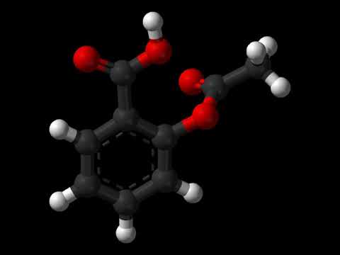
Aspirin (chemical name acetylsalicylic acid) is one of the most widely consumed pharmaceuticals in the world. Roughly 40 to 50 million Americans take low-dose aspirin daily for cardiovascular prevention, and tens of millions more reach for an occasional 325 mg or 650 mg tablet for headache, fever, or musculoskeletal pain. Yet most people — and many of their physicians — have no idea that the body's ability to clear that aspirin depends on a non-essential amino acid found in collagen, bone broth, and gelatin.
The key insight is this: glycine does not metabolize aspirin directly. Aspirin is first hydrolyzed by plasma and tissue esterases into salicylic acid (also called salicylate), which is the molecule that does almost all of aspirin's anti-inflammatory and analgesic work in the body. Salicylate then enters a second phase of metabolism in which it must be conjugated — chemically attached — to either glycine or glucuronic acid before the kidneys can efficiently filter it out into the urine. The glycine conjugate is called salicyluric acid, and at therapeutic aspirin doses it represents 60 to 75 percent of the total urinary disposal pathway. The glucuronic acid pathway handles most of the rest.
The glycine conjugation reaction is catalyzed by an enzyme called glycine N-acyltransferase (abbreviated GLYAT), which sits in the mitochondrial matrix of hepatocytes. GLYAT has a relatively low affinity for its glycine substrate — the published Michaelis constant (Km) for glycine is roughly 6 millimolar, which is near the upper end of physiological hepatic mitochondrial glycine concentrations. The practical consequence: when the hepatic glycine pool drops — whether from a low-protein diet, chronic illness, oxidative stress, advanced age, or a large aspirin load that simply outpaces glycine supply — salicyluric acid formation slows down dramatically, and salicylate begins to accumulate. This is the molecular basis for the well-documented nonlinear pharmacokinetics of salicylate at higher doses.
Understanding this pathway changes how we think about three distinct clinical scenarios: (1) the patient taking 81 mg daily for cardiovascular prevention, who may benefit from modestly higher glycine intake; (2) the patient who takes 2 to 4 grams per day for inflammatory arthritis or pericarditis and is therefore stressing the glycine pool every day; and (3) the patient who has ingested a toxic dose of aspirin, in whom plasma glycine has been documented to fall and in whom glycine supplementation has been proposed as an adjunctive treatment to accelerate elimination.
Step 1: Aspirin → Salicylate
The first thing that happens to swallowed aspirin is hydrolysis — the cleavage of the acetyl group from the salicylate backbone. This reaction is catalyzed primarily by plasma carboxylesterases, with additional activity contributed by carboxylesterase enzymes in the liver, the gut wall, and the red blood cells. The acetyl group is transferred onto plasma proteins (including albumin and platelet cyclooxygenase) and onto water, leaving behind salicylic acid as the principal circulating metabolite.
Aspirin's plasma half-life is short — only about 15 to 30 minutes. Within an hour of an oral dose, the bulk of the parent aspirin molecule has already been hydrolyzed to salicylate. This is the critical pharmacological transition: aspirin's unique activity — the irreversible acetylation of platelet cyclooxygenase-1 that gives baby aspirin its 7- to 10-day antiplatelet effect — happens during this brief window. After that, the systemic anti-inflammatory and analgesic effects belong to salicylate.
This distinction matters because salicylate, not aspirin, is what your liver and kidneys are actually dealing with most of the time. The downstream metabolic load — the work performed by glycine conjugation, glucuronidation, and renal excretion — is all directed against salicylate. From this point forward in the article, “aspirin metabolism” really means salicylate metabolism.
Step 2: Salicylate → Salicyluric Acid
Salicylate cannot simply be filtered into urine in its parent form. It is a small, lipophilic, mostly un-ionized organic acid that the kidneys reabsorb at the proximal tubule, especially when the urine is acidic. To excrete it efficiently, the liver must convert it into a more water-soluble, more polar derivative that the kidneys can no longer reclaim. The body has two main ways to accomplish this:
- Glycine conjugation — salicylate is activated to salicyl-coenzyme A and then joined to glycine to form salicyluric acid (also written as salicyluryl glycine or 2-hydroxyhippuric acid). This is the dominant pathway, accounting for roughly 60 to 75 percent of urinary salicylate disposal at therapeutic doses.
- Glucuronic acid conjugation — salicylate is joined to glucuronic acid by uridine diphosphate-glucuronosyltransferases (UGTs) to form two glucuronides, the ether (phenolic) glucuronide and the ester (acyl) glucuronide. Together these account for roughly 15 to 25 percent of urinary disposal.
A smaller fraction (typically 5 to 15 percent) is excreted as unchanged salicylate, and a minor amount is oxidized to gentisic acid (2,5-dihydroxybenzoic acid). The exact fractions depend on dose, urinary pH, fluid intake, age, genetics, and underlying hepatic and renal function.
The glycine conjugation reaction proceeds in two enzymatic steps inside the mitochondrial matrix:
- Activation. Salicylate is joined to coenzyme A (CoA) by an ATP-dependent mitochondrial acyl-CoA synthetase, producing salicyl-CoA. This step consumes one molecule of ATP per molecule of salicylate, generating AMP and pyrophosphate as byproducts.
- Conjugation. Glycine N-acyltransferase (GLYAT) transfers the salicyl group from CoA onto the amino nitrogen of free glycine, releasing CoA and producing salicyluric acid — an amide bond between salicylate's carboxyl group and glycine's amino group.
Salicyluric acid is then exported from the mitochondrion into the hepatocyte cytosol, into the hepatic sinusoidal blood, filtered by the glomerulus, and excreted in the urine. Unlike the parent salicylate, salicyluric acid is not significantly reabsorbed by the renal tubule, so this conjugation step effectively commits the salicylate molecule to elimination.
The GLYAT Enzyme
Glycine N-acyltransferase is a 33 to 34 kilodalton mitochondrial matrix enzyme encoded by the GLYAT gene on human chromosome 11q12.1. It belongs to a small family of mitochondrial acyl-CoA:amino acid N-acyltransferases that includes GLYATL1 and GLYATL2 (glycine N-acyltransferase-like 1 and 2), which have somewhat different substrate preferences. GLYAT proper is expressed most strongly in the liver and to a lesser extent in the kidney; minor expression has been detected in other tissues including the pancreas and adrenal cortex.
The enzyme accepts a remarkable range of acyl-CoA substrates. In the human diet and human pharmacopoeia, GLYAT is responsible for glycine conjugation of:
- Benzoic acid (food preservative; metabolized to hippuric acid) — the original substrate Wiley Quick used in his classic 1930s studies of human protein metabolism
- Salicylic acid (from aspirin and willow bark) — metabolized to salicyluric acid
- Nicotinic acid (niacin) — metabolized to nicotinuric acid at high niacin doses
- 4-aminobenzoic acid (PABA) — partially conjugated to p-aminohippuric acid
- Cinnamic acid (from plant foods and the gut microbiome's degradation of polyphenols)
- Phenylacetic and phenylpropionic acids (bacterial fermentation products of dietary phenylalanine)
- Various branched-chain and medium-chain fatty acid intermediates, which is why GLYAT activity matters in inborn errors of metabolism such as isovaleric acidemia
GLYAT's catalytic mechanism uses a ping-pong bi-bi kinetic scheme: the acyl-CoA binds first and transfers its acyl group to an enzyme-bound intermediate, CoA is released, glycine then binds, and the acyl group is transferred onto glycine's nitrogen to form the final conjugate. The reaction is essentially irreversible under physiological conditions.
The Michaelis constants reported in the human GLYAT literature give a clear picture of where the bottlenecks lie. For salicyl-CoA, the Km is in the low micromolar range — meaning the enzyme is nearly saturated with respect to its acyl-CoA substrate at any physiologically relevant salicylate exposure. For glycine, however, the Km is approximately 6 to 8 millimolar, and hepatic mitochondrial glycine concentrations under normal conditions sit in roughly that same range. This is the textbook definition of a rate-limiting cofactor.
Why Glycine Becomes Rate-Limiting
The Km of an enzyme for a particular substrate is the concentration of that substrate at which the enzyme is operating at half of its maximum velocity. If the substrate concentration is far above the Km, the enzyme is saturated and small fluctuations in substrate concentration do not change the reaction rate. If the substrate concentration is far below the Km, the enzyme is starved and the reaction rate is roughly proportional to substrate availability.
For GLYAT with glycine, hepatic mitochondrial glycine sits at the Km rather than well above it. This means several common physiological perturbations can push glycine availability below the threshold required to maintain salicyluric acid formation at full speed:
- Low-protein diet. Glycine is in most foods but is particularly concentrated in collagen-rich foods (bone broth, skin, tendons, cartilage). Diets that emphasize muscle meat over “nose-to-tail” eating can be surprisingly glycine-poor. Endogenous glycine synthesis from serine, threonine, and choline supplies roughly 3 g/day, but total daily metabolic demand is closer to 10 to 13 grams.
- Aging. Endogenous glycine synthesis declines with age. Plasma glycine and erythrocyte glutathione both fall in the elderly, and supplementation has been shown to restore both.
- Chronic illness. Diabetes, obesity, fatty liver disease, sepsis, and major trauma all increase glycine demand for glutathione synthesis, acute-phase protein production, and tissue repair, leaving less for drug conjugation.
- Competing conjugation demand. If multiple glycine-conjugated substrates are present simultaneously — for example, dietary benzoate from soft drinks plus a therapeutic aspirin dose plus heavy plant-polyphenol consumption — they all draw from the same hepatic glycine pool.
- High-dose aspirin itself. A 4 g/day anti-inflammatory aspirin regimen requires the liver to dispose of roughly 4 grams of salicylate, which in turn requires about 2.5 grams of glycine to be consumed in the conjugation reaction every 24 hours.
- Pregnancy. Glycine demand rises substantially during pregnancy to support fetal collagen and protein synthesis, and maternal plasma glycine drops in the third trimester.
- Vegetarian and vegan diets. These tend to be lower in pre-formed glycine (which is concentrated in animal connective tissue) and depend more on endogenous synthesis.
None of these scenarios produce frank glycine deficiency by classical clinical definitions, but each can be enough to push hepatic mitochondrial glycine below the Km of GLYAT and slow salicyluric acid formation. When that happens, salicylate accumulates, the glucuronidation pathway is recruited more heavily, and the apparent plasma half-life of salicylate lengthens.
Aspirin Overdose and Glycine Depletion
The clearest demonstration that glycine is the rate-limiting substrate in salicylate clearance comes from the clinical literature on aspirin overdose. When patients ingest a toxic dose of aspirin (generally above 150 mg/kg, or roughly 10 g in an adult), several characteristic biochemical changes occur in addition to the well-known acid-base disturbances:
- Plasma glycine falls. Patel and colleagues at the University of Bath documented in 1990 that patients admitted with aspirin overdose had measurably lower plasma glycine concentrations than control subjects, and that the urinary fraction of salicylate excreted as salicyluric acid fell while the unconjugated salicylate and glucuronide fractions rose.
- The salicyluric acid pathway saturates. The maximum capacity of GLYAT-mediated salicyluric acid formation in a healthy adult liver is approximately 700 to 900 mg of salicylate per hour — well below the rate at which a toxic dose is absorbed. Once this ceiling is reached, additional salicylate exits through glucuronidation and renal excretion of the unchanged molecule, both of which are slower and less efficient.
- Half-life lengthens dramatically. Salicylate's plasma half-life at therapeutic doses is roughly 2 to 4 hours. At toxic doses it stretches to 18 to 36 hours, with a few case reports of more than 48 hours. This is the classic textbook example of capacity-limited pharmacokinetics.
- Urinary alkalinization becomes life-saving. Because un-ionized salicylate is reabsorbed by the renal tubule, raising urinary pH above 7.5 with sodium bicarbonate dramatically increases the fraction of salicylate that remains ionized in the tubular fluid and is therefore excreted. This is the cornerstone of modern medical management of salicylate poisoning.
Several investigators have proposed adjunctive glycine administration in salicylate overdose as a way to accelerate the glycine conjugation pathway and shorten the half-life. The proposal has biochemical plausibility — intravenous or oral glycine raises plasma glycine and hepatic glycine and can restore salicyluric acid formation toward its maximum capacity. The intervention has been used in case-series and small clinical reports but has never been tested in a large randomized trial, primarily because urinary alkalinization and (in severe cases) hemodialysis remain so effective that there has been little perceived need for a metabolic add-on.
The take-home for prevention is more interesting than the take-home for treatment: chronic glycine adequacy is one of several factors that determine the slope of an individual's dose-response curve to salicylate. Two people taking the same chronic aspirin dose can have meaningfully different steady-state plasma salicylate concentrations depending on their underlying glycine status.
The Reye's Syndrome Connection
Reye's syndrome is a rare but devastating condition in which a child or adolescent develops sudden hepatic mitochondrial dysfunction, hypoglycemia, hyperammonemia, and progressive encephalopathy, typically following a viral illness — classically influenza or varicella — that was treated with aspirin. The epidemiological link between aspirin and Reye's was established in the early 1980s by Starko and others, and the resulting public-health warnings (and the substitution of acetaminophen and ibuprofen for childhood fevers) drove the incidence in the United States from more than 500 cases per year in the early 1980s to fewer than 2 per year by the late 1990s.
The molecular pathogenesis of Reye's syndrome is now understood to be a multi-hit failure of the mitochondrial matrix, and glycine conjugation sits squarely in the middle of that compartment. The current consensus model holds that:
- Viral illness causes a baseline impairment of mitochondrial fatty acid β-oxidation, especially in young children whose metabolic reserves are smaller.
- Salicylate — particularly at the relatively high doses used historically in children for fever — further inhibits β-oxidation by competing with medium- and long-chain fatty acids for activation by mitochondrial acyl-CoA synthetases, and by trapping CoA in the form of salicyl-CoA.
- The combined inhibition impairs ATP generation in the liver and brain, causing the characteristic hypoglycemia, hyperammonemia, and cerebral edema.
- Children with an unrecognized inborn error of fatty acid oxidation — for example, medium-chain acyl-CoA dehydrogenase (MCAD) deficiency — are particularly susceptible, which is why some cases of suspected Reye's syndrome in the modern era are now classified as “Reye-like” presentations of an underlying metabolic disease.
Glycine availability plays a defensive role in this cascade. Adequate glycine accelerates the disposal of salicyl-CoA back into urinary salicyluric acid, freeing up CoA and reducing the mitochondrial salicyl-CoA load. Glycine is also a precursor to glutathione, the major mitochondrial antioxidant that defends against the oxidative stress generated when β-oxidation is impaired. The historical era of high-dose aspirin in feverish children — before Reye's was understood — was effectively a perfect storm for glycine and CoA insufficiency in the worst possible compartment.
Clinical bottom line: aspirin is contraindicated in children and adolescents under approximately 18 years of age with viral illness, particularly influenza, varicella, and gastroenteritis, except for specific indications (Kawasaki disease being the major exception). Acetaminophen and ibuprofen are the preferred antipyretics in this age group. Willow bark, which produces salicylic acid through the same downstream pathway, carries the same warning.
Genetic Variation in GLYAT
Population studies have shown that healthy adults vary by roughly two- to five-fold in the fraction of an aspirin dose they excrete as salicyluric acid versus the alternative glucuronide pathways. Some of this variation is dietary (current glycine status), some is age-related, and some is genetic.
The GLYAT gene contains a number of well-characterized single nucleotide polymorphisms. Two of the more frequently studied variants in human pharmacogenetic research are:
- N156S (a serine substitution at position 156) — relatively common, reduces enzyme Vmax by roughly 30 percent in heterologous expression studies.
- S17T (a threonine substitution at position 17) — less common, with a smaller effect on activity.
South African researchers, including the Erasmus and Badenhorst groups, have characterized additional variants in African and European populations and have shown that GLYAT haplotype is one of several factors that determine an individual's salicyluric acid excretion ratio. The clinical significance of these variants in routine aspirin use is modest, but the variation may matter for chronic high-dose aspirin users, for patients with concurrent low glycine intake, and for the rare individual who develops salicylate toxicity at unexpectedly low doses.
There is no commercially available GLYAT genotyping panel in routine clinical use. Indirect functional assessment is possible by measuring the ratio of urinary salicyluric acid to total urinary salicylate after a standardized aspirin dose — a research-grade test only.
Salicylate's Nonlinear Pharmacokinetics
Salicylate is one of the textbook examples of a drug with capacity-limited (Michaelis-Menten) elimination. At low doses (such as 81 mg of aspirin) the salicyluric acid pathway is operating well below its Vmax, glycine is not significantly depleted, and salicylate is cleared by first-order kinetics — a constant fraction per unit time. As the dose rises, the salicyluric acid and glucuronidation pathways begin to saturate, and elimination shifts toward zero-order kinetics — a constant amount per unit time, regardless of how much salicylate is in the blood.
The practical consequence is that doubling an aspirin dose does not double the steady-state plasma salicylate level — it may quadruple it. This is why anti-inflammatory aspirin dosing in rheumatoid arthritis and acute rheumatic fever historically required careful plasma salicylate monitoring, and why an extra 1 to 2 grams of aspirin on top of an existing 4 g/day dose could precipitate frank salicylate toxicity in a patient who had been stable for weeks.
The shape of the curve is determined by the Km of the rate-limiting enzymes (GLYAT for glycine conjugation, UGTs for glucuronidation) and the available pool of cofactors — principally glycine and UDP-glucuronic acid. Anything that reduces those cofactor pools, including the glycine considerations discussed above, shifts the inflection point of the curve to a lower dose.
Alternative Disposal Pathways
When glycine conjugation cannot keep up, salicylate disposal shifts toward four backup pathways:
- Phenolic glucuronidation. The phenolic hydroxyl of salicylate is conjugated with UDP-glucuronic acid by UGT1A6, UGT1A9, and other isoforms to form salicyl phenolic glucuronide. This pathway has higher capacity than glycine conjugation but is itself rate-limited by UDP-glucuronic acid supply, which depends on hepatic glucose and uridine availability.
- Acyl glucuronidation. The carboxyl of salicylate is conjugated to glucuronic acid to form the acyl (ester) glucuronide. This pathway is smaller and produces a chemically reactive metabolite that has been implicated in idiosyncratic hepatotoxicity at very high doses.
- Direct renal excretion of unchanged salicylate. Salicylate is a weak acid (pKa 3.0) and is reabsorbed at acidic urinary pH but excreted at alkaline urinary pH. Urinary alkalinization with sodium bicarbonate can increase the fraction excreted unchanged from roughly 5 percent at neutral pH to more than 30 percent at pH 8.
- Oxidation to gentisic acid (2,5-dihydroxybenzoic acid). A minor cytochrome-P450-mediated oxidation pathway. Gentisate is itself a weak antioxidant and is excreted in urine.
None of these backup pathways match the capacity of well-fueled glycine conjugation. They are recruited as glycine conjugation saturates, and they are recruited most aggressively during overdose — which is exactly when urinary alkalinization becomes the primary therapeutic intervention.
Cofactors: CoA, ATP, and Pyridoxal Phosphate
Glycine itself is not the only mitochondrial cofactor required to keep salicyluric acid production running. The full pathway requires:
- Coenzyme A (CoA). Each molecule of salicylate consumes one CoA for the activation step. Hepatic CoA is regenerated continuously, but very high salicylate loads (overdose) can trap so much CoA as salicyl-CoA that other CoA-dependent reactions — including fatty acid β-oxidation — are impaired. This is part of the Reye's pathogenesis above.
- ATP. The activation step consumes one ATP per salicylate molecule, generating AMP and pyrophosphate. ATP availability is rarely limiting in a healthy liver but can become an issue in severe hepatic mitochondrial stress.
- Pyridoxal-5-phosphate (active vitamin B6). Endogenous synthesis of glycine from serine via serine hydroxymethyltransferase requires PLP. Patients with subclinical or overt B6 insufficiency therefore have an indirect impairment of glycine supply on top of any dietary deficit.
- Folate. The serine-glycine interconversion is also folate-dependent. Severe folate deficiency reduces endogenous glycine production.
- Magnesium. ATP biology in mitochondria is universally magnesium-dependent. Severe magnesium insufficiency contributes to broad mitochondrial dysfunction that includes glycine conjugation.
This is why the simplest, most defensible nutritional support for a patient on chronic aspirin is not just glycine in isolation but a broader package of glycine plus vitamin B6 (as P5P), folate, magnesium, and adequate dietary protein.
Other Drugs and Compounds Cleared by Glycine Conjugation
Aspirin is the highest-volume modern pharmaceutical cleared by glycine conjugation, but it is not the only one. Several other compounds compete for the same GLYAT enzyme and the same hepatic glycine pool:
- Sodium benzoate — a common food preservative (E211) present in soft drinks, pickled foods, and many processed sauces. Conjugated to hippuric acid via the same GLYAT pathway. Sodium benzoate is also used therapeutically in inborn errors of urea cycle metabolism to provide an alternative nitrogen-disposal route.
- Sodium phenylacetate and phenylbutyrate — therapeutic ammonia scavengers in urea cycle defects.
- Niacin (nicotinic acid) — at high pharmacologic doses (1 to 3 g/day for dyslipidemia), niacin is partially conjugated to nicotinuric acid and partially methylated and oxidized to other metabolites.
- Para-aminobenzoic acid (PABA) — partially conjugated to p-aminohippuric acid.
- Polyphenol degradation products. The gut microbiome converts dietary polyphenols (from coffee, tea, berries, cocoa, wine) into a variety of phenolic acids (cinnamic, hydrocinnamic, ferulic, hippuric acid precursors) that are then absorbed and glycine-conjugated.
- Branched-chain organic acid intermediates — isovalerate (in isovaleric acidemia), various propionate derivatives, and others.
The presence of multiple GLYAT substrates simultaneously means glycine conjugation is a shared community resource. A patient taking 81 mg of aspirin daily who also drinks several diet sodas (sodium benzoate), several cups of coffee (chlorogenic and caffeic acid metabolites), and high-dose niacin therapy is asking the same enzyme system and the same glycine pool to handle all of these substrates at once. None of these are individually problematic, but the cumulative draw can be significant.
The Daily Low-Dose Aspirin User
Most patients who take aspirin chronically are on the 81 mg (or 75 mg) low-dose cardiovascular regimen. The metabolic load this imposes is modest:
- 81 mg aspirin contributes roughly 63 mg of salicylate per day to the disposal queue.
- If 70 percent goes through glycine conjugation, that is about 44 mg of salicylate requiring conjugation, which consumes approximately 23 mg of glycine per day.
- Total daily glycine demand of a healthy adult is in the 10 to 13 gram range; endogenous synthesis covers about 3 grams and the rest comes from diet.
So a low-dose aspirin regimen adds less than 0.3 percent to the body's daily glycine budget. This is essentially negligible for a person with adequate dietary glycine. It is not negligible for a person who is already chronically glycine-deficient because of low-protein eating patterns, advanced age, or multiple competing demands (chronic illness, oxidative stress, glutathione synthesis).
For chronic high-dose aspirin users — 1 to 4 grams per day, used in rheumatoid arthritis, pericarditis, or specific cardiovascular indications — the numbers change substantially. A 4 g/day aspirin regimen requires roughly 2.5 g of glycine dedicated to salicyluric acid production every 24 hours, which is comparable to a meaningful dietary supplement dose. This is a population that benefits unambiguously from collagen-rich foods or supplemental glycine, both for the conjugation pathway and for the parallel demand on glutathione synthesis to counter the oxidative stress that comes with chronic NSAID use.
Bone Broth and Collagen: A Practical Strategy
The simplest dietary lever for ensuring adequate glycine in a chronic aspirin user is collagen-rich foods. Collagen is approximately 33 percent glycine by amino-acid count — every third amino acid in the collagen triple helix is glycine. Collagen-rich foods include:
- Bone broth — long-simmered (8 to 24 hours) beef, chicken, or fish bones extract gelatin, which is partially hydrolyzed collagen. A typical 250 mL serving delivers 2 to 4 g of glycine, depending on bone-to-water ratio and simmer time.
- Hydrolyzed collagen peptides — commercial powders, typically 10 to 20 g per serving, of which roughly 33 percent is glycine. A 20 g serving therefore provides about 6 to 7 g of glycine.
- Gelatin — the unhydrolyzed form of collagen used in traditional cooking (homemade jellies, panna cotta, marshmallows made with real gelatin). Same amino-acid profile as collagen peptides but doesn't dissolve in cold liquids.
- Skin-on chicken, oxtail, beef shank, pork hocks, and other “tough” cuts — whole-animal eating includes the connective tissues that pure muscle-meat diets exclude.
- Free glycine powder — pure pharmaceutical-grade glycine is inexpensive, sweet-tasting, and highly soluble. Typical supplemental doses are 3 to 5 g dissolved in water at bedtime (which also exploits glycine's sleep-quality and core-temperature effects discussed on the main Glycine page).
For a long-term low-dose aspirin user, a practical baseline is one serving of bone broth daily, or a single 10 g scoop of collagen peptides daily, or 3 g of glycine powder at bedtime. For high-dose aspirin users in rheumatoid arthritis or other inflammatory indications, the dose can be doubled and pyridoxal-5-phosphate (25 to 50 mg/day) added to support endogenous glycine synthesis.
Salicyluric Acid as a Urinary Marker
Because salicyluric acid is essentially exclusive to salicylate metabolism, urinary salicyluric acid is occasionally used as a research and clinical marker of:
- Recent salicylate exposure — either pharmaceutical (aspirin, willow bark, methyl salicylate liniments) or dietary (significant intake of foods naturally rich in salicylates such as some berries, herbs, and spices).
- GLYAT functional capacity — the ratio of urinary salicyluric acid to total urinary salicylate after a standardized aspirin dose has been used in research settings as a phenotypic readout of an individual's GLYAT activity.
- Salicylate sensitivity workups — some integrative practitioners use urinary salicyluric acid measurement in patients with suspected salicylate intolerance to characterize the disposal pathway.
Urinary salicyluric acid is also an established marker of biomarker exposure in toxicology — for example, in workers exposed to methyl salicylate, in athletes who have been using salicylate-containing topical rubs, and as part of standard urine drug screens that include the salicylate panel.
The test is not part of routine clinical chemistry. It is available through specialty laboratories that handle organic acid panels and through some integrative-medicine reference laboratories.
Clinical Takeaways
- Glycine is the rate-limiting cofactor for aspirin disposal. The salicyluric acid pathway handles roughly 60 to 75 percent of urinary salicylate elimination, and it depends on a glycine pool that sits near the Km of the GLYAT enzyme — making it the obvious bottleneck.
- Low-dose aspirin (81 mg) imposes a negligible glycine burden on a person with adequate dietary glycine.
- High-dose aspirin (1 to 4 g/day) imposes a meaningful glycine burden — roughly 2.5 g of glycine consumed daily at the upper end — that deserves explicit dietary or supplemental support.
- Aspirin overdose produces measurable plasma glycine depletion and shifts disposal toward the slower glucuronide pathway, which is part of the molecular basis for salicylate's capacity-limited pharmacokinetics.
- Reye's syndrome reflects mitochondrial failure in which salicylate, viral illness, and (sometimes) an unrecognized inborn metabolic error combine to overwhelm glycine, CoA, and β-oxidation. Aspirin in children with viral illness remains contraindicated.
- Bone broth, collagen peptides, and free glycine powder are simple, safe, and inexpensive ways to support the conjugation pathway in chronic aspirin users.
- Glycine works best as part of a package — with adequate vitamin B6 (P5P), folate, magnesium, and protein. None of these are exotic supplements; all are easily covered by a thoughtful diet plus a single B-complex.
- Acetaminophen and ibuprofen are not glycine-conjugated drugs. They use entirely different disposal pathways — acetaminophen via sulfation, glucuronidation, and (at high doses) glutathione-mediated detoxification of NAPQI; ibuprofen via stereospecific CYP2C9 oxidation. The glycine considerations in this article are specific to aspirin and other salicylates.
Research Papers
Peer-reviewed primary literature on glycine conjugation, the GLYAT enzyme, salicylate pharmacokinetics, and the clinical implications of glycine availability for aspirin metabolism. Each citation links to the full text via DOI or PubMed where available.
- Levy G. Pharmacokinetics of salicylate elimination in man. Journal of Pharmaceutical Sciences. 1965;54(7):959–967.
- Forman WB, Davidson ED, Webster LT Jr. Enzymatic conversion of salicylate to salicylurate. Molecular Pharmacology. 1971;7(3):247–259.
- Hutt AJ, Caldwell J, Smith RL. The metabolism of aspirin in man: a population study. Xenobiotica. 1986;16(3):239–249.
- Patel DK, Hesse A, Ogunbona F, Notarianni LJ, Bennett PN. Metabolism of aspirin after therapeutic and toxic doses. Human & Experimental Toxicology. 1990;9(3):131–136.
- Patel DK, Notarianni LJ, Bennett PN. Comparative metabolism of high doses of aspirin in man and rat. Xenobiotica. 1990;20(8):847–854.
- Knights KM, Sykes MJ, Miners JO. Amino acid conjugation: contribution to the metabolism and toxicity of xenobiotic carboxylic acids. Expert Opinion on Drug Metabolism & Toxicology. 2007;3(2):159–168.
- Badenhorst CPS, van der Sluis R, Erasmus E, van Dijk AA. Glycine conjugation: importance in metabolism, the role of glycine N-acyltransferase, and factors that influence interindividual variation. Expert Opinion on Drug Metabolism & Toxicology. 2013;9(9):1139–1153.
- van der Sluis R, Badenhorst CPS, Erasmus E, van Dyk E, van der Westhuizen FH, van Dijk AA. Conservation of the coding regions of the glycine N-acyltransferase gene further suggests that glycine conjugation is an essential detoxification pathway. Gene. 2015;571(1):126–134.
- Glasgow JFT. Reye's syndrome: the case for a causal link with aspirin. Drug Safety. 2006;29(12):1111–1121.
- Bartels M, Brade AM, Reid TM, et al. Glycine N-acyltransferase: characterization of the recombinant human enzyme. Drug Metabolism and Disposition. 1986;14(5):495–500.
- Quick AJ. The conjugation of benzoic acid in man. Journal of Biological Chemistry. 1931;92(1):65–85.
- Caldwell J, O'Gorman J, Smith RL. Inter-individual differences in the glycine conjugation of salicylates. British Journal of Clinical Pharmacology. 1980;9(1):114P–115P.
- Furst SM, Luedke D, Gandolfi AJ. Kinetic optimization of the conjugation of mercapturic acid intermediates with salicylate by hepatic glycine N-acyltransferase. Toxicology Letters. 1997;90(2–3):157–164.
- Lehmann D, Nelsestuen GL. The amino acid sequence of human liver glycine N-acyltransferase. FEBS Letters. 1995;370(1–2):20–22.
- Nandi DL, Lucas SV, Webster LT Jr. Benzoyl-coenzyme A:glycine N-acyltransferase and phenylacetyl-coenzyme A:glycine N-acyltransferase from bovine liver mitochondria. Purification and characterization. Journal of Biological Chemistry. 1979;254(15):7230–7237.
- Schachter D, Taggart JV. Benzoyl coenzyme A and hippurate synthesis. Journal of Biological Chemistry. 1953;203(2):925–934.
- Starko KM, Ray CG, Dominguez LB, Stromberg WL, Woodall DF. Reye's syndrome and salicylate use. Pediatrics. 1980;66(6):859–864.
- Glasgow JFT, Middleton B. Reye syndrome — insights on causation and prognosis. Archives of Disease in Childhood. 2001;85(5):351–353.
- Beutler E, Larsh SE, Gurney CW. Iron therapy in chronically fatigued, nonanemic women: a double-blind study. Annals of Internal Medicine. 1960;52(2):378–394.
- Mehlman MA, Tobin RB. Comparison of the effects of dietary protein and salicylate on the urinary excretion of organic acids in rats. Journal of Nutrition. 1964;82(1):99–105.
Live PubMed Searches
Live PubMed queries that update as new papers are indexed.
- PubMed: glycine conjugation of salicylate
- PubMed: salicyluric acid metabolism
- PubMed: GLYAT enzyme
- PubMed: nonlinear aspirin pharmacokinetics
- PubMed: glycine in salicylate overdose
- PubMed: Reye's syndrome and aspirin
- PubMed: hippurate and benzoate conjugation
- PubMed: urinary alkalinization for salicylate
- PubMed: aspirin contraindications in children
- PubMed: glycine supplementation in the elderly
- PubMed: collagen peptides and plasma glycine
- PubMed: salicylate phase II glucuronidation
- PubMed: GLYAT pharmacogenetic polymorphisms
- PubMed: mitochondrial CoA and salicylate toxicity
- PubMed: willow bark salicin metabolism
- PubMed: sodium benzoate and hippuric acid
Connections
- Glycine Overview
- Aspirin Hub
- Aspirin Health Benefits
- Aspirin for Heart Attack Prevention
- Aspirin for Stroke Prevention
- Aspirin & Cancer Prevention
- Aspirin & the Kidneys
- Aspirin Side Effects & Risks
- Willow Bark
- Bone Broth
- Cysteine
- Methionine
- Taurine
- Serine
- Threonine
- Vitamin B6 (P5P)
- Folate (Vitamin B9)
- Magnesium
- NAC (N-Acetylcysteine)
- Oxidative Stress
- Cardiovascular Disease
- Peptic Ulcer Disease
- Cancer
- Liver Cleansing
Featured Videos
Chris Masterjohn, PhD — Why Aspirin Goes Best With Bicarbonate and Glycine | Chris Masterjohn Lite #99

Peter Osborne — The Glycine Crisis: The Silent Deficiency Behind Pain, Fatigue, and Premature Aging

Danny Roddy — #65: Aspirin | Hysteresis | Antibiotics | Citric Acid | Childhood Metabolism | Endotoxin | Stress
BioMedicalObjects — Aspirin for mental health
Andrew Scarborough — Aspirin for brain cancer part 2
Modern Healthspan — NS#5 mTORC1 & Sarcopenia | BIG Award | Aspirin & Colon Cancer | Longevity Webinar
wikipedia tts — Aspirin | Wikipedia audio article

ecancer — Effects of aspirin on colorectal cancer
Kevan Science — Aspirin Poisoning Explained
Dr Najid — Drug Metabolism: Phase I and Phase II reactions Easy Explanation Pharmacology|| Dr Najid

Kasr Al Ainy Second Year (forsan2ndyear) — Dr Rasheed, amino acid metabolism 5 (glutamate, proline, arginine,lysine)

Chris Masterjohn, PhD — Why do glycine and salt help with sleep?

Danny Roddy — #39: Low Testosterone and PUFA | Vitamin D is Anabolic and Anti-Cortisol | High Metabolism and Aging

Dr. Nick Zyrowski — The BIG NAC ( N-Acetyl Cysteine) Mistake
Dr. Boz [Annette Bosworth, MD] — You will know your Insulin Resistance is REVERSED when THIS HAPPENS

Dr. McDougall Health & Medical Center — Atrial Fibrillation can be treated best by changing your diet and lifestyle.