O'Keefe & the Mosers: The Brain's Map, and Why Getting Lost Matters
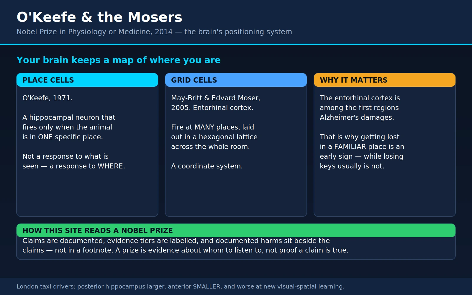
Table of Contents
- The Prize and the Three Scientists
- Place Cells, 1971
- Grid Cells, 2005
- How the System Fits Together
- The Human Evidence
- Why This Matters for Alzheimer's
- What "Brain GPS" Does and Does Not Mean
- Memory, Imagination, and the Wider Role
- Sleep and Replay
- What This Does Not License
- Where Mainstream Science Agrees — and What Remains Debated
- Key Research Papers
- Connections
- Featured Videos
1. The Prize and the Three Scientists
On October 6, 2014, the Nobel Assembly at the Karolinska Institutet awarded the Nobel Prize in Physiology or Medicine in two parts: one half to John O'Keefe, the other half jointly to May-Britt Moser and Edvard I. Moser, "for their discoveries of cells that constitute a positioning system in the brain."
Reduced to a sentence, the discovery is this: there are individual nerve cells in your brain whose job is to keep track of where you are. Not what you see, not what you hear, not what your legs are doing — where you are. Some of them fire only when you occupy one particular spot in a room. Others fire in a repeating triangular pattern spread across the whole floor, like a sheet of graph paper laid over the world. Together they build and maintain an internal map, and that map is stitched into the same brain structure that stores your memories of what happened to you.
That last connection is why this is not merely a beautiful piece of basic neuroscience. It is the anatomical reason that getting lost in a familiar place can be one of the earliest visible signs of Alzheimer's disease — and it is also the reason a great deal of nonsense is now sold on the back of the phrase "train your brain's GPS." This page tries to separate those two things carefully.
John O'Keefe
John O'Keefe was born in New York City on November 18, 1939, to Irish immigrant parents, and holds both American and British citizenship. He built his career in London at University College London, where he is Professor of Cognitive Neuroscience and became the founding director of the Sainsbury Wellcome Centre for Neural Circuits and Behaviour. He made the discovery that eventually won him the prize in 1971, more than four decades before the telephone call from Stockholm — a reminder that in this field, the wait between the finding and the recognition is often measured in careers rather than years.
May-Britt and Edvard Moser
May-Britt Moser was born in Fosnavåg, Norway, in January 1963. Edvard Ingjald Moser was born in Ålesund, Norway, on April 27, 1962. They met as psychology students at the University of Oslo, married in 1985, did their doctoral work there together, and then went abroad as postdoctoral fellows — to Richard Morris's laboratory in Edinburgh in 1995–96, and for a short visiting spell to John O'Keefe's laboratory in London. In 1996 they returned to Norway to take up positions at the Norwegian University of Science and Technology (NTNU) in Trondheim, where they built the research group that became the Kavli Institute for Systems Neuroscience. Both were promoted to full professor in 2000.
They are one of a small number of married couples to share a Nobel Prize. The others in the sciences are Marie and Pierre Curie (Physics, 1903), Irène Joliot-Curie and Frédéric Joliot (Chemistry, 1935), and Carl and Gerty Cori (Physiology or Medicine, 1947); Esther Duflo and Abhijit Banerjee shared the 2019 economics prize, which is administered alongside the Nobels but was established separately. The Mosers divorced in 2016, two years after the prize. They have gone on collaborating and jointly leading the Trondheim laboratory ever since — a detail worth stating plainly rather than tiptoeing around, because the popular telling of the story tends to freeze them permanently as "the married couple who won the Nobel," and the more interesting fact is that the scientific partnership outlasted the marriage.
2. Place Cells, 1971
In the late 1960s, working out how to record electrical activity from single neurons in an animal that was awake and walking around — rather than anaesthetised and immobilised — was itself a technical achievement. O'Keefe was one of the people who did it. He lowered fine electrodes into the hippocampus of freely moving rats and listened to what individual cells had to say.
What he and his collaborator Jonathan Dostrovsky reported in Brain Research in 1971 was strange. Certain hippocampal cells were almost silent most of the time, then fired vigorously whenever the rat entered one specific part of the enclosure. Move the rat to the far corner and that cell went quiet; a different cell, recorded from the same electrode, took over. Each cell had what came to be called a place field — its own patch of the world.
Why this was startling
To understand the shock, you have to know what a cortical neuron was expected to do in 1971. Neurons were understood in terms of inputs and outputs: a cell in the visual cortex fires when a bar of light at a particular angle crosses a particular part of the retina; a cell in the motor cortex fires when a particular muscle group is about to contract. Sensation in, action out. That framework had just produced its own Nobel-calibre work — the receptive-field studies of Hubel and Wiesel.
A place cell fits neither category. Consider what "being in the north-west corner" actually is. It is not a stimulus. There is no north-west-corner-detector on the retina. The same corner looks entirely different depending on which way the animal is facing; two different corners can look, smell and sound nearly identical. To fire reliably for one location and not another, a cell has to be responding to something the brain has computed — an inference about position, assembled from fragmentary sensory evidence and from the animal's own movement, and held against a remembered layout of the room.
In other words, O'Keefe had found a neuron whose firing tracked an internal model rather than an external signal. That was the first time anyone had listened to a single cell and heard something that could reasonably be called a thought about the world.
The cognitive map
This landed in a live argument. The psychologist Edward Tolman had proposed in the 1940s that rats learning a maze do not simply chain together stimulus–response habits but build an internal "cognitive map" of the space — an idea that behaviourist psychology found unwelcome precisely because a map is an unobservable mental representation. O'Keefe's recordings gave that unobservable thing an address.
He and the psychologist Lynn Nadel spent the following years turning the observation into a full theory, published in 1978 as the book The Hippocampus as a Cognitive Map (Oxford: Clarendon Press). Its central claim was that the hippocampus implements an allocentric map — a representation of space organised around the world's own layout rather than around the observer's body — and that this map is the substrate on which certain kinds of memory are built. The book is long, dense, and was substantially ahead of the evidence available to support it. Much of it has held up.
One further property of place cells matters for everything that follows. Move a rat from one room to a different room and the whole population remaps: cells adopt new place fields, in a new arrangement, essentially unrelated to the old one. The hippocampus does not hold one map of everywhere. It holds a distinct map for each environment it knows — which is exactly what you would want from a structure that also has to keep separate memories of what happened here versus what happened there.
3. Grid Cells, 2005
The Mosers set out to answer the obvious next question: where does the place cell get its information? A place cell reports a location. Something upstream must be computing that location.
They went looking one synapse back, in the medial entorhinal cortex — the main cortical gateway into the hippocampus. In 2004 they reported in Science (Fyhn M, Molden S, Witter MP, Moser EI, Moser MB. Spatial representation in the entorhinal cortex. Science 2004;305(5688):1258-64) that cells there also carried sharp spatial signals. Then, by recording over larger enclosures — big enough for the pattern to declare itself — they found what those cells were actually doing.
The hexagonal lattice
The 2005 paper in Nature, with Torkel Hafting as first author, describes cells that fire at many locations at once, and not at random. In the authors' own words, a grid cell "is activated whenever the animal's position coincides with any vertex of a regular grid of equilateral triangles spanning the surface of the environment." A field of equilateral triangles is a hexagonal lattice — the same packing you see in a honeycomb or a stack of oranges. Plot where one grid cell fires as the animal wanders a large box for half an hour, and you get a startlingly regular constellation of dots.
Three further findings from that paper give the system its character:
- Neighbouring cells share orientation and spacing but differ in phase. Their lattices are the same grid, shifted. Between them, a small population of grid cells covers every point in the enclosure.
- Scale varies systematically along the entorhinal cortex. The spacing and the size of individual firing fields increase from the dorsal end toward the ventral end — fine-grained grids at one end, coarse ones at the other, giving the brain the equivalent of several map scales at once.
- The map is anchored to landmarks but does not require them. The paper's closing line is the important one: the map "is anchored to external landmarks, but persists in their absence, suggesting that grid cells may be part of a generalized, path-integration-based map of the spatial environment."
Why that last point is the whole discovery
Put plainly: the grid is self-generated. The animal carries it. If you switch off the lights and remove every cue, the pattern does not collapse — the brain keeps updating position from the animal's own motion, a process called path integration (the formal name for what sailors call dead reckoning).
This is what a coordinate system is. A landmark-based map tells you "I am next to the red chair"; it fails the moment the chair is moved or the room is dark. A metric grid tells you "I have travelled about two metres north-east of where I started," and it keeps working in a featureless field at night. Every animal that can walk out of its burrow, forage in a wandering path, and then return home in a straight line needs something of this kind. Before 2005, nobody knew what it looked like in a brain.
The other components
Grid cells do not work alone. The entorhinal cortex and neighbouring structures also contain:
- Head-direction cells — first described by Jeffrey Taube, Robert Muller and James Ranck in the postsubiculum in 1990 (J Neurosci 1990;10(2):420-35). Each fires when the animal's head points in one particular compass direction, wherever in the room it happens to be standing. This is the brain's internal compass needle, and it is what allows the grid to keep a stable orientation.
- Border cells — reported by the Moser group in 2008 (Solstad T, Boccara CN, Kropff E, Moser MB, Moser EI. Representation of geometric borders in the entorhinal cortex. Science 2008;322(5909):1865-8). These fire when the animal is near a wall or a drop-off, along one particular edge of the enclosure. Borders are what keep an internally generated grid pinned to the real room, correcting the drift that any dead-reckoning system accumulates.
- Conjunctive and speed-modulated cells — cells that combine position, direction and running speed in a single signal (Sargolini F, Fyhn M, Hafting T, McNaughton BL, et al. Science 2006;312(5774):758-62). If you are going to integrate your own motion into a position estimate, you need to know how fast you are going and which way you are pointing.
4. How the System Fits Together
The picture that emerged over the following decade — summarised by the Mosers themselves in a widely read review (Moser EI, Kropff E, Moser MB. Place cells, grid cells, and the brain's spatial representation system. Annu Rev Neurosci 2008;31:69-89) — is roughly a division of labour:
- The entorhinal cortex supplies the metric. Grid cells provide distance and geometry, head-direction cells provide bearing, border cells provide the edges. This is a coordinate frame: abstract, reusable, indifferent to what any particular room contains.
- The hippocampus supplies the meaning. Place cells take that coordinate frame and bind it to this place, with its particular smells, sights, dangers and rewards — and, crucially, to what happened here. Remapping between environments is the signature of that binding: a different room gets a different code.
The reason this second half matters so much is that the hippocampus is not primarily known to medicine as a navigation organ. It is known as the memory organ — and that connection was made by accident, on an operating table, decades before anyone recorded a place cell.
The patient H.M.
In 1953, a young man with severe, drug-resistant epilepsy underwent an experimental operation in which large parts of the medial temporal lobe on both sides — including most of both hippocampi — were removed. His seizures improved. His memory was destroyed. Known during his lifetime only as H.M., and after his death in 2008 as Henry Molaison, he could hold a conversation, retained his childhood memories and his intelligence, and could learn new motor skills — but he could not form new conscious memories of events. The 1957 report by the surgeon William Beecher Scoville and the neuropsychologist Brenda Milner (Loss of recent memory after bilateral hippocampal lesions. J Neurol Neurosurg Psychiatry 1957;20(1):11-21) is one of the most consequential case reports in the history of medicine. It is the reason "hippocampus" and "memory" are linked words at all.
So when O'Keefe found a spatial map inside that same structure, he was not finding it in a neutral location. He was finding it inside the memory system. The modern view is that these are two faces of one mechanism: episodic memory — your record of specific events, with their time and place attached — needs a way to represent where and when, and the spatial map is a large part of the machinery that does it. That is also why almost everybody's autobiographical memories come with a location built in: you rarely remember an event as free-floating, you remember it happening somewhere.
The complementary question — what physically changes inside a neuron when a memory is stored — belongs to a different Nobel. Eric Kandel's work on the cell biology of memory, which traced learning down to specific molecular changes at synapses, is covered on our page for Carlsson, Greengard and Kandel. Read together, the two prizes answer different halves of the same question: Kandel explains how a memory is written into a synapse; O'Keefe and the Mosers explain the coordinate system that says where the remembered thing happened.
5. The Human Evidence
Everything above was found in rats. A reasonable person should ask whether any of it applies to people. The honest answer is that the evidence in humans is real, is convergent, and is thinner than the animal evidence — because you cannot lower an electrode into a healthy human brain.
Direct recordings, in patients who were already having electrodes implanted
The exception is neurosurgery. People with severe epilepsy are sometimes implanted with depth electrodes for a week or two so that surgeons can locate the seizure focus. With consent, those patients can play navigation games while their neurons are recorded.
In 2003, Arne Ekstrom, Michael Kahana, Itzhak Fried and colleagues recorded 317 neurons in the human medial temporal and frontal lobes while patients explored a virtual town (Cellular networks underlying human spatial navigation. Nature 2003;425(6954):184-8). They found cells that responded at specific spatial locations — primarily in the hippocampus — alongside separate cells in the parahippocampal region that responded to views of landmarks, and cells elsewhere that tracked the person's navigational goal. That is a human place cell, recorded directly.
A decade later, Joshua Jacobs and colleagues reported direct recordings of grid-like firing in humans navigating a virtual environment (Direct recordings of grid-like neuronal activity in human spatial navigation. Nat Neurosci 2013;16(9):1188-90).
Grid-like signals in ordinary brains, by fMRI
In 2010, Christian Doeller, Caswell Barry and Neil Burgess found an ingenious way to detect grid coding without electrodes (Evidence for grid cells in a human memory network. Nature 2010;463(7281):657-61). Because neighbouring grid cells share the same orientation, their combined activity should be sensitive to which way you are moving, with a six-fold rotational symmetry inherited from the hexagonal lattice — a pattern large enough for fMRI to see. They looked for exactly that as volunteers explored a virtual environment, and found it: a speed-modulated six-fold directional signal, strongest in the right entorhinal cortex, in a network that also included parietal, temporal and medial prefrontal regions. Notably, how coherent that directional signal was across a person's entorhinal cortex correlated with how well they performed on the spatial memory task.
This measure — the human "grid-like signal" — matters enormously for section 6, so it is worth being clear about what it is. It is a statistical regularity extracted from an averaged blood-flow signal covering millions of neurons. It is strong evidence that grid-like coding exists in humans. It is not a picture of a grid cell, and it is not something that can be read out of one person on one scan.
The London taxi drivers — what the studies actually showed
The most famous human result in this whole field is also the most frequently mangled. Here it is with its real shape.
The original, 2000. Eleanor Maguire and colleagues at UCL scanned licensed London taxi drivers — people who have passed "the Knowledge," a notoriously punishing examination on the layout of 25,000 streets — and compared their brains with those of people who did not drive taxis (Navigation-related structural change in the hippocampi of taxi drivers. Proc Natl Acad Sci U S A 2000;97(8):4398-403). The posterior hippocampi of the taxi drivers were larger than those of controls; a more anterior region was larger in the controls. Hippocampal volume correlated with time spent driving a taxi — positively at the back, negatively at the front.
The limits of that study are not obscure; they are stated in the paper. It was cross-sectional — a single snapshot of two groups — so it cannot distinguish "the job changed their brains" from "people with this brain shape become taxi drivers." The samples were small by any modern imaging standard. And the finding is a trade-off, not a bonus: something at the front was smaller.
The control that ruled out the boring explanations, 2006. Maguire, Katherine Woollett and Hugo Spiers compared taxi drivers with London bus drivers — matched for driving experience and stress, but following fixed routes (London taxi drivers and bus drivers: a structural MRI and neuropsychological analysis. Hippocampus 2006;16(12):1091-101). The same posterior-larger, anterior-smaller pattern appeared, and years of navigation experience correlated with hippocampal grey matter only in the taxi drivers. So the difference tracks spatial knowledge specifically, not stress or self-motion or time behind a wheel. That paper also delivered the finding almost nobody repeats: the taxi drivers were worse than the bus drivers at acquiring new visuo-spatial information. The authors' own speculation is that the elaborate map comes at a cost to new spatial memories.
The longitudinal follow-up, 2011. Woollett and Maguire then did the study the 2000 paper could not (Acquiring "the Knowledge" of London's layout drives structural brain changes. Curr Biol 2011;21(24):2109-14). They followed average-IQ adults across four years of training for the Knowledge, scanning them before and after. Those who qualified showed a selective increase in grey matter volume in the posterior hippocampus, along with changes to their memory profile. Those who trained but failed to qualify showed no structural change, and neither did controls. That design — same people, before and after, with a matched group who did the same training and did not succeed — is what turns a correlation into something much closer to a causal claim.
What this licenses, and what it does not. It licenses saying that years of extreme, effortful, real-world spatial learning are associated with measurable structural change in the adult human hippocampus. It does not license the popular version — that navigating without a phone will grow your hippocampus, that satnav use shrinks it, or that hippocampal volume is a meaningful health metric for any individual. Those claims have not been tested by these studies. What was tested was four years of full-time study of one of the most complex street layouts on Earth, in a self-selected group, with a real qualifying examination at the end.
6. Why This Matters for Alzheimer's
This is the section with practical consequences, so it is the one that most needs to be stated precisely — both about what the evidence supports and about what it does not.
The anatomical coincidence that is not a coincidence
In 1991, the neuroanatomists Heiko and Eva Braak published the staging scheme that still organises how pathologists think about Alzheimer's disease (Neuropathological stageing of Alzheimer-related changes. Acta Neuropathol 1991;82(4):239-59). Their central observation was that the neurofibrillary changes do not begin everywhere at once. They begin in a specific place: the transentorhinal and entorhinal cortex, spreading from there into the hippocampus and only later across the wider cortex.
Read that against sections 3 and 4. The entorhinal cortex is where the grid cells are. It is the first stop for the brain's metric coordinate system, and it is among the first tissue that Alzheimer's disease damages. The hippocampus — place cells, and the binding of location to memory — is next in line.
This gives a genuine mechanistic explanation for something families notice long before anyone uses the word dementia: a person begins to get lost. Not lost in the sense of taking a wrong turn in an unfamiliar city, but lost on the route they have driven for twenty years. Historically this was filed under "memory problems" or dismissed as ordinary ageing. The Nobel-winning work says it deserves to be taken seriously in its own right, because the disease starts in the navigation hardware.
What the research actually shows
Three strands of evidence, each with its own boundaries:
Navigation is impaired early, and may be impaired before memory tests notice. The 2018 review by Gillian Coughlan, Michael Hornberger and colleagues (Spatial navigation deficits — overlooked cognitive marker for preclinical Alzheimer disease? Nat Rev Neurol 2018;14(8):496-506) makes the case that diagnostics have leaned on episodic memory tests as the gold standard despite their limited sensitivity for identifying people at risk, and that spatial navigation is a candidate to fill the gap — partly because the brain's navigation system overlaps the regions the disease attacks first, and partly because navigation tasks carry fewer verbal, cultural and educational biases than standard cognitive tests. Note the question mark in that paper's own title. It is a review that appraises evidence and names research gaps; it is not a validation study.
Grid-like signals are measurably altered decades before any symptoms, in people carrying a risk gene. In 2015, Lukas Kunz, Nikolai Axmacher, Christian Doeller and colleagues scanned young, healthy adults who carry APOE-ε4, the most common genetic risk factor for late-onset Alzheimer's (Reduced grid-cell-like representations in adults at genetic risk for Alzheimer's disease. Science 2015;350(6259):430-3). The carriers showed reduced grid-cell-like fMRI representations and altered navigational behaviour in a virtual arena, both associated with poorer spatial memory. They also showed increased hippocampal activity, which the authors suggest may be compensation that keeps overt performance intact. The phrase in the abstract is "decades before potential disease onset."
Now hold two things in mind at once. This is a striking result. And APOE-ε4 is a risk allele, not a diagnosis — many carriers never develop Alzheimer's disease, and most people who do develop it are not carriers. These were group averages in healthy young volunteers, not predictions about any one of them.
Game data can separate risk groups that memory tests cannot. Coughlan and colleagues used data from Sea Hero Quest, a mobile game built as a research instrument, to benchmark a small, deeply characterised laboratory cohort against 27,108 players (Toward personalized cognitive diagnostics of at-genetic-risk Alzheimer's disease. Proc Natl Acad Sci U S A 2019;116(19):9285-9292). Benchmarked that way, high-genetic-risk participants could be reliably distinguished from low-risk ones — while standard neuropsychological episodic memory tests could not tell them apart. The paper's own framing is careful and future-tense: it describes this as a "steppingstone" toward individualised diagnostics, something that "in the future" could classify spatial impairment at a more individual level.
What the research does not show — stated flatly
No navigation app or game can diagnose Alzheimer's disease, or predict it, in an individual person. Nothing in this literature supports that, and the researchers who built these tools do not claim it. Three specific reasons:
- Group differences are not individual predictions. Two groups can differ reliably on average while their score distributions overlap so heavily that any one person's score tells you almost nothing.
- Navigation ability varies enormously for reasons that have nothing to do with disease. The same Sea Hero Quest dataset, analysed across more than 2.5 million players worldwide, found that navigation ability clusters by country, that a nation's economic wealth predicted its inhabitants' average performance, and that a country's gender inequality predicted the size of the gap between men's and women's scores (Coutrot A, Silva R, Manley E, de Cothi W, et al. Global Determinants of Navigation Ability. Curr Biol 2018;28(17):2861-2866.e4). Age, sex, education, culture, wealth, video-game familiarity and how well you see the screen all move the score. A raw result is not a reading of your entorhinal cortex.
- None of these tasks has been validated as a clinical diagnostic. They are research instruments. Diagnosis of Alzheimer's disease rests on clinical assessment, and increasingly on imaging and fluid biomarkers — not on a game score.
Ordinary navigation decline versus the kind that warrants assessment
This is the distinction that actually helps people, and it is worth being calm and concrete about it, because the alternative is a lot of frightened people who are perfectly well and a smaller number of people who quietly avoid getting help.
Common, and usually nothing. Sense of direction declines gradually with age in most people — that is a normal population pattern, visible in the huge datasets above, not a disease. Ordinary examples:
- Taking longer than you used to to learn the layout of a new hospital, airport or shopping centre.
- Losing your bearings in an unfamiliar city, or coming out of a subway station facing the wrong way.
- Forgetting which level of the car park you left the car on, and having to walk around to find it.
- Relying on satnav in places you don't know, and feeling less confident without it.
- Being worse at this than your partner, and having always been worse at it.
The common thread is that these involve unfamiliar environments, or details rather than layouts, and that you know you are momentarily lost and can work out what to do.
Worth mentioning to a doctor. The pattern that matters is disorientation in familiar places, or a loss of the sense that a place is familiar at all:
- Getting lost on a route you have travelled for years — the way home, the drive to a regular shop, the walk to a friend's house.
- Not recognising a familiar street or building, or feeling that your own neighbourhood has become strange.
- Becoming confused about where you are inside your own home — for example, which door leads where.
- Setting out somewhere and, mid-journey, not being able to recall where you were going or why.
- Being found some distance from home, unable to explain how you got there.
- Navigation trouble arriving alongside other changes — repeating questions, difficulty with familiar multi-step tasks, word-finding problems, misplacing things in odd places, withdrawal, or a change in personality.
One more signal deserves its own line, because people close to the person are usually right: if someone who knows you well is worried, that is worth acting on, even when formal testing looks normal.
Not a memory-clinic question — an urgent one. Disorientation that comes on suddenly, over hours or a day or two, especially with confusion, drowsiness or fluctuating alertness, is not the pattern described above. That is the pattern of delirium or of a stroke, and it needs same-day medical attention rather than a referral letter.
And being assessed is not the same as being diagnosed. A proper assessment starts by looking for the many treatable things that impair orientation and memory: thyroid disease, vitamin B12 deficiency, medication side effects (sedatives and anticholinergics in particular), depression, sleep apnoea, alcohol, uncorrected hearing and vision loss, and infection. A good number of people who go in worried about dementia come out with something else, and some of it is fixable. Our page on Alzheimer's disease covers diagnosis and management in more depth, and the other dementias — vascular, Lewy body and frontotemporal — have their own pages, because they do not all begin the same way.
7. What "Brain GPS" Does and Does Not Mean
"The brain's inner GPS" is a good headline and a poor description. It got attached to this work in 2014 and it has been quietly misleading people ever since, so it is worth taking apart.
What GPS actually is: a receiver that compares timing signals from satellites and computes an explicit numerical position on the surface of the Earth, which it can display, transmit or log. Coordinates exist in it as readable values.
What the brain has instead: populations of neurons whose firing rates correlate with position, heading, distance travelled and proximity to boundaries. There is no register anywhere holding your latitude. There is a distributed pattern of activity across many thousands of cells, from which an experimenter with electrodes in an animal, a lot of trials and a decoding algorithm can estimate where the animal is — approximately, and after the fact.
Several things follow that the metaphor obscures:
- It locates you within a remembered environment, not on the planet. Place cells remap completely between rooms. There is no global frame. Your brain does not know where Trondheim is; it knows the layout of places you have been.
- The human evidence is coarser than the animal evidence. The fMRI grid-like signal is a six-fold statistical regularity averaged over millions of neurons across many trials. Calling it "reading someone's grid cells" overstates it by a wide margin.
- Nobody has read a person's felt sense of location out of their brain. The step from "these cells fire when the animal is here" to "this is what it is like to know where you are" is not solved. It is one instance of the general unsolved problem of how neural activity relates to conscious experience, and this field has no special exemption from it.
- The system may not be only about space. A substantial line of current research asks whether grid-like coding is a general-purpose way of representing any structured, continuous space of relationships — conceptual "maps," social hierarchies, sequences of sounds — with physical space merely the version that evolution built first and that experimenters find easiest to measure. That is genuinely unsettled, and it is one of the more interesting open questions in the field.
The practical consequence of all this is section 10: because "GPS" sounds like a device, it sounds like something that could be tuned up, recalibrated or exercised. That framing is where the marketing gets in.
8. Memory, Imagination, and the Wider Role
If the hippocampus were only a map, damage to it would only cause people to get lost. H.M. showed that it does much more than that, and later work showed something stranger still.
Episodic memory — the memory of specific events you personally experienced — requires binding several things into one retrievable package: what happened, where it happened, when it happened, and what it felt like. The spatial map contributes the "where," and possibly the scaffolding on which the rest is hung. This is why place is so hard to strip out of a memory: try to recall a conversation from last year and you will almost certainly find yourself recalling a room.
The stranger finding concerns the future. In 2007, Demis Hassabis, Dharshan Kumaran, Seralynne Vann and Eleanor Maguire asked people with hippocampal amnesia to imagine new experiences — not to recall anything, simply to describe a plausible scene they had never encountered, such as lying on a white sandy beach (Patients with hippocampal amnesia cannot imagine new experiences. Proc Natl Acad Sci U S A 2007;104(5):1726-31). The patients' imagined scenes were impoverished and fragmentary. They could produce isolated details but struggled to assemble them into a coherent spatial whole.
The interpretation that grew out of this is that remembering the past and imagining the future are not opposite operations but the same one, run in two directions: both require constructing a scene — putting objects and yourself into a coherent spatial context — and the hippocampus is the machine that does the constructing. That reframes the structure. It is not an archive. It is a scene generator, used for recall, for planning, and for imagining what has never happened.
Two honest caveats. Human lesion studies use small numbers of patients whose damage is rarely confined to the hippocampus. And the "scene construction" interpretation is one reading of these findings; it is influential but it is not the only account in the literature.
9. Sleep and Replay
One of the most evocative results in modern neuroscience came out of the place-cell literature, and it happens during sleep.
In 1994, Matthew Wilson and Bruce McNaughton recorded large numbers of hippocampal place cells in rats running a track, then kept recording while the animals slept (Reactivation of hippocampal ensemble memories during sleep. Science 1994;265(5172):676-9). During subsequent sleep, the same cells that had fired together during the run fired together again — the sequences of the waking journey recurring in the sleeping brain. The phenomenon became known as replay. Later work showed it runs compressed in time, sometimes backwards, and occurs during quiet waking as well as sleep, often riding on brief high-frequency bursts of hippocampal activity called sharp-wave ripples.
The leading proposal is that replay is part of how experience is consolidated — rehearsed within the hippocampus and gradually distributed to the neocortex for long-term storage — and that the same mechanism supports recall and the simulation of routes not yet taken. The review by H. Freyja Ólafsdóttir, Daniel Bush and Caswell Barry lays out the range of functions replay has been proposed to serve, and how much of that is established (The Role of Hippocampal Replay in Memory and Planning. Curr Biol 2018;28(1):R37-R50).
What is established, and where inference begins. Replay itself is directly observed in animals, with electrodes, in many laboratories — that is not in doubt. Interventions that disrupt sharp-wave ripples in rodents impair memory on subsequent tests, which is real evidence that replay does something rather than merely happening. In humans, the picture is thinner and mostly indirect: it comes from decoding patterns in MEG and fMRI, and from rare intracranial recordings in patients being monitored for epilepsy. The general claim that sleep supports memory consolidation in people is well supported from behavioural studies. The specific claim that human hippocampal replay is the mechanism doing it is a reasonable inference from animal work, not a demonstrated fact in humans. Anyone who tells you otherwise is skipping a step.
What this does justify, comfortably, is treating sleep as part of memory health rather than as time subtracted from the day. The machinery that builds the map appears to keep working on it overnight. Our pages on Hall, Rosbash and Young (the molecular clock that schedules sleep in the first place), insomnia and circadian rhythm sleep–wake disorders cover the practical side.
10. What This Does Not License
Every genuinely exciting neuroscience result grows a commercial shadow, and this one grew a large one. Two claims in particular are worth naming.
"Brain training" and navigation games sold for dementia prevention
The best single assessment of the brain-training evidence is a long systematic review commissioned by Psychological Science in the Public Interest, by Daniel Simons and colleagues (Do "Brain-Training" Programs Work? Psychol Sci Public Interest 2016;17(3):103-186). Its method was pointed: the authors evaluated the studies that the brain-training companies themselves cite on their own websites — presumably the strongest evidence available for the products.
The conclusion comes in three descending steps, and the shape of it is what matters:
- Extensive evidence that training improves performance on the trained task.
- Less evidence that it improves performance on closely related tasks.
- Little evidence that it improves performance on distantly related tasks, or that it improves everyday cognitive performance.
The review also found that many of the studies had major design or analysis shortcomings, and that none of the cited studies met all of the best practices the authors set out as necessary for clear conclusions. Our page on Hubel, Wiesel and Sperry covers the brain-training evidence and the plasticity claims behind it in more detail.
Play a navigation game for six weeks and you will get better at that navigation game. That is the first bullet, and it is real. It is also exactly what you would expect, and it is not evidence that your hippocampus is protected. Sea Hero Quest in particular was designed as a measuring instrument for research, not as a treatment; nothing in the papers that use it claims that playing it reduces anyone's risk of dementia.
What genuinely is supported
The contrast is sharp and worth ending on, because the honest message is not "nothing helps." Quite a lot appears to help — it is just not sold in an app store.
The Lancet standing Commission on dementia prevention, intervention and care (Livingston G, Huntley J, Liu KY, Costafreda SG, et al. Lancet 2024;404(10452):572-628) periodically reviews the evidence on modifiable risk factors for dementia. Its list — assembled across successive editions in 2017, 2020 and 2024, each of which revised the previous one — includes:
- Hearing loss, and treating it. Untreated hearing loss is one of the more prominent factors on the list — a reason our page on Georg von Békésy and on hearing loss belongs in this conversation.
- Physical inactivity — see Exercise.
- High blood pressure, particularly in midlife.
- Smoking.
- Less education in early life.
- Social isolation and low social contact.
- Alongside others the Commission has assessed, including depression, diabetes, obesity, excessive alcohol, traumatic brain injury, air pollution, and — added in the most recent edition — untreated vision loss and high LDL cholesterol.
How to read that list without over-reading it. The Commission's headline figure is a population attributable fraction: a modelled estimate of how much dementia in a whole population is statistically associated with these factors, computed under the assumption that the associations are causal and that the exposures could be eliminated. It is a public-health planning number. We deliberately do not quote the percentage here, for two reasons: it has been revised between editions as factors were added, and — more importantly — it is not a probability that applies to you. Treating your hearing loss does not reduce your personal dementia risk by a fixed percentage. It removes one contributor from a picture with many, some of which (age, genetics) nobody can modify.
What can fairly be said: these are real associations, several are supported by trial as well as observational evidence, every item on the list is worth addressing on its own merits regardless of dementia, and the whole list is more solidly grounded than any product that promises to exercise your inner GPS.
11. Where Mainstream Science Agrees — and What Remains Debated
Settled
- Place cells and grid cells exist. Both have been replicated in many laboratories, in multiple species, for decades. This is not a contested finding.
- The entorhinal–hippocampal system is central to spatial and episodic memory. The convergence of lesion evidence (H.M. and the many patients since), single-cell recording, and imaging is about as strong as evidence gets in systems neuroscience.
- Grid coding does not require landmarks. The pattern persists in darkness, which is the core evidence that the system performs path integration.
- The entorhinal cortex is among the earliest regions affected in Alzheimer's disease. This is standard neuropathology, established independently of the navigation work.
- Humans have place-responsive cells and grid-like signals. Shown by direct intracranial recording and by fMRI, in independent laboratories.
- Cognitive training improves the trained task, with weak transfer beyond it. This is a robust and repeatedly replicated pattern, not a fringe skeptical position.
Genuinely open
- How grid cells are generated. Competing computational accounts — continuous attractor networks, oscillatory interference, learning-based schemes — have been argued for two decades without resolution.
- Whether the grid code is really about space at all, or is a general format for representing structured relationships of any kind, with physical space as the special case we happen to be able to measure.
- What exactly the human fMRI grid-like signal reflects. That it exists is well replicated; that it is a direct proxy for grid-cell activity is an inference.
- Whether spatial navigation testing will become a clinical screening tool, and if so with what sensitivity and specificity, in whom, and at what age. This is an active research programme with no validated clinical product at its end yet.
- The causal role of replay in consolidation, especially in humans.
- What structural MRI differences in expert navigators physically are. "More grey matter" is a measurement, not a mechanism; neurogenesis, dendritic and synaptic change, and glial or vascular change are all candidates, and the question is not settled.
- Whether habitual satnav use affects the navigation system in any lasting way. This is frequently asserted in popular coverage and thinly evidenced.
12. Key Research Papers
- O'Keefe J, Dostrovsky J. The hippocampus as a spatial map. Preliminary evidence from unit activity in the freely-moving rat. Brain Res 1971;34(1):171-5 — the discovery of place cells.
- O'Keefe J, Nadel L. The Hippocampus as a Cognitive Map. Oxford: Clarendon Press; 1978 — the theoretical framework built on the 1971 finding. A book, not a journal article, so it has no PubMed record.
- Hafting T, Fyhn M, Molden S, Moser MB, Moser EI. Microstructure of a spatial map in the entorhinal cortex. Nature 2005;436(7052):801-6 — the discovery of grid cells and their hexagonal lattice.
- Solstad T, Boccara CN, Kropff E, Moser MB, Moser EI. Representation of geometric borders in the entorhinal cortex. Science 2008;322(5909):1865-8 — border cells, which pin the internal grid to the real room.
- Scoville WB, Milner B. Loss of recent memory after bilateral hippocampal lesions. J Neurol Neurosurg Psychiatry 1957;20(1):11-21 — the patient H.M., and the origin of the link between hippocampus and memory.
- Hassabis D, Kumaran D, Vann SD, Maguire EA. Patients with hippocampal amnesia cannot imagine new experiences. Proc Natl Acad Sci U S A 2007;104(5):1726-31 — the hippocampus as a scene-construction machine, not an archive.
- Ekstrom AD, Kahana MJ, Caplan JB, Fields TA, et al. Cellular networks underlying human spatial navigation. Nature 2003;425(6954):184-8 — place-responsive cells recorded directly in the human brain.
- Doeller CF, Barry C, Burgess N. Evidence for grid cells in a human memory network. Nature 2010;463(7281):657-61 — the six-fold symmetric fMRI signature of grid-like coding in people.
- Maguire EA, Gadian DG, Johnsrude IS, Good CD, et al. Navigation-related structural change in the hippocampi of taxi drivers. Proc Natl Acad Sci U S A 2000;97(8):4398-403 — the famous study. Cross-sectional, small, and showing a front/back trade-off rather than a straightforward enlargement.
- Woollett K, Maguire EA. Acquiring "the Knowledge" of London's layout drives structural brain changes. Curr Biol 2011;21(24):2109-14 — the four-year longitudinal follow-up, with trainees who failed to qualify as the control that the 2000 study lacked. Cite these two together or neither.
- Braak H, Braak E. Neuropathological stageing of Alzheimer-related changes. Acta Neuropathol 1991;82(4):239-59 — why the disease reaches the navigation system first.
- Kunz L, Schröder TN, Lee H, Montag C, et al. Reduced grid-cell-like representations in adults at genetic risk for Alzheimer's disease. Science 2015;350(6259):430-3 — altered entorhinal coding in healthy young APOE-ε4 carriers.
- Coughlan G, Laczó J, Hort J, Minihane AM, Hornberger M. Spatial navigation deficits — overlooked cognitive marker for preclinical Alzheimer disease? Nat Rev Neurol 2018;14(8):496-506 — the case for navigation testing, stated as a question by its own authors.
- Ólafsdóttir HF, Bush D, Barry C. The Role of Hippocampal Replay in Memory and Planning. Curr Biol 2018;28(1):R37-R50 — what replay is proposed to do, and how much of that is demonstrated.
- Livingston G, Huntley J, Liu KY, Costafreda SG, et al. Dementia prevention, intervention, and care: 2024 report of the Lancet standing Commission. Lancet 2024;404(10452):572-628 — the modifiable risk factors, as population-level estimates rather than personal guarantees. Earlier editions appeared in 2017 and 2020 and differ from this one; check which edition any figure comes from.
Further papers cited in the text above, each linked at first mention: Fyhn et al. (Science 2004, entorhinal spatial coding); Taube, Muller and Ranck (J Neurosci 1990, head-direction cells); Sargolini et al. (Science 2006, conjunctive coding); Moser, Kropff and Moser (Annu Rev Neurosci 2008, the synthesis); Jacobs et al. (Nat Neurosci 2013, human grid-like recordings); Maguire, Woollett and Spiers (Hippocampus 2006, taxi versus bus drivers); Coughlan et al. (PNAS 2019, Sea Hero Quest benchmarking); Coutrot et al. (Curr Biol 2018, global determinants of navigation ability); Wilson and McNaughton (Science 1994, replay); and Simons et al. (Psychol Sci Public Interest 2016, brain training).
Live PubMed Searches
- Place cells and the hippocampal spatial map
- Grid cells in the entorhinal cortex
- Spatial navigation in preclinical Alzheimer's disease
- Hippocampal replay and memory consolidation
- Modifiable risk factors for dementia
13. Connections
- All Notable Doctors
- Nobel Prize in Medicine — the wing this page belongs to: every laureate from 1901 onward, and what each prize actually established
- Carlsson, Greengard & Kandel — the cell biology of memory: how a memory is physically written into a synapse, the other half of the question this page asks
- Cajal & Golgi — the neuron doctrine: the reason it made sense to record from a single cell at all
- Hubel, Wiesel & Sperry — receptive fields, critical periods and the real limits of "brain training"
- Hall, Rosbash & Young — the molecular clock that schedules the sleep in which replay happens
- Georg von Békésy — how hearing works, and why untreated hearing loss appears on the dementia risk-factor list
- Levi-Montalcini & Cohen — nerve growth factor: the signals that build and maintain the circuits described here
- Carl & Gerty Cori — the 1947 laureates, and the married couple the Mosers are most often compared with
- Alzheimer's Disease — diagnosis, treatment and what the entorhinal starting point means in practice
- Neurology — the full section: brain and nervous-system conditions
- Vascular Dementia — the dementia that begins with blood vessels rather than the entorhinal cortex
- Lewy Body Dementia — a different early pattern: visual hallucinations, fluctuating alertness, movement changes
- Frontotemporal Dementia — where personality and language change first and navigation is often spared
- Epilepsy — the condition whose surgical monitoring made every human single-cell recording on this page possible
- Hearing Loss — one of the modifiable dementia risk factors, and one of the most treatable
- Insomnia — when the sleep that consolidation depends on is the thing that is broken
- Brain Fog — the far more common complaint, and the reversible causes worth ruling out first