Hubel, Wiesel & Sperry: How the Brain Builds Vision, and the Critical Period
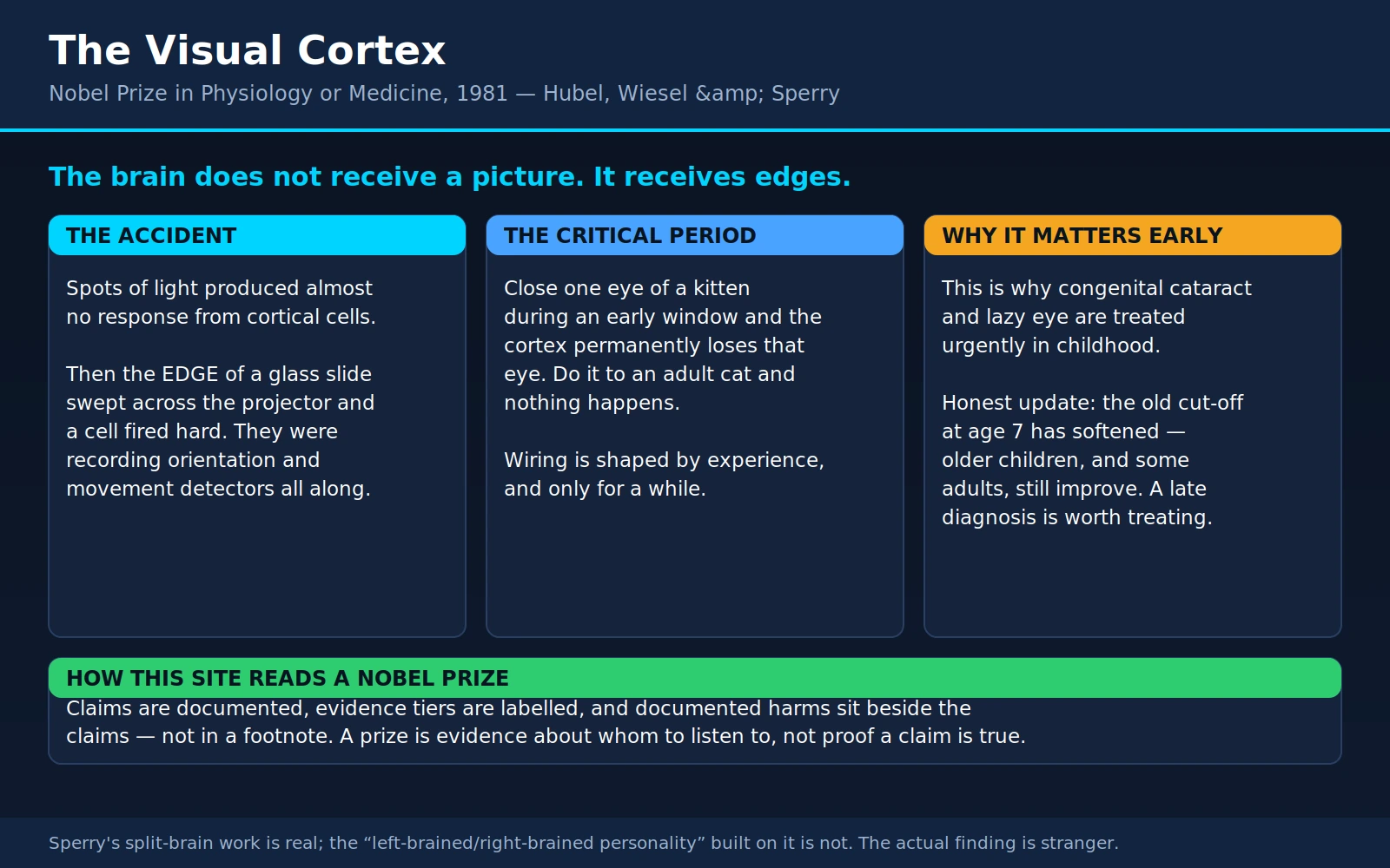
Table of Contents
- The Prize and the Three Men
- Before Hubel and Wiesel: Kuffler's Spots
- The Accident That Started It
- What They Found: Simple Cells, Complex Cells, Columns
- Why This Shaped Artificial Intelligence
- The Critical Period
- Amblyopia: What Parents Need to Know
- Sperry's Split Brains
- What Split-Brain Research Does Not Show
- Chemoaffinity: Sperry's Earlier and Arguably Larger Idea
- What This Means for You Today
- Where Mainstream Science Agrees — and What Remains Debated
- Key Research Papers
- Connections
- Featured Videos
1. The Prize and the Three Men
The 1981 Nobel Prize in Physiology or Medicine is really two prizes wearing one label, and that is why this page covers both halves. One half went jointly to David H. Hubel and Torsten N. Wiesel "for their discoveries concerning information processing in the visual system." The other half went, entirely separately, to Roger W. Sperry "for his discoveries concerning the functional specialization of the cerebral hemispheres." The three men were not collaborators. They worked on opposite coasts of the United States, on different questions, with different animals and different patients. What the Nobel committee recognised in 1981 was a single larger idea arriving from two directions at once: that the cerebral cortex is not a general-purpose porridge but a machine with parts, and that those parts can be identified, mapped, and — crucially — disturbed.
David Hunter Hubel (1926–2013) was born in Windsor, Ontario, to American parents, grew up in Montreal, and took his medical degree at McGill. He came to the United States in the 1950s, spent time at the Walter Reed Army Institute of Research, and there built the tool that would make his career: a tungsten microelectrode fine enough and mechanically stable enough to hold a conversation with a single neuron in a living, breathing animal for hours at a stretch. Without that electrode, and the hydraulic microdrive that positioned it, the experiments below were not physically possible.
Torsten Nils Wiesel (born 1924) was born in Uppsala, Sweden, and grew up on the grounds of the Beckomberga psychiatric hospital outside Stockholm, where his father was chief psychiatrist. He trained at the Karolinska Institute, came to Johns Hopkins in 1955, and later served as president of Rockefeller University and as an unusually effective scientific diplomat — he spent decades on human-rights work for scientists and on building research capacity in countries that had none.
The two met in 1958 in the Baltimore laboratory of Stephen Kuffler, and the partnership that followed is one of the most productive in the history of biology: roughly twenty-five years of joint work, moving with Kuffler's group to Harvard Medical School, where their department became the model for neurobiology departments everywhere. They published together with a rhythm most laboratories never manage — and, notably, they alternated first authorship rather than fighting over it.
Roger Wolcott Sperry (1913–1994) was a very different figure. Born in Hartford, Connecticut, he took an undergraduate degree in English literature at Oberlin before switching to psychology and then zoology, earning his doctorate under Paul Weiss at the University of Chicago. He spent most of his career at Caltech. He was a paleontologist's kind of experimentalist — patient, mechanical, willing to spend years rotating the eyes of newts — and he had a philosopher's appetite for the biggest question in his field: what happens to the unity of a mind when you cut the brain in half.
This page tells both stories because both are unusually useful to a reader who is not a neuroscientist. Hubel and Wiesel's work is the reason your child's eye check at age four is not a formality. Sperry's work is the origin of one of the most abused ideas in popular psychology, and correcting that abuse is worth doing carefully — because the real finding is stranger and more interesting than the myth built on top of it.
2. Before Hubel and Wiesel: Kuffler's Spots
To understand why the accident in section 3 mattered, you need to know what everybody expected to find, and why they expected it.
By the 1950s the retina was beginning to make sense. Haldan Keffer Hartline, Ragnar Granit and George Wald — who would share their own Nobel Prize in 1967 — had established how light is converted into a nerve signal and how single optic-nerve fibres behave. Then in 1953, Stephen Kuffler recorded from single retinal ganglion cells in the cat and described something beautifully simple. Each cell watched a small circular patch of the visual world, and that patch was organised as a centre and a surround with opposite signs. An "on-centre" cell fired hard when a small spot of light landed in the middle of its patch, was silenced when light landed in the ring around it, and responded rather feebly to diffuse light covering both — because centre and surround cancelled. "Off-centre" cells did the reverse.
This is already a profound result. It means the eye does not send the brain a picture. It sends a running commentary about local contrast: where in the image things get brighter or darker, and by how much. Uniform brightness is barely reported at all, which is why the retina can work over an enormous range of illumination without saturating, and why the optic nerve — about a million fibres per eye — can carry a useful signal at all. It is compression by discarding what does not change.
So the reasonable expectation, going into the cortex, was more of the same, only bigger. If retinal cells like spots, cortical cells should like spots too — perhaps larger spots, perhaps spots in cleverer arrangements. That expectation is what Hubel and Wiesel took into the laboratory in 1958, and it is what nearly cost them the discovery.
3. The Accident That Started It
The setup was this. An anaesthetised cat faced a screen. Its eyes were held open and focused, so a specific point on the screen mapped to a specific point on the retina. A microelectrode sat in the primary visual cortex — the striate cortex, area 17, at the very back of the brain — with its tip close enough to a single neuron to hear that neuron's action potentials, which were fed to a loudspeaker so the experimenters could listen rather than stare. Onto the screen they projected spots of light using glass slides slotted into a modified ophthalmoscope: black dots on clear glass, various sizes, various positions.
Cell after cell said nothing. Hours went by. A cortical neuron would sit there ticking over at its background rate while spot after spot was pushed across its patch of visual field, and the loudspeaker gave back an indifferent, occasional pop. It is worth pausing on how demoralising this is: the electrode was in the right place, the animal was fine, the cells were alive, and the stimulus that had worked so cleanly one synaptic stage earlier did essentially nothing.
Then, in Hubel's own retelling, the machine gun. As they slid a glass slide into its holder, a cell suddenly fired in a hard, unmistakable burst. It was not responding to the dot on the slide. It was responding to the faint but perfectly sharp shadow cast by the edge of the glass slide itself as that edge swept across the screen — a straight line, at a particular angle, moving in a particular direction.
Two things had to happen for that moment to become a discovery rather than a footnote, and both are worth naming, because they are the transferable lesson.
First, they had to notice. A burst of firing that coincides with the experimenter's hand moving is, on any ordinary day, an artefact. It is the sort of thing that gets muttered about and ignored, or written off as movement noise in the recording. Hubel and Wiesel had spent long enough listening to that loudspeaker to know exactly what noise sounded like, and this was not noise.
Second, they had to be willing to abandon the model they had walked in with. The cell was not a bad spot-detector. It was an excellent detector of something else entirely, and the something else was an oriented edge in motion. Within days the whole experimental programme had been rebuilt around bars and edges and slits at every angle, and the results started pouring in. The first full description, Receptive fields of single neurones in the cat's striate cortex, was published in the Journal of Physiology in 1959.
It is one of the great accidents in neuroscience, and it deserves the same caveat every great accident deserves: it was not luck alone. Louis Pasteur's line about chance favouring the prepared mind is a cliché precisely because it keeps being true. The preparation here was a purpose-built electrode, a stable animal preparation, months of patient failure, and two people who had listened to enough cortical neurons to recognise a real answer when one arrived by mistake.
4. What They Found: Simple Cells, Complex Cells, Columns
Over the following decade Hubel and Wiesel turned that accident into an anatomy. Their 1962 paper — Receptive fields, binocular interaction and functional architecture in the cat's visual cortex — is the one most often called the founding document of modern visual neuroscience, and their 1968 study extended the picture to the monkey, whose visual system is close enough to ours to be the standard model for human vision.
Simple cells
A simple cell responds best to a straight edge or bar of a particular orientation, at a particular position in the visual field. Rotate the bar twenty degrees away from the cell's preferred angle and the response falls off sharply; rotate it ninety degrees and the cell essentially stops. Move the bar a small distance sideways and, again, the response collapses. The receptive field can be mapped as elongated excitatory and inhibitory strips lying side by side — which is exactly what you would get by lining up a row of Kuffler's centre-surround cells and adding their outputs together. That is not a metaphor; it was Hubel and Wiesel's proposed wiring diagram, and it is still the textbook one.
Complex cells
A complex cell also wants a particular orientation — but it no longer cares much where in its receptive field the edge is. Slide a correctly angled bar anywhere across a sizeable patch and the cell keeps firing. Many complex cells are also direction-selective: they respond to the bar sweeping left-to-right and are silent for right-to-left, even though the bar is identical.
Notice what has just happened. The complex cell has thrown away information — specifically, exact position — and in exchange has gained something the simple cell did not have: it can report "there is an edge at this angle around here" regardless of small movements of the image, of the eye, or of the object. That trade is called invariance, and it is the single most important idea in the whole story. Hubel and Wiesel later described a further class, end-stopped or hypercomplex cells, which respond best when the bar stops inside the field — detectors for corners, line endings and curvature.
The hierarchy
Stack those observations and you get a hierarchy of increasing abstraction: retinal cells report local contrast; simple cells assemble contrast into oriented edges at specific places; complex cells pool simple cells to report oriented edges anywhere in a region; end-stopped cells report where edges terminate. Each level discards some specificity and buys some generality. Nobody in 1962 could see the top of that ladder, and to be honest nobody can today — but the shape of the ladder has held up for sixty years.
The single most important thing on this page for a general reader is this: the visual cortex does not receive a picture. There is no screen in your head and nobody watching it. What arrives at the cortex is a set of measurements — contrast here, an edge at 40 degrees there, motion leftward in this patch, a line ending over there — and everything you experience as seeing is constructed from those measurements. The vividness and completeness of your visual world is a reconstruction, produced so fluently that it feels like reception. This is not philosophy; it is the direct implication of what a microelectrode hears in area 17.
Columns: the physical architecture
Then came the architecture, which is what elevated the work from a catalogue of cell types to a description of how cortex is built.
When Hubel and Wiesel drove an electrode straight down through the cortex, perpendicular to its surface, every cell along the track preferred the same orientation. When they drove it in obliquely, the preferred orientation shifted in small, orderly steps as the electrode advanced. The cortex is therefore organised into orientation columns: slender vertical modules, each devoted to one angle, laid out side by side so that the full 180 degrees is covered again and again across the surface.
Overlaid on this is a second, independent map. In monkeys, the input from the two eyes stays segregated in layer 4 of the primary visual cortex, forming alternating stripes about half a millimetre wide — ocular dominance columns — one set fed by the left eye, the next by the right, interleaved like the stripes of a zebra. Above and below layer 4, cells combine the two inputs and become binocular, which is where stereoscopic depth begins. These stripes were later made visible anatomically, and photographs of them are among the most reproduced images in neuroscience.
Put the two maps together and you get what Hubel and Wiesel called a hypercolumn: roughly a millimetre square of cortex containing a complete set of orientations, for both eyes, analysing one small patch of the visual field. The whole of primary visual cortex is a tiled sheet of these modules, each doing the same computation on a different piece of the image. It is the closest thing biology has offered to a circuit diagram of a piece of cerebral cortex, and it was obtained by listening to one cell at a time.
5. Why This Shaped Artificial Intelligence
One honest paragraph, and then a caveat, because this is a place where overclaiming is easy.
In 1980 the Japanese engineer Kunihiko Fukushima published a pattern-recognition architecture he called the neocognitron. Its design was explicitly borrowed from Hubel and Wiesel: it was built of alternating layers that Fukushima named S-cells and C-cells after simple and complex cells. The S-layers detected local features at specific positions; the C-layers pooled S-cell outputs to become tolerant of small shifts in position — exactly the specificity-for-invariance trade described above. Stack those pairs and each successive stage responds to larger, more abstract patterns. Fukushima's stated goal was a recogniser "unaffected by shift in position," which is precisely the property the complex cell buys.
That architecture — local feature detectors, followed by pooling, repeated in layers — is the skeleton of the convolutional neural network, the design that from the late 1980s onward took over machine vision and, by the 2010s, made computers better than untrained humans at certain image-classification tasks. The line of descent from a cat's striate cortex, through a 1980 engineering paper, to the software that reads handwriting on cheques and flags tumours on scans is real and documented, not a retrospective flourish.
Now the caveat, and it matters. A convolutional network is not a model of the visual cortex, and it should not be described as one. It learns by backpropagation, a procedure with no established biological counterpart. It has no equivalent of the massive feedback projections that run backwards from higher visual areas to lower ones in a real brain — and in cortex those descending connections often outnumber the ascending ones. It has no attention that redirects processing, no neuromodulators, no separate cell types with distinct inhibitory roles, no eye movements, and no body. It can be fooled by adversarial images that a human would classify correctly without hesitation, and it fails in ways that no animal fails. The honest statement is that Hubel and Wiesel supplied engineers with an organising principle — hierarchical local feature detection with pooling — that turned out to be extraordinarily powerful in silicon. That is a genuine and large debt. It is not the same as saying the machines see the way we do.
6. The Critical Period
This is the part of the work with the most direct consequences for human medicine, and it began as a simple question: how much of this exquisite cortical architecture is built by genes, and how much by experience?
To find out, Hubel and Wiesel closed one eyelid of a kitten for a period early in life, then reopened it and recorded from the visual cortex. The eye itself was healthy. The retina worked. The optic nerve was intact. But the cortex had changed, and it had changed in one direction only.
In a normal cat, the great majority of cells in the visual cortex can be driven by either eye. After early monocular deprivation, that is destroyed: in their 1970 study, three months of eyelid closure from birth reduced the proportion of cortical cells the deprived eye could influence from about 85 per cent to about 7 per cent. Those cells had not died. They had been captured by the open eye.
Three further findings turned this from an interesting result into a principle.
First, the effect is confined to a window. Susceptibility in the kitten began abruptly near the start of the fourth week of life, stayed high until somewhere between the sixth and eighth weeks, then declined and had disappeared by the end of the third month. During the peak, damage was startlingly fast: as little as three to four days of closure in the fourth or fifth week produced a sharp drop in the number of cells driveable by both eyes, and six days reduced the closed eye's influence to a fraction of normal.
Second, the adult brain is immune. The same procedure in an adult cat — eyelid closure for more than a year — produced no detectable effect on cortical responses at all. The vulnerability is not a property of eyelid closure. It is a property of a particular age.
Third, it is a competition, not a disuse. This is the finding people most often get wrong. In their 1965 comparison, Wiesel and Hubel closed both eyes rather than one, and the damage to binocular cortical responses was far less severe than after closing one. If deprivation simply caused cells to wither from lack of use, closing both eyes should have been twice as bad. Instead, an eye is harmed chiefly by being out-competed by its partner for cortical territory. A related experiment in kittens reared with an artificial squint — both eyes open, both seeing, but pointing in different directions so their images never agree — produced a different but equally telling result: the cells that vanished were the binocular ones, the very cells that compare the two images and generate stereoscopic depth.
Recovery, when the deprived eye was reopened, was limited. In the 1970 experiments, reopening an eye that had been closed for three months produced only very restricted physiological recovery even over years, though behaviourally some vision returned, and shorter closures left more room for improvement.
The human consequence, stated plainly
The kitten and the child are not identical, and the human critical period for the visual cortex is longer and more graded than the kitten's sharp few weeks. But the principle transfers, and it is the scientific basis for an entire branch of paediatric medicine:
- A young brain wires itself according to the input it actually receives. An eye that sends a blurred, misaligned or absent image during the sensitive years does not merely see badly — it loses cortical representation, and that loss is the hard part to reverse.
- Therefore timing determines outcome. A dense congenital cataract is not a problem you schedule for a convenient month. In a study of children with dense congenital cataract in one eye, acuity outcomes were essentially flat for treatment delays up to about six weeks, and then declined steeply for every week of delay after that — a bilinear pattern, with an early window of near-full opportunity followed by a real and rising cost. That is what "surgical urgency" means here: not that the eye is in danger, but that the cortex is.
- And the recovery, once input arrives, can begin astonishingly fast. Infants treated for dense cataracts had newborn-level acuity immediately after surgery — and then improved measurably over the following month, with some improvement detectable after as little as one hour of patterned visual input. The visual system is not waiting on a clock. It is waiting on a signal.
7. Amblyopia: What Parents Need to Know
This is the practical heart of the page. If you read one section, read this one.
What amblyopia is
Amblyopia — the old name is "lazy eye," which is unhelpful in two ways at once, since neither the eye nor the child is lazy — is reduced vision in an eye that is anatomically normal, caused by that eye having been at a disadvantage during the developmental window. It is the commonest cause of reduced vision in one eye in children: among children younger than six, one to six per cent have amblyopia or one of its risk factors.
It is a brain problem, not an eye problem. That single sentence, which comes straight out of Hubel and Wiesel's kittens, explains almost everything that follows. If you put a strong enough spectacle lens in front of an amblyopic eye, the retinal image becomes sharp — and the child still cannot read the letters, because the cortex never built the machinery to make use of that image. It also explains why the treatment is not aimed at the eye. It is aimed at the competition.
The three causes
- Strabismus — the eyes are not aligned. The two images cannot be fused, so the brain suppresses one of them to avoid double vision, and the suppressed eye loses the competition. This is the human version of the artificial-squint kittens, right down to the loss of stereoscopic depth.
- Anisometropia — the two eyes need very different spectacle prescriptions, so one eye's image is chronically blurrier than the other's. This is the sneaky one. The eyes look perfectly straight, the child never complains, and because the good eye works fine there is nothing for anybody to notice. It is routinely picked up only at a screening.
- Deprivation — something physically blocks the image: a congenital cataract, a drooping eyelid covering the pupil, a corneal opacity. Rarest, most severe, most time-critical.
Why screening exists
Amblyopia is the reason preschool vision screening is not bureaucratic box-ticking. A child with anisometropic amblyopia has no symptoms, no complaint, and no visible sign; the condition is silent by construction, and the only way to catch it is to look for it. The United States Preventive Services Task Force recommends vision screening at least once in all children aged 3 to 5 years to detect amblyopia or its risk factors, grading that recommendation "B" — meaning there is moderate certainty of moderate net benefit — on the reasoning that untreated amblyopia causes permanent, uncorrectable vision loss and the benefit of catching it is enjoyed for a lifetime. For children under three, the same review concluded the evidence was insufficient to judge, which is a statement about the evidence, not permission to ignore an eye that is visibly turning.
Screening is not the same as a threshold for concern. If you notice an eye drifting, a persistent head turn or tilt, a white or absent reflection in one pupil in flash photographs, or a child who consistently objects to having one particular eye covered, that is a reason to be seen now rather than at the next scheduled check.
What treatment actually looks like
Modern amblyopia treatment has three steps, and they run in order.
Step one is glasses, and it is genuinely a treatment. Any significant refractive error is corrected first, and then the child is simply followed in spectacles, because a substantial fraction improve on optical correction alone — improvement can continue for up to six months before it plateaus, and rushing past this step means patching children who did not need patching.
Step two is penalising the stronger eye, and here parents have a real choice. The Pediatric Eye Disease Investigator Group randomised 419 children under seven with moderate amblyopia to patching or to atropine drops in the stronger eye. Both worked: acuity in the amblyopic eye improved by about 3.2 lines with patching and 2.8 lines with atropine. Patching worked faster at first, but by six months the difference between the groups was, in the authors' own words, "small and clinically inconsequential" — and parents rated atropine slightly more acceptable. Either is a reasonable first-line choice, which means the choice can be made on what your family can actually sustain.
Step three is that the regimens are lighter than the folklore. Later trials found that successful patching can be as little as one to two hours a day, and successful atropine as little as one drop twice a week. If you have been told your child needs the eye covered for most of waking life, that is worth a second conversation.
The honest update: older children are still worth treating
For decades the teaching was that amblyopia treatment is futile after about age seven or eight — the window has closed, the cortex has set, do not bother. That teaching has genuinely softened, and the evidence for the softening is good.
The Pediatric Eye Disease Investigator Group randomised 507 patients aged 7 to 17 to optical correction alone, or to optical correction plus two to six hours a day of patching with near visual activities (with atropine added for the 7-to-12-year-olds). Response was defined as an improvement of two lines or more at 24 weeks. The results, stated exactly:
- Among 7- to 12-year-olds (404 children), 53 per cent of the treated group responded, against 25 per cent on glasses alone. Treatment helped even in children who had already been patched before.
- Among 13- to 17-year-olds (103 patients), the overall responder rates were 25 per cent versus 23 per cent — no significant difference. But among teenagers who had never been treated for amblyopia before, the rates were 47 per cent versus 20 per cent, a significant benefit.
- About a quarter of all patients aged 7 to 17 improved on optical correction alone — glasses, and nothing else.
And now the part that keeps this honest: most patients, including the responders, were left with a residual visual acuity deficit. Treating a nine-year-old is not the same as treating a three-year-old, and it usually will not produce a normal eye. The authors were also explicit that at the time of publication they did not yet know whether the gains would be sustained after treatment stopped.
So the correct message to a parent who has just been given a late diagnosis is neither "it's too late" nor "it will be fine." It is this: a late diagnosis is worth treating rather than abandoning. Roughly half of previously-untreated older children and teenagers gain two lines or more, some gain it from spectacles alone, the treatment is low-risk, and two lines of vision in a second eye is not a trivial thing to own for the rest of a life. What you should not do is accept a shrug.
Congenital cataract: the timing point again
It bears repeating in this section because it is the one situation where days matter. A dense cataract present at birth blocks patterned light from reaching the retina at the exact moment the cortex is most plastic. The evidence above points to a window of roughly the first six weeks in which outcomes are near-flat, followed by a steep decline. The urgency is not about the lens — a lens can be removed at any age. It is about what the cortex will have become by the time the lens is removed. If a newborn has a white pupil, an absent red reflex, or a cataract identified on examination, "we will review in a few months" is the wrong answer.
8. Sperry's Split Brains
Now the other half of the 1981 prize, and a different kind of experiment entirely.
The two cerebral hemispheres are joined by the corpus callosum, a bundle of roughly 200 million nerve fibres — the largest fibre tract in the human brain. In the early 1960s, neurosurgeons Joseph Bogen and Philip Vogel began cutting it in a small number of patients with severe, drug-resistant epilepsy, on the reasoning that a seizure starting in one hemisphere could not generalise into a whole-brain convulsion if the bridge were removed. It is important to be clear that this was a treatment for desperate cases, not an experiment on volunteers, and for several patients it worked well.
What made these patients scientifically extraordinary is that, in ordinary conversation, they seemed unchanged. Personality, intelligence, language, memory, walking, working — all essentially intact. Sperry, at Caltech, together with his graduate student Michael Gazzaniga, designed the tests that revealed what had actually happened.
The trick that made it visible
The method exploits how the visual system is wired. Everything to the left of where you are fixating — the left visual field, from both eyes — is routed to the right hemisphere; everything to the right goes to the left hemisphere. In an intact brain the callosum shares it instantly and you never notice the split. Cut the callosum, flash an image very briefly to one side while the patient holds fixation — too briefly for the eyes to move — and that information is now trapped in one hemisphere.
Touch is arranged similarly: the left hand reports to, and is controlled by, the right hemisphere. And in most people speech is produced by the left hemisphere. Put those three facts together and you have a way to ask each half of a brain a question separately.
What the patients did
Flash a picture of an object to the right visual field, and it reaches the left, speaking hemisphere: the patient names it without difficulty. Flash the same object to the left visual field, and it reaches the right hemisphere: the patient says they saw nothing at all — and yet the left hand, reaching under a screen into a pile of objects, selects the correct one by touch. One half of the brain knows something the other half cannot say.
Sperry and Gazzaniga's first reports of these effects appeared in 1962, and Sperry's own synthesis, Hemisphere deconnection and unity in conscious awareness, in 1968. Across many such experiments a picture emerged of two hemispheres with different aptitudes: the left typically dominant for speech, writing, and grammatical language; the right better at certain spatial and constructional tasks — copying a drawing, arranging blocks, recognising faces, judging spatial relations — and able to comprehend a fair amount of language even when it cannot produce any.
Michael Gazzaniga deserves explicit credit here. He ran a large share of these studies, first as Sperry's graduate student and then over a career of his own, and much of the detailed characterisation of what each hemisphere can and cannot do is his. He also contributed the single most interesting observation to come out of the whole programme: the left-hemisphere interpreter. When the right hemisphere causes a behaviour — the left hand selects something in response to an instruction the speaking hemisphere never saw — the left hemisphere does not say "I don't know why I did that." It produces a confident, fluent, entirely fabricated explanation, and believes it. That finding should unsettle anyone who trusts their own account of why they did something.
9. What Split-Brain Research Does Not Show
This section exists because Sperry's work is the origin of one of the most heavily monetised misreadings in popular psychology, and correcting it is a duty this site takes seriously.
The claim: that people come in two types — "left-brained" people, who are logical, analytical, verbal and orderly, and "right-brained" people, who are creative, intuitive, emotional and artistic — and that education, career choice, product design and self-understanding should follow from which type you are.
The status of that claim: it is not supported, and the specific version involving personality types was never a finding at all. Three separate points, each independent of the others.
First, hemispheric specialisation is real but partial, and it is about processing, not personality. Language production is genuinely lateralised in most people. Certain spatial and attentional functions are genuinely right-weighted. But these are biases in how particular operations are performed, not two different minds with two different temperaments. There is no established route from "your left hemisphere handles syntax" to "you are therefore an unimaginative person," and none was ever demonstrated.
Second, large imaging studies find no whole-brain dominance to be a type of. The decisive test is straightforward: if some people are globally left-dominant and others globally right-dominant, then in a large sample the strengths of all the left-lateralised connections should rise and fall together within individuals, and likewise for the right. Nielsen and colleagues tested exactly this in resting-state functional MRI from 1,011 people aged 7 to 29, comparing every pair of 7,266 brain regions. They found the lateralised hubs you would expect — left-sided ones including Broca's and Wernicke's language areas, right-sided ones including attention-control regions. What they did not find was any global phenotype. Lateralisation behaved as a local property of individual networks, not a whole-brain trait, and the data were, in their words, "not consistent with a whole-brain phenotype of greater 'left-brained' or greater 'right-brained' network strength across individuals." Being strongly lateralised for language tells you nothing about how lateralised you are for anything else.
Third, the educational products built on top of it do not work. The broader "learning styles" idea — that each learner has a style, that teaching should be matched to it, and that matching improves outcomes — was reviewed by Pashler and colleagues, who found that although the belief is enormously popular, almost no studies used the design capable of testing it, and the credible evidence that existed largely contradicted the hypothesis. Belief in this family of ideas is remarkably durable among people who want it to be true: a survey of 242 teachers in the United Kingdom and the Netherlands, all of them specifically interested in the neuroscience of learning, found they endorsed on average 49 per cent of a list of neuromyths, with the myths tied to commercial educational programmes doing best of all. Notably, greater general knowledge about the brain did not protect against believing them.
So: "right-brain training," hemispheric-dominance questionnaires, whole-brain-balancing courses, and curricula sold on the promise of teaching to a child's dominant hemisphere are marketing. Not fraud in every case — many of the people selling them believe it — but not science either. If a programme's claim rests on which half of a brain you use, that claim is doing no work.
The real finding is stranger than the myth
And here is what makes the popular version such a poor bargain. It took a genuinely astonishing discovery — one that reaches directly into the question of what a self is — and traded it for a personality quiz.
What Sperry actually showed is that you can divide the machinery of consciousness with a scalpel. In a split-brain patient, one hemisphere can know something the other hemisphere sincerely denies knowing. One hand can be doing something the speaking half of the person cannot explain and will cheerfully invent a reason for. Two goals can be pursued at once by the same body. Sperry's own conclusion, in 1968, was that each hemisphere appeared to have its own private sensations, perceptions, and memories — a conscious system, of a sort, running in parallel with another one in the same skull.
That interpretation is still argued over, and the argument is live and good. In 2017 a team led by Yair Pinto tested two patients with complete, radiologically confirmed callosal transection across a wide variety of tasks, and reported that the canonical textbook picture — that a split-brain patient can respond to left-field stimuli only with the left hand and to right-field stimuli only with the right hand or verbally — is not universally true. Their patients showed awareness of stimuli throughout the entire visual field, and above-chance recognition of location, orientation and identity, regardless of which hand or voice reported it, including on high-confidence trials. Their conclusion: cutting the callosum splits perception but does not create two independent conscious perceivers in one head.
Whether the correct picture is two minds, one mind with divided perception, or something our vocabulary is not yet equipped to describe, remains open. That is a far better thing to find in a fifty-year-old body of work than a tidy answer — and it is incomparably more interesting than being told you are a right-brained person.
10. Chemoaffinity: Sperry's Earlier and Arguably Larger Idea
Sperry won his Nobel for the split-brain work, but a good case can be made that his most consequential contribution came two decades earlier, from a much smaller animal.
The question was one of the oldest in neuroscience: when a nerve fibre grows to its target, how does it know where to go? The dominant answer in the 1940s, associated with Sperry's own doctoral supervisor Paul Weiss and comfortable with the behaviourist temper of the times, was that it largely did not need to know. Connections were thought to form somewhat indiscriminately and then be sorted out by function — useful connections strengthened by use, useless ones pruned. Structure was to be explained by learning.
Sperry devised a test with an almost brutal elegance. Amphibians — newts and frogs — can regenerate a cut optic nerve, which mammals cannot. He cut the optic nerve of a newt and rotated the eye 180 degrees in its socket, then let the nerve regrow and waited to see how the animal behaved.
If the "sorted out by use" theory were right, the regenerating fibres would eventually find whatever connections produced correct behaviour, and the animal would learn to see the world the right way up. That is not what happened. The regenerated fibres reconnected to their original targets in the optic tectum — the map was restored exactly as it had been before, except that the eye was now upside down. The animal therefore struck at prey with a systematic 180-degree error: shown a fly above and behind it, it lunged below and in front. And it never corrected. It went on making the same inverted error indefinitely, no matter how much practice and failure it accumulated.
The conclusion was inescapable and, at the time, deeply unfashionable: the fibres were not finding their targets by trial, error and use. They were finding them by chemical matching — each fibre and each target carrying identity tags that let a growing axon recognise its correct partner. Sperry set this out formally in 1963 as the chemoaffinity hypothesis, proposing gradients of such cytochemical labels across the retina and the tectum whose intersection specified each connection.
He was right, and it took thirty years to prove it in molecular detail. The graded labels he predicted turned out to be real families of guidance molecules — the ephrins and their Eph receptors are the textbook example of the retinotectal gradient he described — alongside netrins, semaphorins and slits, the modern vocabulary of axon guidance. That entire field is the descendant of a rotated newt eye.
The connection to the rest of this page should be visible. Chemoaffinity says the coarse wiring is laid down by molecular instruction, independent of experience. Hubel and Wiesel's kittens say the fine tuning of that wiring — who gets which cortical territory — is settled by experience during a limited window. Neither is the whole story and neither cancels the other. The genome builds the scaffolding; the world decorates it, on a deadline.
For the complementary discovery of how growing nerve cells are attracted and sustained by chemical signals from their targets, see Rita Levi-Montalcini and Stanley Cohen and nerve growth factor; for the cellular anatomy that made any of these questions askable, see Cajal and Golgi.
11. What This Means for You Today
If you have a young child
Get the eyes checked, on schedule, even though nothing seems wrong — because in the commonest silent form of amblyopia, nothing will seem wrong. At least one screening between ages three and five is the formal recommendation. Add an unscheduled visit for any eye that turns, any persistent head tilt or face turn used to look at things, any white or missing red reflex in flash photographs, or strong objection to covering one particular eye. If glasses are prescribed, treat them as the first stage of a medical treatment and not as an accessory: a meaningful fraction of amblyopia improves on spectacles alone, and that improvement can take several months to complete.
If your child was diagnosed late
Do not accept "too late" as the end of the conversation. Around half of previously-untreated 7-to-12-year-olds and of previously-untreated teenagers gained two lines or more in the trial described above, and a quarter of everyone aged 7 to 17 improved from optical correction alone. Expect a residual deficit, expect it to take months, and take the gain.
If you are an adult wondering about your own amblyopia
The picture is genuinely more optimistic than it was in 1990 and genuinely less optimistic than the marketing. A degree of neural plasticity does persist well beyond the sensitive period, and a range of newer approaches — perceptual learning, video-game-based training, and binocular methods designed to reduce the stronger eye's suppression of the weaker one rather than simply covering it — have produced real improvements in laboratory settings. Those same approaches have also had a string of disappointments in randomised clinical trials, which is exactly the pattern you should expect from something real but weak. Adult plasticity is real; it is slower, smaller and harder to elicit than the critical-period kind. Treat any product promising to restore adult stereo vision in a few weeks with the scepticism you would apply to any other confident claim about the brain.
An honest tiering of "brain training" and vision therapy
These are frequently sold together and frequently confused, so here they are separated by how good the evidence actually is.
- Reasonably good evidence — office-based vergence therapy for convergence insufficiency. This is the one that survives scrutiny. Convergence insufficiency is a specific, measurable difficulty turning the eyes inward for near work, producing eye strain, blur and headaches while reading. In the Convergence Insufficiency Treatment Trial, 221 children aged 9 to 17 were randomised across four arms, including a genuine placebo office-based therapy — an unusually rigorous design for this field. Office-based vergence and accommodative therapy with home reinforcement produced a successful or improved outcome in 73 per cent, against 43 per cent for home pencil push-ups, 35 per cent for placebo office therapy and 33 per cent for home computer therapy. That is a real effect on a real, narrowly-defined condition.
- Weak or absent evidence — general "vision therapy" for reading, dyslexia or learning difficulties. This is a much broader set of claims, and it does not hold up. The American Academy of Pediatrics, together with the American Academy of Ophthalmology and the American Association for Pediatric Ophthalmology and Strabismus, has restated across several decades that learning disabilities and dyslexia are language-based, not visual, and that eye exercises, behavioural vision therapy and tinted lenses or filters have not been shown to improve reading in these children. Convergence insufficiency can and does coexist with dyslexia, and treating it may make reading more comfortable — but comfort is not decoding, and treating the eyes does not treat dyslexia.
- No support — hemispheric "brain balancing," right-brain training, and learning styles. Covered in section 9. There is no whole-brain dominance for a product to correct.
- Mixed and evolving — commercial cognitive "brain training." Practising a task reliably makes you better at that task. Whether it makes you better at anything else is where the field's difficulties lie, and general claims about improved intelligence, memory or dementia prevention run well ahead of the evidence.
The general principle worth taking away
Experience physically builds nervous systems, and it does its heaviest work early, on a schedule you do not control. That is why paediatric screening exists at all — not only for eyes but for hearing, where the same logic applies with the same urgency, as the mapping work of Georg von Békésy on the cochlea's orderly frequency map first made clear. It is also why the honest version of adult neuroplasticity is neither "the adult brain is fixed" nor "you can rewire your brain in thirty days," but something less quotable and more useful: the adult brain changes, slowly, in response to sustained and specific practice, and much less freely than a child's.
12. Where Mainstream Science Agrees — and What Remains Debated
Firmly established
- Neurons in the primary visual cortex are selective for stimulus orientation, and many are selective for direction of motion. This has been replicated in cats, monkeys, mice, ferrets and humans, by microelectrode, by optical imaging and by functional MRI, for more than sixty years.
- The visual cortex is organised into orientation columns and, in primates, ocular dominance columns. The stripes have been demonstrated physiologically, anatomically and by imaging.
- Visual cortical wiring is shaped by early visual experience during a limited developmental window, and monocular deprivation during that window causes lasting loss of the deprived eye's cortical influence.
- The deprivation effect is driven by competition between the two eyes, not by simple disuse.
- Amblyopia is a cortical disorder, and treating it early produces better outcomes than treating it late.
- Cutting the corpus callosum prevents the direct transfer of information between the hemispheres, and each hemisphere can then be tested separately.
- Hemispheric specialisation is real — most notably left-hemisphere dominance for speech production in the large majority of people.
- Axon guidance is molecular. Growing nerve fibres find targets by chemical recognition, as Sperry proposed; the ephrin/Eph gradients are the worked example.
Genuinely open
- Where the human critical periods actually begin and end. The kitten's window is sharp; the human one is longer, graded, probably different for different visual functions, and not sharply bounded. The old teaching that amblyopia treatment fails after seven or eight is now known to be too pessimistic — but nobody can tell you precisely how much plasticity a given twelve-year-old has.
- Whether critical periods can be reopened pharmacologically. This is an active and serious research programme. Critical-period timing in the visual cortex is controlled by the maturation of local inhibitory circuits and consolidated by changes in the extracellular matrix around neurons, which suggests intervention points. In one landmark experiment, chronic fluoxetine reinstated ocular dominance plasticity and promoted recovery of vision in adult amblyopic rats, apparently by reducing intracortical inhibition. Whether any of this becomes a human treatment is unsettled; a recent systematic review of serotonergic drugs for amblyopia in people exists precisely because the question is unresolved. Treat "reopen the critical period" as an interesting hypothesis under test, not an available therapy.
- How much recovery is achievable in adult amblyopia, and by what method. Real laboratory gains, repeated clinical-trial disappointments, and no consensus on which approach deserves scale-up.
- How far split-brain findings generalise. This is the largest caveat on Sperry's half of the prize, and it deserves stating plainly. The classic conclusions rest on a very small number of patients — a few dozen worldwide, and the most-studied handful account for most of the published data. Every one of them had severe, long-standing, drug-resistant epilepsy, which itself reorganises brains; most had years of medication and, in some cases, pre-existing damage; and all had undergone major neurosurgery. They are not a random sample of humanity, and they were never intended to be one. The 2017 Pinto findings above, showing awareness across the whole visual field regardless of response type, are a direct challenge to the textbook account, and other groups have proposed that surviving subcortical routes explain such results. The core observation — that severing the callosum blocks direct interhemispheric transfer — is not in doubt. The grand interpretation, about divided consciousness, is exactly where careful people still disagree.
- How closely artificial vision systems resemble biological ones. Convolutional networks descend architecturally from Hubel and Wiesel, and some of their intermediate layers do resemble cortical responses. Whether that resemblance is deep or superficial is contested, and their distinctive failure modes are evidence for caution.
13. Key Research Papers
- Hubel DH, Wiesel TN. Receptive fields of single neurones in the cat's striate cortex. J Physiol 1959;148(3):574-91 — the paper that came out of the glass-slide accident.
- Hubel DH, Wiesel TN. Receptive fields, binocular interaction and functional architecture in the cat's visual cortex. J Physiol 1962;160(1):106-54 — simple and complex cells, and the first description of cortical columns.
- Wiesel TN, Hubel DH. Single-cell responses in striate cortex of kittens deprived of vision in one eye. J Neurophysiol 1963;26:1003-17 — the first monocular deprivation experiment. (Two companion papers appear in the same issue; this is the cortical one.)
- Hubel DH, Wiesel TN. Receptive fields and functional architecture of monkey striate cortex. J Physiol 1968;195(1):215-43 — extends the account to the primate, and so to us.
- Hubel DH, Wiesel TN. The period of susceptibility to the physiological effects of unilateral eye closure in kittens. J Physiol 1970;206(2):419-36 — the critical period defined: onset in week four, gone by month three, and no effect at all in the adult cat.
- Fukushima K. Neocognitron: a self organizing neural network model for a mechanism of pattern recognition unaffected by shift in position. Biol Cybern 1980;36(4):193-202 — the engineering paper that carried simple and complex cells into computer science.
- Sperry RW. Chemoaffinity in the orderly growth of nerve fiber patterns and connections. Proc Natl Acad Sci U S A 1963;50(4):703-10 — the hypothesis that founded the field of axon guidance.
- Gazzaniga MS, Bogen JE, Sperry RW. Some functional effects of sectioning the cerebral commissures in man. Proc Natl Acad Sci U S A 1962;48(10):1765-9 — the first split-brain report.
- Sperry RW. Hemisphere deconnection and unity in conscious awareness. Am Psychol 1968;23(10):723-33 — Sperry's own synthesis, and the source of the "two consciousnesses" interpretation.
- Pinto Y, Neville DA, Otten M, et al. Split brain: divided perception but undivided consciousness. Brain 2017;140(5):1231-7 — a direct modern challenge to the textbook account.
- Nielsen JA, Zielinski BA, Ferguson MA, Lainhart JE, Anderson JS. An evaluation of the left-brain vs. right-brain hypothesis with resting state functional connectivity magnetic resonance imaging. PLoS One 2013;8(8):e71275 — 1,011 people, no whole-brain "left-brained" or "right-brained" phenotype.
- Pashler H, McDaniel M, Rohrer D, Bjork R. Learning Styles: Concepts and Evidence. Psychol Sci Public Interest 2008;9(3):105-19 — the review that found the popular idea essentially untested and, where tested, unsupported.
- Pediatric Eye Disease Investigator Group. A randomized trial of atropine vs. patching for treatment of moderate amblyopia in children. Arch Ophthalmol 2002;120(3):268-78 — 419 children; the difference between the two treatments at six months was small and clinically inconsequential.
- Scheiman MM, Hertle RW, Beck RW, et al. (Pediatric Eye Disease Investigator Group). Randomized trial of treatment of amblyopia in children aged 7 to 17 years. Arch Ophthalmol 2005;123(4):437-47 — 507 patients; the trial that broke the "too late after seven" teaching. (Note: a 2006 letter in the same journal carries an almost identical title; this is the trial itself.)
- Birch EE, Stager DR. The critical period for surgical treatment of dense congenital unilateral cataract. Invest Ophthalmol Vis Sci 1996;37(8):1532-8 — outcomes flat to about six weeks, then declining steeply; Hubel and Wiesel's kittens, in a human clinic.
Live PubMed Searches
- Visual cortex receptive fields (Hubel & Wiesel)
- Amblyopia treatment in older children
- Critical period and ocular dominance
- Split brain and hemispheric specialization
- Left brain / right brain myth
14. Connections
- All Notable Doctors
- Nobel Prize in Physiology or Medicine — the complete roll of laureates, 1901 to the present
- Wald, Hartline & Granit — the eye end of the same system: how photoreceptors convert light into a nerve signal, and what a single optic-nerve fibre reports
- Cajal & Golgi — the neuron doctrine and the staining method that made single nerve cells visible in the first place
- Levi-Montalcini & Cohen — nerve growth factor: the chemical signals that keep growing neurons alive, alongside Sperry's guidance tags
- Hodgkin, Huxley & Eccles — the action potential and the synapse: the signal Hubel's electrode was actually listening to
- Carlsson, Greengard & Kandel — how experience changes synapses, the molecular counterpart of experience-dependent cortical wiring
- Georg von Békésy — the auditory equivalent: an orderly tonotopic map in the cochlea, mirroring the retinotopic and orientation maps of visual cortex, and the reason newborn hearing screening exists for the same reason preschool vision screening does
- Ophthalmology — the full index of eye conditions covered on this site
- Amblyopia (Lazy Eye) — the condition this research explains, in clinical detail: causes, screening, patching, atropine and outcomes
- Strabismus (Crossed Eyes) — eye misalignment, the commonest route into amblyopia and into lost stereo vision
- Cataracts — including the congenital form, where the timing of surgery decides the visual outcome
- Nystagmus — involuntary eye movement, often with its own early-onset sensory causes
- Epilepsy — the condition that made split-brain surgery necessary, and so made the research possible
- Neurology — the full index of brain and nervous-system conditions