Neher and Sakmann: Listening to a Single Molecule
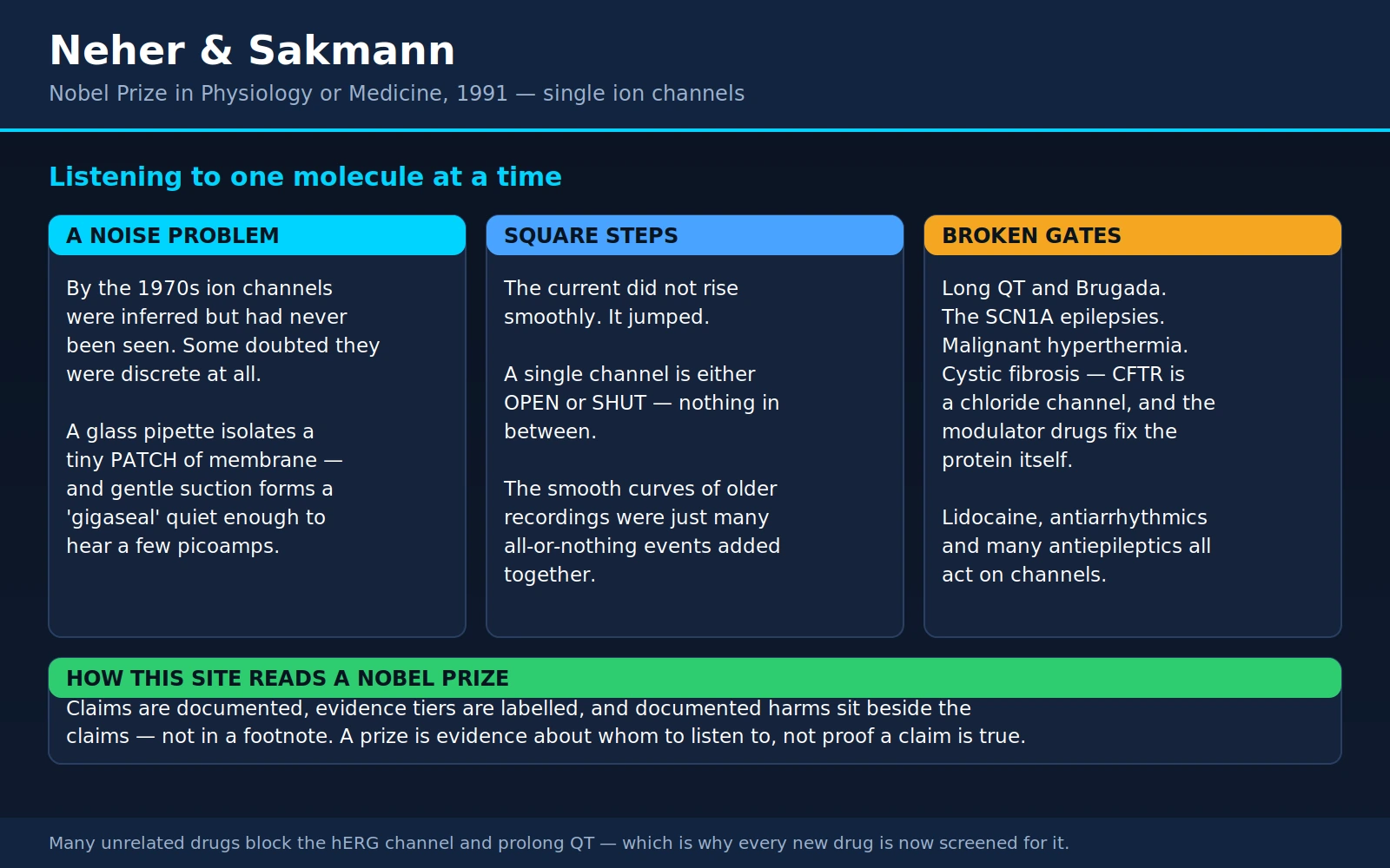
Almost everything your body does quickly — a heartbeat, a thought, a flinch away from a hot pan, the release of insulin after a meal — happens because charged particles move across cell membranes through protein tunnels called ion channels. For most of the twentieth century those tunnels were a reasonable inference. Nobody had ever seen one work.
In 1976, two researchers at the Max Planck Institute for Biophysical Chemistry in Göttingen recorded the electrical current flowing through a single one of them. Erwin Neher, born in 1944 and trained as a physicist, and Bert Sakmann, born in 1942 and trained as a physician, shared the 1991 Nobel Prize in Physiology or Medicine "for their discoveries concerning the function of single ion channels in cells." The instrument they built to do it — the patch clamp — is still, half a century later, the standard tool of cellular neuroscience, cardiac electrophysiology and ion-channel drug discovery.
This page is about what they did, why the result was beautiful, and what it means for anyone living with cystic fibrosis, an inherited arrhythmia, a channel-related epilepsy, or a prescription that carries a warning about the heart's QT interval.
Table of Contents
- The Prize and the Two Men
- The Problem: Currents Without Visible Channels
- The Patch Clamp: A Noise Problem, Solved
- What They Saw: Square Steps
- What It Made Possible
- Channelopathies: When a Single Gate Breaks
- Cystic Fibrosis and the CFTR Modulators
- Medicines That Act on Ion Channels
- hERG, the QT Interval, and Why Drugs Get Withdrawn
- Anaesthesia, and the Honest Gap
- What the Method Still Cannot Tell You
- Where Mainstream Medicine Agrees, and What Remains Debated
- Key Research Papers
- Connections
- Featured Videos
1. The Prize and the Two Men
Neher and Sakmann were an unusually well-matched pair, and the pairing was the point. Neher came from physics. His instincts were about signal and noise, amplifier design, glass, seals, shielding — the unglamorous engineering that decides whether a measurement is possible at all. Sakmann came from medicine and physiology, and had spent time in Bernard Katz's laboratory in London, where the statistical behaviour of the neuromuscular junction was being worked out. He knew which biological question was worth building an instrument for.
They began working together in Göttingen in the early 1970s at the Max Planck Institute for Biophysical Chemistry. The 1976 recording came out of that collaboration. By the time the prize was awarded in 1991, Sakmann had moved to the Max Planck Institute for Medical Research in Heidelberg; Neher remained in Göttingen.
The Nobel citation is worth reading closely, because it is oddly modest. It does not say they discovered ion channels — ion channels had been postulated for decades. It says they made discoveries concerning the function of single ion channels. The prize was for turning a category of thing that had to be argued about into a category of thing you could simply watch.
2. The Problem: Currents Without Visible Channels
By the 1970s, electrophysiology had a magnificent theory and no picture to go with it.
Twenty years earlier, Alan Hodgkin and Andrew Huxley had done something extraordinary with the giant axon of the squid. By clamping the membrane at a fixed voltage and measuring the current that flowed, they showed that the nerve impulse is produced by two separate, voltage-dependent movements of ions — sodium rushing in, then potassium flowing out — and they wrote down equations that predicted the shape and speed of the impulse with startling accuracy. Those equations still work. They earned the 1963 Nobel Prize, shared with John Eccles.
But the Hodgkin–Huxley equations describe the membrane as a whole. They speak of conductances that rise and fall smoothly over milliseconds, like a tap being opened and closed. Hodgkin and Huxley were entirely candid that they did not know what physical object those conductances corresponded to. Their model was a description of behaviour, not of machinery.
Two rival pictures of the machinery were live at the time:
- Pores. The membrane contains a fixed number of discrete protein tunnels. Each one is either open or shut. The smooth conductance curve is a population statistic — the average of many tunnels flicking open and closed at different moments.
- Carriers. There are no fixed tunnels. Mobile molecules pick ions up on one side of the membrane, shuttle across, and drop them on the other. Conductance is continuous because the ferrying is continuous.
These are not small differences. If channels are real discrete objects, then a single mutation in a single gene can break one, and the resulting disease should have a characteristic electrical signature. If transport is a continuous carrier process, that framing does not apply. Everything in the second half of this page — long QT syndrome, cystic fibrosis, the safety testing of every new drug — depends on which picture is correct.
There was strong indirect evidence for pores. Bernard Katz and Ricardo Miledi, working on the frog neuromuscular junction, had noticed that when acetylcholine is applied to the endplate the membrane current does not merely increase — it becomes noisier. From the statistics of that noise they could estimate how big an individual elementary event must be and how long it must last, without ever seeing one. It was a beautiful argument, and Neher and Sakmann used the same noise-analysis approach themselves in a companion study published in the Journal of Physiology in 1976.
But an estimate extracted from statistics is not the same as a recording. What was missing was the direct observation: one channel, one trace, watched in real time.
3. The Patch Clamp: A Noise Problem, Solved
The obstacle was not conceptual. It was electrical noise.
A single ion channel passes a current measured in picoamps — trillionths of an ampere. To give that a sense of scale: one picoamp is roughly six million elementary charges per second. That sounds like a lot until you compare it to the current flowing through the rest of an entire cell membrane, which is many thousands of times larger and constantly fluctuating. Trying to hear one channel against the whole cell is like trying to hear one cricket from the middle of a stadium.
Isolate a patch
The first idea was to stop listening to the whole membrane. Neher and Sakmann pulled a glass pipette down to a very fine tip — an opening of a few micrometres, small enough that the piece of membrane it covers might contain only one or two channels — and pressed it against the surface of a muscle fibre. Now the amplifier is only connected to that little patch. Everything happening elsewhere on the cell is outside the circuit.
They also chose their preparation carefully: frog muscle fibres whose nerve supply had been cut. Denervated muscle spreads acetylcholine receptors across its whole surface instead of concentrating them at the endplate, which makes it far easier to find a patch containing a manageable number of channels. And they cleaned the membrane surface enzymatically so the glass could sit flat against it.
The leak that would not go away
It half worked. The 1976 recordings were real, but they sat on a hiss of background noise, because the seal between glass and membrane leaked. Current that should have gone through the amplifier instead crept around the rim of the pipette. In electrical terms the seal resistance was tens of megaohms — respectable, but not nearly enough. The signal was there; it was just barely above the noise floor, and every trace had to be coaxed.
This is worth saying plainly, because it is the part usually skipped: the invention was not a biological insight. It was a noise problem, solved. Everything the patch clamp went on to make possible followed from making one number — the resistance of the seal between a piece of glass and a piece of cell membrane — about a thousand times bigger.
The gigaseal
The fix arrived around 1980, partly by accident. Working with very clean pipettes on clean membrane, the Göttingen group found that applying gentle suction through the pipette — a slight negative pressure, nothing violent — sometimes caused the membrane to leap into intimate contact with the glass. When it did, the seal resistance jumped by orders of magnitude, into the range of 109 to 1011 ohms: a gigaohm or better. They called it the gigaseal.
Nobody had predicted this. Glass and lipid membrane are not supposed to bond. To this day the physical chemistry of the gigaseal is not fully settled — it is treated in practice as a reliable trick rather than a fully understood phenomenon. What mattered was the consequence. Background current noise falls as the seal resistance rises, so a thousandfold better seal took the hiss far below the signal. Currents of a few picoamps stopped being something you had to extract and became something you could simply look at on a screen.
The 1981 paper in Pflügers Archiv that described how to do it — how to pull and fire-polish the pipettes, how to build the amplifier, how to get the seal — became one of the most heavily cited papers in the whole of biology. Its five authors were Owen Hamill, Alain Marty, Erwin Neher, Bert Sakmann and Fred Sigworth.
4. What They Saw: Square Steps
Here is the result, and it is one of the loveliest in modern biology.
When the trace came up clean, the current through a single acetylcholine-activated channel did not drift, ramp, wobble or swell. It jumped. The trace sat flat at zero, then snapped down to a fixed level, stayed there for a millisecond or a few, then snapped back to zero. Down, flat, up. Down, flat, up. Rectangular pulses of essentially identical height, of varying and unpredictable duration, scattered irregularly in time.
Square steps.
You are looking at the behaviour of one protein molecule. It is open, or it is shut. There is no halfway. The height of the step is fixed because it is set by the physics of the pore — how wide it is, how it handles the ion — and the pore does not change size. What varies is when the molecule opens and how long it stays open, and those are random, governed by thermal jostling and by whether a neurotransmitter molecule happens to be bound.
Why the squareness was the whole answer
The carrier hypothesis dies here. A shuttle service would produce a current that rises and falls with the rate of shuttling. It would not produce a two-state rectangle. The step said, unambiguously: this is a gate, and it has exactly two positions.
And the second implication is the one that reorganised the field. If a single channel is a two-state device, then the smooth, graceful, elegantly modelled conductance curves that Hodgkin and Huxley measured across a whole membrane were never smooth at all. They were the sum of thousands of all-or-nothing events. The smoothness was an artefact of averaging — the same way a photograph of a crowd looks like a continuous grey wash until you zoom in and find individual faces.
Two decades of beautiful mathematics were not overturned by this. They were explained. The Hodgkin–Huxley conductance term, which had been a fitted description of behaviour, was revealed to be a probability: the fraction of a large population of independent gates that happen to be open at a given voltage and moment. The equations kept working, and now they meant something physical.
This is the reason the result deserves to be dwelt on. Most experiments add a fact. This one changed what the previous twenty years of facts had been about. And it did it with an image simple enough to explain to a child: a switch, flicking.
5. What It Made Possible
The gigaseal turned out to be far more than a way to record single channels. Once the glass is sealed that tightly to the membrane, you can do things to the patch that were previously unthinkable. The 1981 paper laid out the family of configurations that are still the working vocabulary of the field:
- Cell-attached patch — the original. The patch stays part of the intact cell; you watch its channels while the cell carries on living.
- Inside-out patch — pull the pipette away and the patch tears free, with the cell's inner face now exposed to the bath. You can now change the solution on the inside of the membrane at will and see what the channel does about it.
- Outside-out patch — a different manoeuvre leaves the outer face exposed, so you can apply drugs or transmitters to the channel's external side with nothing else in the way.
- Whole-cell recording — rupture the membrane under the pipette without breaking the seal. The pipette interior now connects directly to the cell interior. You control the cell's voltage and dialyse its internal chemistry, while recording every channel it has.
That last one, whole-cell recording, is arguably the bigger practical legacy. Almost every modern measurement of what a neuron, a heart cell, a beta cell or a smooth-muscle cell does electrically is a whole-cell patch-clamp recording. It is how synaptic currents are measured. It is how the electrical fingerprint of a cardiac muscle cell is characterised. It is the workhorse behind essentially all of cellular neuroscience and cardiac electrophysiology since roughly 1985.
From a bench craft to a robot
Patch clamping by hand is a skill. An experienced operator manoeuvres a pipette onto a cell under a microscope, applies suction at the right moment, and hopes. It is slow, and good data take years of practice.
Modern automated patch clamp systems replaced the human hand and the microscope with a planar chip: a plate with microscopic holes, into which cells are drawn by suction, sealed and recorded — many at once, under robotic control, unattended. This is what made ion-channel work compatible with industrial drug screening, where you need to test thousands of compounds rather than dozens.
The most consequential application is drug safety testing. Every new drug candidate is now screened for its effect on cardiac ion channels before it ever reaches a person, and that screen is a patch-clamp recording performed by machine. Regulators have moved from a single blunt assay towards a panel of human ion-channel measurements combined with computer reconstructions of the heartbeat and recordings from stem-cell-derived human heart cells — a deliberate shift towards mechanism, and away from a pass/fail number. Section 9 explains why that screen exists at all.
6. Channelopathies: When a Single Gate Breaks
Once channels were established as discrete proteins encoded by specific genes, a whole disease category came into focus. A channelopathy is an illness caused by a channel that opens too much, too little, too early, too late, or not at all. By 2006 mutations in more than sixty ion-channel genes were known to cause human disease, and the list has grown since.
These conditions share a signature that makes sense once you know the mechanism. They are often episodic — the person is completely well between attacks, because a channel that misbehaves under particular conditions behaves normally the rest of the time. They are often triggered by something specific, because the trigger is whatever pushes the faulty gate past its limit. And they are often invisible on scans and biopsies, because nothing is structurally wrong; the fault is in the timing of a molecule.
Long QT syndrome
Every heartbeat has two halves: the cells depolarise (the electrical squeeze) and then repolarise (the reset). The reset is done mainly by potassium flowing out. If the potassium channels that do the resetting are sluggish, the reset takes too long. On an ECG that shows up as a lengthened QT interval.
A heart that resets slowly is vulnerable in a specific way. If an extra beat lands during the extended vulnerable window, it can trigger torsades de pointes, a twisting polymorphic ventricular tachycardia that may stop on its own — producing a faint — or may degenerate into ventricular fibrillation, which is fatal within minutes without defibrillation. This is why long QT syndrome kills young, apparently healthy people: nothing is wrong with the heart's structure, its pumping, or its arteries. The failure is a timing fault in a potassium gate.
A landmark screen of 262 unrelated affected individuals found a causative mutation in 68% of them, and the distribution is lopsided. Two potassium-channel genes account for the overwhelming majority: KCNQ1 (42% of identified mutations) and KCNH2, better known as hERG (45%). The cardiac sodium-channel gene SCN5A accounted for 8%, with KCNE1 (3%) and KCNE2 (2%) making up the rest. Clinically these are called LQT1, LQT2 and LQT3.
Which gene it is matters enormously, and this is the part worth knowing if it is in your family. In a study of 670 genotyped patients with symptoms, the circumstances of dangerous events were strongly gene-specific:
- LQT1 (KCNQ1) — 62% of events occurred during exercise; only 3% during rest or sleep. Swimming is a classic trigger. Beta-blockers work well here: 81% of LQT1 patients were free of recurrence on beta-blocker therapy.
- LQT2 (KCNH2/hERG) — only 13% of events during exercise, 29% during rest or sleep. Sudden loud noises — an alarm clock, a telephone — are a recognised trigger. 59% were recurrence-free on beta-blockers.
- LQT3 (SCN5A) — 13% during exercise, 39% during rest or sleep, so events cluster at night. This group did worst: 50% recurrence-free on beta-blockers, and a death rate of 17% compared with 4% in both LQT1 and LQT2.
The corrected QT interval itself barely differed between the three groups (498, 497 and 506 ms), which is the point: the ECG number alone does not tell you the risk — the gene does. That is a direct clinical consequence of knowing that channels are discrete proteins with individual genes.
There is also a recessive form, Jervell and Lange-Nielsen syndrome, caused by mutations in KCNQ1 or KCNE1, in which congenital deafness accompanies the arrhythmia risk — because the same potassium channel is needed to maintain the ionic environment of the inner ear.
And critically, long QT can be acquired rather than inherited. Many ordinary medicines lengthen the QT interval by blocking the hERG channel, and low blood potassium or magnesium makes that far more dangerous. That story is section 9.
Brugada syndrome
In 1992 Pedro and Josep Brugada described eight patients with a striking and consistent picture: right bundle branch block with persistent ST-segment elevation in the right precordial ECG leads (V1–V3), a normal QT interval, structurally normal hearts on biopsy and imaging, and aborted sudden death from rapid polymorphic ventricular tachycardia. Their paper ends with the honest sentence: "Its causes remain unknown."
Six years later the cause was found. Mutations in SCN5A, the cardiac sodium-channel gene, were identified in affected families — some producing channels that recover from inactivation abnormally fast, one producing a channel that does not function at all. Brugada syndrome is a sodium-channel disease, and where long QT is usually a failure of the reset, Brugada is a failure of the initial electrical push in one region of the heart.
Practical detail that matters to patients: the diagnostic ECG pattern in Brugada is intermittent. It can be absent one day and obvious the next, and it is characteristically unmasked by fever — which is why a febrile illness is treated seriously in someone with a known or suspected diagnosis. SCN5A is found in only a minority of cases, so a negative genetic test does not exclude the syndrome.
Cystic fibrosis
The flagship example, and important enough to have its own section — see section 7. In brief: the CFTR protein is a chloride channel, and cystic fibrosis is what happens when it is missing or broken. Thick secretions, recurrent lung infection, pancreatic insufficiency and salty sweat all follow from chloride, and the water that travels with it, failing to cross a membrane.
Some epilepsies
Neurons fire because sodium channels open. If a sodium channel is faulty, the balance between excitation and inhibition can tip.
The clearest example is Dravet syndrome, historically called severe myoclonic epilepsy of infancy. In 2001 it was shown that de novo mutations in SCN1A — the gene for the neuronal sodium channel NaV1.1 — cause it. More than 90% of people with Dravet syndrome carry an SCN1A mutation. It typically begins in the first year of life with prolonged seizures often provoked by fever, followed by drug-resistant seizures of several types, developmental impairment, gait problems, sleep disturbance and a raised risk of early death.
Here is where knowing the mechanism changes the prescription pad, and it is counter-intuitive enough that it is worth stating as plainly as possible. Many standard epilepsy drugs work by blocking sodium channels. In Dravet syndrome the underlying fault is usually loss of sodium-channel function — and critically, the affected channel sits disproportionately in inhibitory interneurons, the cells whose job is to suppress firing. Blocking sodium channels further silences the brakes. In practice, sodium-channel-blocking anti-seizure medicines are generally minimally effective in Dravet syndrome and can make seizures worse. A drug class that is first-line for many epilepsies is the wrong choice here, and the reason is a fact about one protein.
Malignant hyperthermia
Not every ion channel sits in the outer membrane of a cell. RYR1 encodes the ryanodine receptor, a calcium-release channel in the sarcoplasmic reticulum — the internal calcium store of skeletal muscle. It is the gate that releases calcium to make a muscle contract.
In malignant hyperthermia, that gate can be pushed open uncontrollably by potent volatile anaesthetic gases (halothane, sevoflurane, desflurane, isoflurane) and by the depolarising muscle relaxant succinylcholine. Calcium floods the muscle cytoplasm and cannot be recovered fast enough. Every calcium-driven process in muscle runs at once, generating an enormous metabolic load: rising temperature, racing heart, fast breathing, a sharp rise in carbon dioxide production and oxygen consumption, acidosis, high blood potassium, muscle rigidity and rhabdomyolysis. Untreated it is usually fatal.
The numbers are worth knowing. Reactions occur in roughly 1 in 10,000 to 1 in 250,000 anaesthetics, but the underlying genetic susceptibility may be as common as 1 in 400 people — most of whom will never encounter a triggering drug and will never know. Over 400 variants have been described in RYR1, on chromosome 19q13.1, of which at least 34 are established as causal; under 1% of cases involve CACNA1S, and not all of those variants are causal. Inheritance is autosomal dominant.
The earliest reliable warning sign in theatre is a rise in end-tidal carbon dioxide despite increased ventilation. The specific antidote is dantrolene, which acts on the ryanodine receptor itself, and it should be stocked wherever general anaesthesia is given. With recognition and dantrolene, mortality fell from around 80% three decades ago to under 5%. If anyone in your family has had an unexplained severe reaction, a very high fever, or a death under general anaesthesia, that is information your anaesthetist genuinely needs before an operation.
Periodic paralysis
The periodic paralyses are rare inherited muscle channelopathies in which attacks of flaccid weakness come and go while strength is normal in between. They are caused by mutations in skeletal-muscle ion channels — the calcium channel CaV1.1 (CACNA1S), the sodium channel NaV1.4 (SCN4A), and the potassium channels Kir2.1 (KCNJ2) and Kir3.4 — all of which destabilise the muscle fibre's resting voltage. Most are missense changes that alter how a channel works rather than abolishing it.
- Hypokalaemic periodic paralysis — attacks accompanied by low blood potassium, usually from CACNA1S or SCN4A. Classic triggers are a high-carbohydrate meal and rest after exercise, which is why attacks often strike in the early morning after a heavy evening meal or a hard training session.
- Hyperkalaemic periodic paralysis — attacks with normal or high potassium, from SCN4A. Fasting, cold and rest after exertion are typical triggers.
- Andersen–Tawil syndrome — KCNJ2, combining periodic paralysis with ventricular arrhythmia and characteristic skeletal features.
Inheritance is usually autosomal dominant and onset is typically in the first or second decade. Diagnosis rests on the clinical pattern confirmed by genetic testing; where no mutation is found, documented potassium levels during an attack or a decrement on long exercise testing support the diagnosis. Management has two halves — treating attacks and preventing them — using trigger avoidance, adjustment of potassium, diuretics and carbonic anhydrase inhibitors. Because the mechanism differs between subtypes, the correct treatment for one form can be actively wrong for another; getting the subtype right is not academic.
Congenital myasthenic syndromes
There is a satisfying circularity here. The very channel Neher and Sakmann first recorded — the acetylcholine receptor at the muscle endplate — is the one most often at fault in the congenital myasthenic syndromes, a group of inherited disorders of signal transmission at the junction between nerve and muscle. Most arise from molecular defects in the muscle nicotinic acetylcholine receptor itself; others involve presynaptic proteins, the synaptic basal lamina, endplate development, or protein glycosylation.
They cause fatigable weakness that can look like myasthenia gravis but is genetic rather than autoimmune, so immunosuppression is not the answer. Treatments include cholinergic agonists, long-lived open-channel blockers of the acetylcholine-receptor channel, and adrenergic agonists. Most are treatable — but the same warning applies as in Dravet and the periodic paralyses: a drug that is beneficial in one congenital myasthenic syndrome can be harmful in another, depending on whether the mutant channel stays open too long or does not open enough. This is a field where the specific genetic diagnosis is the treatment plan.
7. Cystic Fibrosis and the CFTR Modulators
If you want a single story that shows what the ion-channel era actually delivered to patients, it is this one.
What is broken
The gene identified in 1989 as the cause of cystic fibrosis encodes CFTR — the cystic fibrosis transmembrane conductance regulator — and CFTR turned out to be a chloride channel sitting in the surface of the cells that line the airways, the pancreatic ducts, the gut, the sweat glands and the reproductive tract.
Chloride carries water with it. When CFTR does not move chloride onto the airway surface, the thin watery layer that lets cilia sweep mucus out of the lungs is not maintained. Mucus becomes thick and static, bacteria colonise it, and a cycle of infection, inflammation and irreversible airway damage begins in childhood. The same failure blocks pancreatic ducts, causing the digestive enzyme deficiency that stunts growth. And because the sweat duct cannot reabsorb chloride, sweat is abnormally salty — which is why the sweat chloride test, a measurement that predates any of the molecular biology, remains the diagnostic standard, and why it is such a useful direct read-out of whether a treatment is fixing the channel.
The mutation classes, and why they decide everything
More than two thousand CFTR variants are known, and they break the protein in different ways. Conventionally they are grouped into classes, and the class determines which drug can possibly help:
- Class I — no protein made. Nonsense or frameshift variants stop production. There is no channel at the membrane at all.
- Class II — protein made but misfolded. It is caught by the cell's quality control and destroyed before it reaches the surface. F508del (also written Phe508del) is the overwhelmingly common example.
- Class III — protein reaches the surface but the gate will not open properly. A gating defect. G551D is the classic case.
- Class IV — the gate opens but the pore conducts poorly.
- Class V — too little normal protein is made.
- Class VI — the protein is unstable at the surface and is removed too quickly.
Two kinds of drug follow directly from that list. A potentiator makes a channel that is present at the surface open more readily — the fix for a class III gating defect. A corrector helps a misfolded protein fold well enough to survive quality control and reach the surface — the fix for class II. Neither can do anything about a class I variant, because there is nothing there to potentiate or correct.
Ivacaftor: the proof of concept
Ivacaftor is a potentiator. In a phase 3 randomised, double-blind, placebo-controlled trial published in 2011, 161 people aged 12 or over with at least one G551D mutation received ivacaftor or placebo for 48 weeks. Over 24 weeks, lung function measured as percent-predicted FEV1 was 10.6 percentage points higher on ivacaftor than placebo. Effects appeared within two weeks. Over 48 weeks, participants on ivacaftor were 55% less likely to have a pulmonary exacerbation, scored 8.6 points higher on the respiratory-symptoms scale, gained an average of 2.7 kg more weight, and had sweat chloride fall by 48.1 mmol/L — direct evidence that the channel itself was working better. Serious adverse events were less common on the drug than on placebo (24% versus 42%).
That last number is the tell. This was not a symptom treatment producing a marginal statistical win. Something upstream had changed.
But G551D is carried by only a small minority of people with cystic fibrosis. Ivacaftor alone was a spectacular result for a few thousand people worldwide and no help at all to the great majority, whose problem is F508del — a folding failure that a potentiator cannot touch.
Triple therapy, and who it reaches
The answer was to combine correctors with the potentiator. Elexacaftor–tezacaftor–ivacaftor pairs two correctors with ivacaftor, and it is the reason cystic fibrosis care changed shape at the end of the 2010s.
The pivotal trial randomised 403 patients aged 12 and over who had one F508del allele and one minimal-function allele — explicitly the group in whom every previous modulator regimen had failed. Against placebo, the triple combination produced percent-predicted FEV1 13.8 points higher at 4 weeks and 14.3 points higher through 24 weeks, a pulmonary exacerbation rate 63% lower, a respiratory quality-of-life score 20.2 points higher (against a minimum clinically important difference of 4 points), and sweat chloride 41.8 mmol/L lower. Only 1% discontinued for adverse events. A companion trial in people carrying two F508del alleles showed further improvement even against active treatment with tezacaftor–ivacaftor alone.
Because nearly 90% of people with cystic fibrosis carry at least one copy of F508del, this brought the great majority within reach of a drug that acts on the underlying defect.
Does it change the long run, not just the numbers?
Trial endpoints over 24 or 48 weeks do not by themselves prove that a disease has been slowed. The most useful evidence on that comes from national registries. An observational post-approval study compared 1,256 ivacaftor-treated patients in the United States with 6,200 matched untreated comparators, and 411 treated against 2,069 comparators in the United Kingdom. In the US cohort the treated group had significantly lower rates of death (0.6% versus 1.6%), transplantation (0.2% versus 1.1%), hospitalisation (27.5% versus 43.1%) and pulmonary exacerbation (27.8% versus 43.3%), with similar trends in the UK, fewer CF-related complications, less colonisation with problem organisms, and better-preserved lung function.
This is observational, not randomised, and the authors say so plainly — matched comparison cannot fully exclude the possibility that people who started the drug differed from those who did not. But the direction and size are consistent with the trial data, and they support the claim that this is disease modification rather than symptom relief.
Being honest about who is left out
This is the part most write-ups skate over, and it is the part that matters most if it applies to you or your child.
People with two minimal-function variants and no F508del are not helped by any currently approved modulator. If both copies of the gene are class I — a nonsense or frameshift variant that means no CFTR protein is produced at all — there is nothing at the membrane for a potentiator to open or a corrector to rescue. These are typically around 10% of people with cystic fibrosis, and the fraction is higher in some populations and ancestries than in others, which means the benefits of the modulator era have not been distributed evenly. For this group the active research directions are different in kind — read-through agents, gene therapy, gene editing, mRNA delivery — and none is yet an approved treatment. It is entirely reasonable to feel that the celebration of this era has left your family out, because in a real sense it has.
Several further caveats apply even to those who do benefit:
- Modulators do not cure cystic fibrosis. They must be taken every day, for life, and the effect stops when the drug stops.
- They do not reverse damage already done. Bronchiectasis and scarred airways stay scarred. This is a strong argument for treating early, and a genuine grief for adults who reached the drug after decades of damage.
- Response varies between individuals with the same genotype, and a minority tolerate the drugs poorly — liver enzyme rises, rash, and mental-health effects including low mood and anxiety have been reported often enough to be worth raising with your team rather than assuming it is coincidence.
Cost and access
Triple modulator therapy carries a list price in the hundreds of thousands of US dollars per patient per year. A 2022 analysis estimated the minimum cost of production of elexacaftor–tezacaftor–ivacaftor at about US$5,676 per year (range $4,628–6,723), derived from published synthetic routes and global import/export prices for the active ingredients — over 90% below the US list price. Treating every eligible diagnosed person with cystic fibrosis worldwide would cost about US$489 million a year at production cost, against roughly US$31.2 billion at the US list price.
The consequence is that access is decided by geography and by which health system you happen to live under. Many countries with substantial cystic fibrosis populations still have limited or no access, and the resulting inequality is the subject of active campaigning — including proposals for voluntary licensing to generic manufacturers, an approach with precedent in other disease areas. None of this is a criticism of the science, which is remarkable. It is a statement about what happens between a working drug and the person who needs it.
8. Medicines That Act on Ion Channels
Ion channels are one of the most heavily drugged protein families in medicine. Most people reading this have taken at least one channel drug, usually without being told that is what it was. Here is the recognisable list.
Local anaesthetics
When a dentist numbs your jaw with lidocaine, the mechanism is a direct consequence of everything above. Pain signals travel from the tissue to the spinal cord as a chain of action potentials, and every action potential requires voltage-gated sodium channels to open in sequence along the nerve fibre. Lidocaine enters the nerve, binds inside the pore of those sodium channels, and physically obstructs it.
Two features make it work as well as it does. First, the binding site is inside the channel — the drug reaches it through the membrane or through the open pore, and the relevant residues lie in the inner lining of the pore. Second, the block is state-dependent: local anaesthetics bind far more tightly to channels that are open or recently inactivated than to resting ones. Since a firing nerve cycles through those states constantly and a quiet nerve does not, the busier the nerve, the more thoroughly it is blocked. The drug preferentially silences exactly the fibres that are carrying the pain.
The sensation of numbness is therefore not a blunting of pain. It is the complete failure of a signal to leave the area. Nothing arrives at the brain to be interpreted.
The same mechanism explains the systemic warning: local anaesthetics that reach the bloodstream in quantity block sodium channels in the heart and brain too, which is why dose limits are taken seriously and why a metallic taste, tingling round the mouth or ringing in the ears during an injection is something to mention immediately.
Antiarrhythmic drugs
The oldest drug classification in cardiology is essentially a classification of ion-channel targets:
- Sodium-channel blockers — flecainide, propafenone, lidocaine, quinidine. They slow the electrical upstroke.
- Beta-blockers — not channel drugs themselves, but they change how channels are regulated (see James Black, who designed them).
- Potassium-channel blockers — amiodarone, sotalol, dofetilide. They deliberately lengthen repolarisation. Note the irony: these drugs prolong the QT interval on purpose, which is exactly the effect that gets other drugs withdrawn, and it is why they are started with monitoring.
- Calcium-channel blockers — verapamil, diltiazem. They slow conduction through the AV node.
Anti-seizure medicines
Most epilepsy drugs are channel drugs. Carbamazepine, oxcarbazepine, phenytoin, lamotrigine and lacosamide act mainly on neuronal sodium channels, damping repetitive firing. Ethosuximide targets T-type calcium channels in the thalamus, which is why it works specifically for absence seizures. Gabapentin and pregabalin bind a subunit of voltage-gated calcium channels. Benzodiazepines and barbiturates act on the GABAA receptor, which is itself a chloride channel.
As section 6 explained, this is precisely why the genetic diagnosis can matter: in Dravet syndrome the sodium-channel blockers are usually the wrong class.
Calcium-channel blockers for blood pressure
Amlodipine, nifedipine and felodipine relax the smooth muscle in artery walls by blocking L-type calcium channels, so less calcium enters, the muscle relaxes, the vessel widens and blood pressure falls. The characteristic side effects follow from the mechanism: ankle swelling, because dilating the arterial side raises pressure in the capillary beds, and flushing or headache from vasodilation. If you take amlodipine and have puffy ankles by evening, that is the ion channel talking.
Sulfonylureas and the KATP channel
Gliclazide, glipizide and glibenclamide (glyburide) treat type 2 diabetes through one of the most elegant channel mechanisms in pharmacology. The pancreatic beta cell has an ATP-sensitive potassium channel, KATP, that is open when the cell has little fuel. Open potassium channels hold the cell electrically quiet. When glucose arrives and is metabolised, ATP rises, the KATP channel closes, the cell depolarises, calcium enters, and insulin is released. The channel is the glucose sensor. Sulfonylureas close that channel directly, bypassing the glucose signal — which is exactly why they can cause hypoglycaemia in a way that metformin does not: they tell the beta cell to release insulin whether or not there is glucose to justify it.
The most remarkable consequence appears in neonatal diabetes. Some babies diagnosed with diabetes in the first six months of life have activating mutations in KCNJ11, the gene for the Kir6.2 subunit of that channel: the gate will not close in response to ATP, so insulin is never released. In a study of 49 such patients, 44 (90%) were able to stop insulin injections entirely after switching to sulfonylurea tablets, with glycated haemoglobin improving from 8.1% to 6.4% at 12 weeks and control sustained at one year. The degree to which each mutant channel responded to the drug in the laboratory predicted the response in the patient. A lifetime of daily injections, replaced by tablets, because someone knew which gate was stuck.
Amiloride
The potassium-sparing diuretic amiloride blocks the epithelial sodium channel (ENaC) in the kidney's collecting duct. Less sodium is reabsorbed, so more is excreted with water — and because sodium reabsorption there is electrically coupled to potassium secretion, blocking it also retains potassium, which is what "potassium-sparing" means. It is also the reason amiloride and potassium supplements together need care.
9. hERG, the QT Interval, and Why Drugs Get Withdrawn
This is one of the better untold stories in modern medicine, and it explains a warning many people have seen on a pharmacy label without knowing where it came from.
An unusually promiscuous channel
The hERG channel — encoded by KCNH2, and named, memorably, for the human ether-à-go-go-related gene — carries the potassium current that does most of the work of resetting the heart after each beat. It is essential for normal cardiac electrical activity, and inherited mutations in it cause LQT2.
It also has an inconvenient structural quirk: an unusually large and accommodating inner cavity. Where most ion channels are fussy about what binds them, hERG is not. A surprisingly diverse group of drugs — chemically unrelated, developed for entirely different purposes, with no shared pharmacology whatsoever — block it as an unintended side effect.
Block hERG and the heart resets more slowly. The QT interval lengthens. In most people, most of the time, nothing happens. But the same vulnerable window that makes inherited long QT syndrome dangerous now exists in a person who does not have a genetic channelopathy at all, and if an extra beat lands in it, torsades de pointes can follow.
The 1990s reckoning
Through the 1990s, reports of drug-induced torsades de pointes associated with non-cardiac drugs rose sharply, as one new medicine after another turned out to carry a proarrhythmic liability that had not been recognised before marketing. Several drugs were withdrawn or had their labels heavily restricted as a result. The clearest documented examples:
- Terfenadine and astemizole — early second-generation antihistamines, taken by enormous numbers of people for hay fever, and associated with cardiotoxic deaths. Their withdrawal is the founding case of the whole story.
- Cisapride — a gastrointestinal prokinetic drug for reflux, withdrawn after QT prolongation attributed to hERG block. Later 5-HT4 agonists were deliberately designed to be selective and free of hERG affinity, and have not shown the same problem.
The response reshaped drug development. Every new molecule is now screened against hERG — by automated patch clamp, the direct descendant of the Göttingen experiment — long before it reaches a human. Regulators went further and began moving from a single hERG number towards a mechanistic package: a panel of human ion-channel assays, computer reconstructions of the cardiac action potential, and recordings from human stem-cell-derived heart cells. The aim is both to catch genuinely dangerous compounds and to stop discarding safe ones, because a crude hERG cut-off also rejects useful drugs that block hERG but are counterbalanced by effects on other channels. Verapamil is the standard example: it blocks hERG at clinically relevant concentrations and yet does not prolong QT in people, because it blocks calcium channels at the same time.
What this means for you, practically
Do not read this section as a reason to stop a medicine. Read it as a reason to keep one list.
A long roster of ordinary drugs carries a QT caution: some antibiotics (notably macrolides such as clarithromycin, and fluoroquinolones), some antifungals, several antipsychotics, methadone, ondansetron for nausea, some antimalarials, some cancer drugs, and the antiarrhythmics that lengthen QT deliberately. Any one of them, in a healthy person with a normal baseline QT and normal blood chemistry, is usually a non-event.
The real risk is stacking, and it is almost always a combination of factors rather than one drug. The recognised risk factors are well characterised:
- Two or more QT-prolonging drugs at once — the single commonest scenario, and it often happens because different prescribers each added one.
- A drug that blocks the metabolism of another — this was the actual mechanism behind the antihistamine deaths. Terfenadine was largely harmless until an interacting drug or grapefruit juice stopped the liver clearing it and blood levels climbed.
- Low potassium or low magnesium — which is why vomiting, diarrhoea, an eating disorder, or a loop diuretic changes the calculation. This is a genuinely actionable point: the electrolytes are checkable and correctable.
- Older age, existing heart disease, and inherited long QT syndrome, including undiagnosed carriers.
Practical steps that are worth taking and cost nothing: keep one complete list of everything you take, including over-the-counter medicines and supplements, and show it to every prescriber and to your pharmacist — pharmacists catch these interactions routinely and it is exactly what they are for. If you are told you have a long QT interval, ask for it in writing. If a new drug is started and you begin fainting, having palpitations, or feeling your heart race, that is not something to wait out. And if you already know you have long QT syndrome, curated lists of drugs to avoid exist and your cardiology team will point you to one.
10. Anaesthesia, and the Honest Gap
General anaesthesia is administered tens of millions of times a year, with a safety record that is one of medicine's genuine triumphs. It is also, at the level that matters most, unexplained.
What is known is real and specific. General anaesthetics interact with ion channels: they enhance inhibitory GABAA receptors (chloride channels), inhibit excitatory NMDA receptors, and activate certain two-pore-domain potassium channels that quiet neurons by holding them hyperpolarised. Different agents have different profiles — propofol and the volatile agents lean heavily on GABAA, ketamine and xenon on NMDA blockade. Much of this molecular detail was worked out with patch-clamp recordings.
What is not known is how any of that produces the loss of consciousness. Enhancing inhibition at a synapse is a molecular event. Consciousness is a property of a brain. The path from one to the other — which circuits, which networks, in what order, and why a modest shift in inhibitory tone abolishes subjective experience rather than merely slowing it — has not been established. Research has moved towards thinking of anaesthesia as the hijacking of the brain's own sleep and arousal circuitry rather than a simple global dampening, and towards network-level measures of how brain regions stop communicating with one another. These are promising directions, not settled answers.
We are stating this plainly because pages on this subject often do not. Anaesthesia is empirically excellent and mechanistically unresolved. Doctors can reliably render you unconscious, keep you safely there, monitor the depth, and bring you back; they cannot fully explain why the drug removes the experience. Both halves of that sentence are true, and the first half is the one that matters when you are being wheeled into theatre.
The honest gap is worth naming for a second reason: it is a good calibration point for the rest of this page. Patch clamping told us what individual molecules do with enormous precision. It did not, on its own, tell us what a brain does. Knowing the parts is not the same as knowing the system.
11. What the Method Still Cannot Tell You
Since this page has spent ten sections admiring an instrument, it is worth being clear about its edges.
- It measures current, not structure. Patch clamp reveals that a gate opens and for how long. It does not show the shape of the protein. That came later, from X-ray crystallography and cryo-electron microscopy, and the two approaches are complementary rather than interchangeable.
- Most recordings are not made in a living human. They come from cultured cells, animal tissue, brain slices, or cell lines engineered to express a single human channel. Each step towards convenience is a step away from the real thing — which is precisely why drug-safety testing has moved towards human stem-cell-derived heart cells.
- A channel in isolation behaves differently from a channel in a cell. Excise a patch and you remove the channel from its native lipid environment, its partner subunits and its regulatory machinery. Some of the resulting differences are informative; some are artefacts.
- Whole-cell recording washes the cell out. Connecting the pipette interior to the cell interior lets you control the internal solution — and also dilutes away the cell's own soluble contents, which can change the very signalling you are trying to measure. Workarounds exist, but the trade-off is permanent.
- Knowing the channel does not always give you the drug. Cystic fibrosis is the success story precisely because it is unusual. Many channelopathies have had their gene identified for twenty or thirty years and still have no treatment aimed at the defect itself.
12. Where Mainstream Medicine Agrees, and What Remains Debated
Settled
- Ion channels are discrete membrane proteins that open and close in an all-or-nothing manner. This is not a model or an interpretation; it is a direct observation, reproduced in thousands of laboratories for fifty years.
- Patch clamp, in its whole-cell form, is the standard method for measuring cellular electrical activity, and its single-channel form remains the reference method for characterising channel behaviour.
- Channelopathies are a real and well-defined disease category, with more than sixty ion-channel genes implicated in human disease.
- CFTR is a chloride channel, and CFTR modulators improve lung function, reduce exacerbations and improve nutrition in people with responsive mutations. The randomised evidence is strong and consistent.
- hERG block by non-cardiac drugs causes QT prolongation and can cause torsades de pointes. Pre-clinical cardiac safety screening exists because of this, and it is not controversial.
- In long QT syndrome, the causative gene — not the QT interval alone — predicts both the triggers of events and the response to beta-blockers.
Genuinely debated or unresolved
- How the gigaseal works. Fifty years on, the physical chemistry of the glass–membrane bond is still not fully explained. It is used daily and understood incompletely.
- How anaesthetics abolish consciousness. The molecular targets are known; the route from those targets to unconsciousness is not.
- How well QT prolongation predicts actual arrhythmia risk. A drug can lengthen QT without being dangerous, as verapamil shows. Regulators are actively moving away from QT as a single surrogate marker, and what should replace it is an area of ongoing work.
- Long-term outcomes of CFTR modulators. The drugs are new relative to a lifetime. Registry data are encouraging on mortality and transplantation, but the shape of the survival curve for a child who starts triple therapy at two years old is genuinely not yet known — nobody has been on them long enough.
- Optimal management of the milder channelopathies. When and whether to implant a defibrillator in an asymptomatic person with a Brugada pattern, or how to treat a symptom-free carrier of a long QT variant found incidentally on a genetic panel, are decisions with real disagreement between expert centres.
- Whether animal and cell-line data translate. The move to human stem-cell-derived cardiomyocytes exists precisely because the field is not fully confident that they do.
Where the page draws a line
Ion channels attract a certain amount of loose talk — devices that claim to "rebalance cellular voltage", supplements that claim to "open your channels", and the general idea that because cells are electrical, electricity is therapeutic. Nothing in the science above supports any of that. What Neher and Sakmann established was that a specific protein has two states and that specific molecules change the odds of which state it is in. Everything therapeutic that has come from it — ivacaftor, lidocaine, amlodipine, dantrolene, sulfonylureas — works because a defined molecule binds a defined site on a defined protein. Precision is the whole point of the discovery, and vagueness is its opposite.
13. Key Research Papers
Every citation below was verified against PubMed for authorship, journal, year, volume and pages, and its abstract was read where one exists. Where a paper has no indexed abstract — the 1976 Nature letter — the text above does not quote figures from it. Note also that the Nature and Journal of Physiology papers of 1976 are a same-author, same-year pair and are easily confused with one another in a literature search.
- Neher E, Sakmann B. Single-channel currents recorded from membrane of denervated frog muscle fibres. Nature 1976;260(5554):799-802 — the founding observation. No abstract is indexed for this paper.
- Hamill OP, Marty A, Neher E, Sakmann B, Sigworth FJ. Improved patch-clamp techniques for high-resolution current recording from cells and cell-free membrane patches. Pflugers Arch 1981;391(2):85-100 — the gigaseal, the pipette, the amplifier, and the inside-out, outside-out and whole-cell configurations. One of the most-cited papers in biology.
- Ashcroft FM. From molecule to malady. Nature 2006;440(7083):440-7 — the overview of channelopathies as a disease category; the source for "more than sixty ion-channel genes".
- Splawski I, Shen J, Timothy KW, et al. Spectrum of mutations in long-QT syndrome genes. KVLQT1, HERG, SCN5A, KCNE1, and KCNE2. Circulation 2000;102(10):1178-85 — the gene-by-gene breakdown quoted in section 6.
- Schwartz PJ, Priori SG, Spazzolini C, et al. Genotype-phenotype correlation in the long-QT syndrome: gene-specific triggers for life-threatening arrhythmias. Circulation 2001;103(1):89-95 —670 patients; exercise, startle and sleep as gene-specific triggers, and gene-specific beta-blocker response.
- Brugada P, Brugada J. Right bundle branch block, persistent ST segment elevation and sudden cardiac death: a distinct clinical and electrocardiographic syndrome. A multicenter report. J Am Coll Cardiol 1992;20(6):1391-6 — the original description, ending "Its causes remain unknown."
- Chen Q, Kirsch GE, Zhang D, et al. Genetic basis and molecular mechanism for idiopathic ventricular fibrillation. Nature 1998;392(6673):293-6 — the answer to the previous paper: SCN5A mutations, six years later.
- Claes L, Del-Favero J, Ceulemans B, Lagae L, Van Broeckhoven C, De Jonghe P. De novo mutations in the sodium-channel gene SCN1A cause severe myoclonic epilepsy of infancy. Am J Hum Genet 2001;68(6):1327-32 — the genetic basis of Dravet syndrome.
- Myers KA. SCN1A as a therapeutic target for Dravet syndrome. Expert Opin Ther Targets 2023;27(6):459-467 — SCN1A in over 90% of cases, and the warning that sodium-channel-blocking anti-seizure medicines can worsen seizures here.
- Engel AG, Shen XM, Selcen D, Sine SM. Congenital myasthenic syndromes: pathogenesis, diagnosis, and treatment. Lancet Neurol 2015;14(4):420-34 — disorders of the acetylcholine receptor itself, and the warning that a drug helpful in one syndrome may harm in another. (A published erratum carries a separate PubMed record; this is the review.)
- Rosenberg H, Pollock N, Schiemann A, Bulger T, Stowell K. Malignant hyperthermia: a review. Orphanet J Rare Dis 2015;10:93 — RYR1, the triggering agents, dantrolene, and the fall in mortality from about 80% to under 5%.
- Fialho D, Griggs RC, Matthews E. Periodic paralysis. Handb Clin Neurol 2018;148:505-520 — the channel genes behind the periodic paralyses and how they destabilise the resting potential.
- Statland JM, Fontaine B, Hanna MG, et al. Review of the Diagnosis and Treatment of Periodic Paralysis. Muscle Nerve 2018;57(4):522-530 — triggers, diagnosis when genetic testing is negative, and the treatment approach.
- Ramsey BW, Davies J, McElvaney NG, et al. A CFTR potentiator in patients with cystic fibrosis and the G551D mutation. N Engl J Med 2011;365(18):1663-72 — the ivacaftor trial. Indexed under a study group as well as named authors, which is worth knowing if an author search fails.
- Middleton PG, Mall MA, Dřevínek P, et al. Elexacaftor-Tezacaftor-Ivacaftor for Cystic Fibrosis with a Single Phe508del Allele. N Engl J Med 2019;381(19):1809-1819 — 403 patients with one F508del and one minimal-function allele; the trial that extended modulators to the majority.
- Heijerman HGM, McKone EF, Downey DG, et al. Efficacy and safety of the elexacaftor plus tezacaftor plus ivacaftor combination regimen in people with cystic fibrosis homozygous for the F508del mutation: a double-blind, randomised, phase 3 trial. Lancet 2019;394(10212):1940-1948 — the homozygous companion trial, compared against active tezacaftor–ivacaftor rather than placebo.
- Bessonova L, Volkova N, Higgins M, et al. Data from the US and UK cystic fibrosis registries support disease modification by CFTR modulation with ivacaftor. Thorax 2018;73(8):731-740 — observational registry outcomes on death, transplantation, hospitalisation and exacerbations.
- Guo J, Wang J, Zhang J, Fortunak J, Hill A. Current prices versus minimum costs of production for CFTR modulators. J Cyst Fibros 2022;21(5):866-872 — the production-cost estimate and the global access argument.
- Fozzard HA, Lee PJ, Lipkind GM. Mechanism of local anesthetic drug action on voltage-gated sodium channels. Curr Pharm Des 2005;11(21):2671-86 — where lidocaine binds, and why the block is state-dependent.
- Pearson ER, Flechtner I, Njølstad PR, et al. Switching from insulin to oral sulfonylureas in patients with diabetes due to Kir6.2 mutations. N Engl J Med 2006;355(5):467-77 — 44 of 49 patients off insulin; the KATP channel as a drug target and a diagnosis.
- Sanguinetti MC, Tristani-Firouzi M. hERG potassium channels and cardiac arrhythmia. Nature 2006;440(7083):463-9 — why a "surprisingly diverse group of drugs" blocks one channel.
- Cataldi M, Maurer M, Taglialatela M, Church MK. Cardiac safety of second-generation H1-antihistamines when updosed in chronic spontaneous urticaria. Clin Exp Allergy 2019;49(12):1615-1623 — the astemizole and terfenadine deaths, the modern antihistamines' clean record, and the risk-factor list quoted in section 9.
- Tack J, Camilleri M, Chang L, et al. Systematic review: cardiovascular safety profile of 5-HT4 agonists developed for gastrointestinal disorders. Aliment Pharmacol Ther 2012;35(7):745-67 — cisapride's QT liability attributed to hERG, and the selective successors that avoid it.
- Gintant G, Sager PT, Stockbridge N. Evolution of strategies to improve preclinical cardiac safety testing. Nat Rev Drug Discov 2016;15(7):457-71 — the move from a single hERG assay to human ion-channel panels, in-silico reconstruction and stem-cell-derived cardiomyocytes.
- Rogers M, Obergrussberger A, Kondratskyi A, Fertig N. Using automated patch clamp electrophysiology platforms in ion channel drug discovery: an industry perspective. Expert Opin Drug Discov 2024;19(5):523-535 — what the Göttingen bench technique became once a robot did it.
Live PubMed Searches
- Patch clamp single-channel recording
- Ion channelopathies
- CFTR modulators and elexacaftor outcomes
- Drug-induced QT prolongation and hERG
- SCN1A and Dravet syndrome treatment
14. Connections
- All Notable Doctors
- Nobel Prize in Physiology or Medicine — every laureate from 1901 to 2025, including the 1963 and 1991 prizes that bookend this story
- Hodgkin, Huxley & Eccles — the equations that described the membrane current mathematically and left the molecule unexplained; the page this one answers
- Katz, von Euler & Axelrod — the synapse these channels serve: Katz showed transmitter is released in discrete packets, and his noise analysis of the endplate was the direct precursor to the patch clamp
- Black, Elion & Hitchings — drug design aimed at a defined receptor, the same logic that produced the CFTR modulators
- Julius & Patapoutian — the 2021 prize for the temperature and touch receptors, which are themselves ion channels, found with the tools described here
- Willem Einthoven — the ECG, and therefore the QT interval that sections 6 and 9 depend on
- Cystic Fibrosis — the flagship channelopathy: symptoms, diagnosis, daily management and the modulator era in full
- Long QT Syndrome — the inherited and drug-induced forms, testing, and living with the diagnosis
- Brugada Syndrome — the sodium-channel arrhythmia, its intermittent ECG pattern, and why fever matters
- Arrhythmia — the wider picture of abnormal heart rhythm, including the drugs that treat it by acting on channels
- Sudden Cardiac Death — what channelopathies contribute to sudden death in the young, and who should be screened
- Epilepsy — seizure types, treatment, and why the genetic diagnosis can change which drug class is right
- Myasthenia Gravis — the autoimmune attack on the same acetylcholine receptor that the congenital myasthenic syndromes break genetically
- Potassium — the ion that resets the heartbeat; low levels are a genuine and correctable QT risk factor
- Magnesium — the other electrolyte on the QT risk list, and the standard emergency treatment for torsades de pointes
- Covered in depth on its own page: Calcium