Bishop & Varmus: Cancer Genes Were Ours All Along
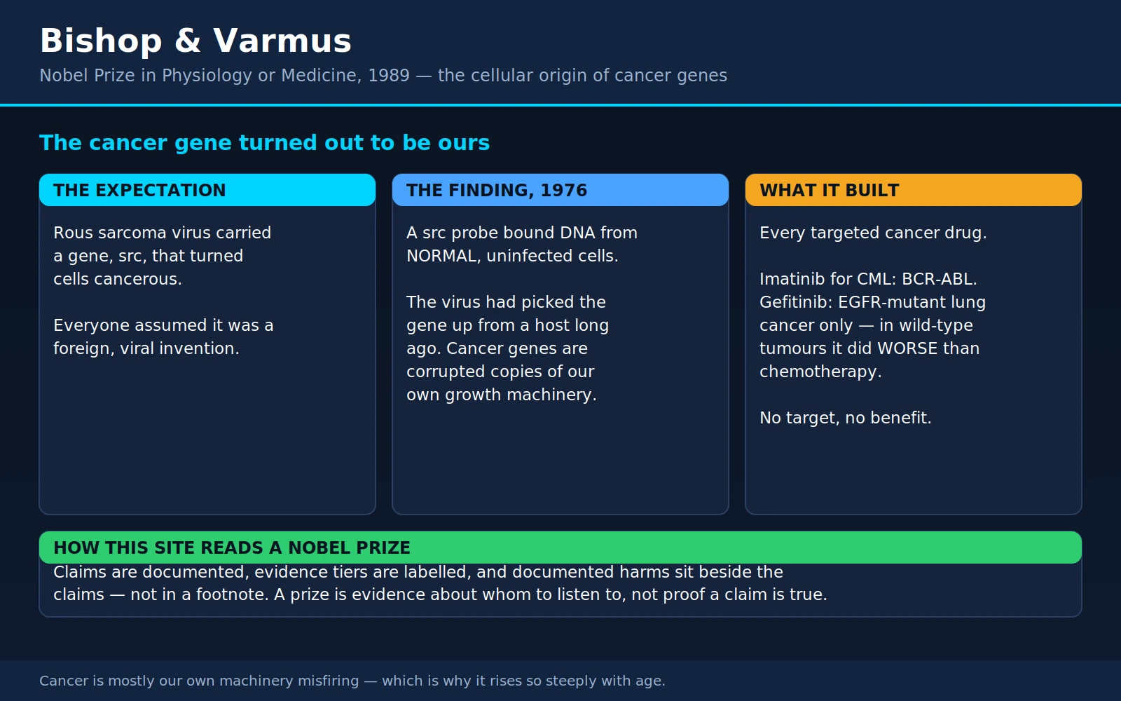
Table of Contents
- The Prize and the Two Men
- The Question They Inherited
- The 1976 Experiment
- Proto-Oncogenes and Oncogenes
- From src to a Catalogue: RAS, MYC, HER2, ABL
- The Other Half: Tumor Suppressors and the Two-Hit Rule
- What This Means for You
- Targeted Therapy: The Direct Payoff
- Tumor Genomic Testing: What a Molecular Profile Is For
- Liquid Biopsy and Early Detection
- What This Finding Does Not License
- Where Mainstream Medicine Agrees — and What Remains Debated
- Key Research Papers
- Connections
- Featured Videos
1. The Prize and the Two Men
In 1989 the Nobel Assembly gave the Prize in Physiology or Medicine to two men working in the same San Francisco laboratory, J. Michael Bishop and Harold E. Varmus, "for their discovery of the cellular origin of retroviral oncogenes." That sentence is opaque unless you already know the field. Translated, it says something that changed how the entire world thinks about cancer: the genes that cause cancer are not foreign. They are our own genes, doing our own jobs, broken.
Before their work, the most exciting hypothesis in cancer research was that tumors were caused by viruses carrying alien cancer genes — that somewhere out there was a set of malignant instructions belonging to the virus world, which a human cell could catch the way it catches a cold. After their work, the picture inverted. The cancer gene the virus carried turned out to be a stolen copy of a gene already living in normal cells, present in chickens, in mice, in cattle, in salmon and in us. The virus had not invented anything. It had picked our pocket.
J. Michael Bishop was born on February 22, 1936, in York, Pennsylvania, the son of a Lutheran minister, and grew up in rural Pennsylvania at a distance from anything resembling a research career. He took a chemistry degree at Gettysburg College in 1958 and a medical degree at Harvard in 1962, discovered in medical school that he wanted the laboratory rather than the ward, and joined the faculty at the University of California, San Francisco in 1968, becoming a full professor in 1972. He spent essentially his entire scientific life at UCSF and served as its chancellor from 1998 until 2009, a period in which the university built out its Mission Bay research campus. He received the National Medal of Science in 2003. He died in San Francisco on March 20, 2026, at the age of 90.
Harold E. Varmus, born in 1939 in Oceanside, New York, arrived at biology by a route almost nobody takes. He read English literature at Amherst College, then took a master's degree in English at Harvard in 1962, working on Anglo-Saxon and metaphysical poetry, and only then turned to medicine — rejected by Harvard Medical School, accepted at Columbia's College of Physicians and Surgeons, where he earned his MD in 1966. He came to UCSF in 1970 and began working alongside Bishop; the collaboration lasted decades and was, by both accounts, unusually equal. What followed the Nobel is nearly as remarkable as what preceded it: Varmus served as Director of the National Institutes of Health from 1993 to 1999, as President and CEO of Memorial Sloan Kettering Cancer Center from 2000 to 2010, and as Director of the National Cancer Institute from 2010 to 2015. He is, as of 2026, the Lewis Thomas University Professor of Medicine at Weill Cornell Medicine and still runs a laboratory. Few scientists have had one career at that level; Varmus has had three, and he came to all of them from Anglo-Saxon poetry.
The literature background is not a charming footnote. Varmus has said in interviews that what a humanities education gave him was the habit of arguing from evidence in prose — of building a case, noticing what a text does not say, and distrusting a conclusion that sounds better than its support. The 1976 experiment was in the end an act of interpretation as much as of chemistry: the data were a set of hybridization curves, and the finding was what those curves meant.
Dominique Stehelin, Peter Vogt, and a contested omission
The Nature paper that won the prize has four authors: Dominique Stehelin, Harold E. Varmus, J. Michael Bishop, and Peter K. Vogt. That fact matters, and this site's record of Nobel omissions gets another entry here.
Dominique Stehelin was a young French postdoctoral fellow in the Bishop–Varmus laboratory, and he did much of the bench work — the painstaking preparation of the radioactive probe and the hybridization experiments that produced the result. He is first author on the paper. He was not included in the prize, and in France the omission was contested loudly and publicly. When the award was announced in October 1989 the Centre National de la Recherche Scientifique, where Stehelin had by then built his own career in Lille, reacted with open dismay and considered a formal protest; its director general subsequently issued a statement that congratulated Bishop and Varmus while pointedly praising Stehelin's role. Stehelin himself told reporters that he had done the work "all by myself, from A to Z," over three years in the San Francisco lab.
We record this factually and without adjudicating it, because both readings are defensible and the Nobel committee's deliberations are sealed for fifty years. On one side: the person whose hands produced the decisive data was left out, and the prize's three-laureate ceiling was not even reached — there was room. On the other: the question, the strategy, the interpretation and the decade of follow-up that turned one result into a theory of cancer were Bishop's and Varmus's, and Peter Vogt — the virologist who supplied the mutant viruses without which the experiment was not possible — was equally uncredited, which complicates any simple story about a wronged junior author.
What is not in dispute is the general pattern. The Nobel statutes cap a prize at three living people, science is done by groups, and the arithmetic guarantees casualties. This site notes the same fault line on the pages for Watson, Crick and Wilkins — where Rosalind Franklin's data were essential and Franklin, dead by 1962, was ineligible — and in the complete roll of laureates from 1901 to 2025. The prize is a spotlight, not a census.
2. The Question They Inherited
To see why 1976 was a shock, you have to know what everybody expected the answer to be.
The story starts in 1911 with Peyton Rous at the Rockefeller Institute, who ground up a chicken sarcoma, pushed the material through a filter fine enough to hold back every cell and bacterium, injected the cell-free liquid into healthy chickens, and grew new tumors. Something submicroscopic and transmissible caused cancer. The finding was so far outside the thinking of the time that it was largely dismissed; Rous received his Nobel Prize in 1966, fifty-five years later, at the age of eighty-seven. That story, and the parallel story of Charles Huggins and the first hormonal cancer therapy, is told on our page for Rous and Huggins. The agent Rous had found is now called Rous sarcoma virus (RSV), and it is the direct ancestor of everything on this page.
By the early 1970s, RSV had been dissected far enough to expose the specific culprit. It is a retrovirus — an RNA virus that copies its genome into DNA and inserts it into the host chromosome, a trick discovered by Howard Temin and David Baltimore and covered on our page for Baltimore, Temin and Dulbecco. RSV's genome is tiny, and researchers including Peter Vogt and Steve Martin had isolated mutants of it that could still replicate but had lost the ability to transform cells — they infected happily and caused no tumor. Comparing the mutants with the wild-type virus localized the cancer-causing capacity to a single gene, named src (for sarcoma, and pronounced "sarc"). One gene. Delete it and the virus is harmless; restore it and cells pile up in disordered heaps and grow without restraint.
That was the state of play, and it pointed in an obvious direction. If a single viral gene could convert a normal cell into a cancer cell, then cancer genes were viral genes — a distinct category of malignant instructions belonging to the virus world. The corollary was irresistible: find the human tumor viruses, and you find the causes of human cancer. Vaccines and antivirals would follow. Cancer would become an infectious disease with an infectious-disease solution.
Enormous institutional weight sat behind that hope. The Special Virus Cancer Program had been pouring federal money into the hunt for human tumor viruses since the 1960s, and the National Cancer Act of 1971 — the "War on Cancer" — arrived with a widely shared expectation that the war would be won the way polio had been won, by finding the virus. It was not a stupid expectation. Viruses genuinely do cause some human cancers, as later work would prove decisively; our pages on Harald zur Hausen (HPV and cervical cancer) and Barry Marshall and Robin Warren (Helicobacter pylori and stomach cancer) document two of the biggest. But as a general theory of cancer, it was wrong, and the experiment that showed it was wrong was designed to test something else entirely.
3. The 1976 Experiment
The question Bishop and Varmus actually asked was narrower and stranger than "what causes cancer." It was: where did the virus get src?
The reasoning behind the question is worth following, because it is the whole discovery in miniature. Retroviruses are small and streamlined; every gene they carry costs them replication speed. A gene like src, which the virus does not need in order to reproduce, is expensive baggage. Retroviruses also insert themselves into host chromosomes as part of their normal life cycle, and imprecise excision from the chromosome is a plausible way for a virus to leave carrying a piece of the host. Temin had already proposed something along these lines in his "protovirus" hypothesis. So the question had a testable form: is there anything in the DNA of a normal, uninfected chicken that looks like src?
Answering it required a probe — a piece of radioactive DNA that would stick specifically to src sequences and to nothing else in the genome. Making that probe was the hard part, and it is the part Stehelin did. The method, in outline:
- Take the RNA genome of wild-type Rous sarcoma virus, which contains src, and use reverse transcriptase to copy it into radioactively labeled DNA. This gives labeled DNA representing the whole viral genome.
- Take the RNA of the transformation-defective deletion mutants — the crippled viruses from Vogt's collection, which are identical to the wild type except that they are missing src.
- Mix the two. Every labeled DNA fragment that corresponds to an ordinary viral gene finds its partner in the mutant RNA and pairs up. The fragments corresponding to src find nothing to pair with, because src is exactly what the mutant lacks.
- Separate the paired material from the unpaired. What is left over is labeled DNA representing src alone. Bishop and Varmus called it cDNA-sarc.
This is subtraction as an experimental technique, and in the mid-1970s, before cloning and sequencing were routine, it was a serious piece of craft. The reagent that came out of it was a molecular question in a tube: show me everything in this genome that resembles the cancer gene.
Then they asked the tube about normal chickens — birds with no infection, no tumor, nothing unusual about them at all. The probe should have found nothing.
It lit up. Normal, uninfected chicken DNA contained a sequence closely related to src. Not a viral remnant sitting in the genome as a passenger, but a gene, present at one or a few copies per cell, in the DNA of every normal bird they tested. The paper that reported it — Stehelin, Varmus, Bishop and Vogt, Nature, March 1976 — carries a title that is the finding stated flatly: "DNA related to the transforming gene(s) of avian sarcoma viruses is present in normal avian DNA."
The obvious objection was that chickens are the natural host of this virus, so perhaps chickens alone carry the sequence as an ancient viral scar. The laboratory closed that door in a 1978 follow-up in PNAS. Under conditions permissive enough to allow imperfectly matched sequences to pair, the src probe bound DNA from chicken, human, calf, mouse and salmon — but not from sea urchin, fruit fly or E. coli. The melting behavior of those hybrids put the mismatch at roughly 3–4 percent against chicken DNA and 8–10 percent against the other vertebrates: close relatives, diverging gently with evolutionary distance, exactly as a real conserved gene does. Meanwhile the probes for the rest of the viral genome bound chicken DNA and nothing else.
That asymmetry is the argument. The genuinely viral genes were confined to the virus's own host. The cancer gene was everywhere in the vertebrate lineage, and had been for something like five hundred million years. A gene that a salmon and a human both still carry, in recognizable form, is not a piece of viral wreckage. It is a gene the body needs.
Which reversed the arrow. The virus had not given the cell a cancer gene. At some point in its history, an ancestral retrovirus replicating inside a bird had accidentally scooped up a normal cellular gene, carried it away, and — in the copying and re-copying that follows — damaged it into a permanently switched-on form. The transformation of that virus into a carcinogen was a mistake made at the cell's expense, using the cell's own parts.
4. Proto-Oncogenes and Oncogenes
The vocabulary that came out of this is now used everywhere in cancer medicine, and it is worth learning properly because it is not difficult and it demystifies an enormous amount.
A proto-oncogene is a normal gene in your normal cells. Its job is growth: it tells cells when to divide, when to move, when to survive rather than die. You cannot heal a cut, mount an immune response, grow from an embryo, or replace the lining of your gut without these genes doing their work. The cellular version of src — written c-src, "c" for cellular — is a proto-oncogene. It encodes an enzyme that attaches phosphate groups to other proteins, a switch in the signaling chains that tell a cell to grow. Under normal control it flips on when growth is called for and off when it is not.
An oncogene is that same gene after something has gone wrong with it. The viral version, v-src, is the copy the virus carried off and mangled: truncated at one end in a way that removes its regulatory brake, so the enzyme is stuck permanently in the "on" position. A cell receiving that version is being screamed at to grow, continuously, forever, with nobody's finger on the switch.
The analogy most people find useful is a car's accelerator. A proto-oncogene is a working accelerator: you press it to go, release it to stop. An oncogene is that accelerator jammed to the floor. Nothing exotic has been added to the car. A normal part has failed in one specific direction.
And it does not take a virus to jam it. Once you know the accelerator is already in the cell, every ordinary source of genetic damage becomes a plausible cause of cancer:
- Point mutation — a single letter of DNA changed, altering one amino acid, leaving a protein that can no longer be switched off. This is what a KRAS mutation is.
- Amplification — the gene is duplicated over and over, so the cell carries ten or fifty copies instead of two and the growth signal is proportionally louder. This is what HER2-positive breast cancer is.
- Translocation — two chromosomes break and are stitched back together wrongly, fusing one gene to another and creating a hybrid protein with no off switch, or moving a growth gene next to a permanently active control region. This is what the Philadelphia chromosome of chronic myeloid leukemia is.
- Insertion — a virus lands next to a proto-oncogene and its own powerful control sequences drive the neighbor. This is how some retroviruses cause tumors without carrying an oncogene at all, a mechanism Bishop and Varmus's laboratory also helped establish.
Bishop set out the mature version of this framework in a 1987 review in Science written for exactly the reader this page is written for. His thesis was simple and, by then, well supported: cancer is a genetic disease of somatic cells — a disease of the genes in the tissue, not necessarily of the genes you inherited — and the genes involved are a limited, identifiable set that the body uses every day.
It is hard to overstate how much this reframed the field. Almost every subsequent advance in cancer biology is downstream of it. Before, cancer was a mysterious loss of order with a hundred competing theories — a virus, a chemical, a metabolic derangement, a failure of immune surveillance, a reversion to embryonic behavior. After, cancer had a mechanism you could name, a molecule you could purify, a gene you could clone, and eventually a target you could design a drug against. The whole of what is now called precision oncology begins here. So does the modern understanding of why carcinogens are carcinogens: they damage DNA, and damaged DNA is how a proto-oncogene becomes an oncogene.
There is also a quieter consequence that matters for a reader who has been frightened by cancer. The disease stopped being an invasion. Cancer is not something that gets into you. It is something your own cells start doing. That is grimmer in one sense — there is no foreign body to expel — and enormously more tractable in another, because the machinery involved is machinery we can study, measure and in a growing number of cases interrupt.
5. From src to a Catalogue: RAS, MYC, HER2, ABL
Once the search image existed, proto-oncogenes turned up rapidly. Other cancer-causing retroviruses were pulled apart the same way, and each one turned out to be carrying its own stolen cellular gene. Within about a decade the field had assembled a working catalogue, and the names on it are the names on modern pathology reports.
- RAS (from a rat sarcoma virus) — a family of switch proteins sitting just inside the cell membrane, relaying growth signals inward. Mutated KRAS is among the most common driver mutations in human cancer, prominent in pancreatic, colorectal and lung tumors. For thirty years it was considered undruggable; the first inhibitors of a specific mutant form reached patients in the 2020s.
- MYC (from an avian myelocytomatosis virus) — a master control protein that switches on hundreds of growth genes at once. Deregulated MYC is central to Burkitt lymphoma and appears widely across other cancers.
- ERBB2 / HER2 — a growth-factor receptor on the cell surface, whose gene is amplified in a subset of breast and stomach cancers. Its viral cousin was found in a rat neural tumor virus.
- ABL (from Abelson murine leukemia virus) — another phosphate-attaching switch enzyme, closely related to src. Fused to a gene called BCR by the chromosome swap that produces the Philadelphia chromosome, it drives chronic myeloid leukemia.
- SRC itself — the founder, and still a target of active drug development.
Two features of this catalogue are worth noticing. First, the entries are not exotic. They are the ordinary hardware of growth signaling: receptors on the surface, switches inside, enzymes relaying the message, transcription factors reading it out in the nucleus. Second, they are not random. Cancer does not arise from mutations anywhere; it arises from mutations in specific nodes of specific pathways, which is precisely why the disease is targetable at all.
Modern tumor sequencing has firmed this into numbers. The large synthesis by Vogelstein and colleagues in Science in 2013, drawing on thousands of sequenced tumors, described a landscape in which a typical adult solid tumor carries a few dozen coding mutations, of which only a handful — commonly two to eight — are actual drivers; the rest are passengers, damage that came along for the ride and does nothing. Those drivers concentrate in a modest number of signaling pathways. The catalogue Bishop and Varmus opened turned out to be roughly the right size.
6. The Other Half: Tumor Suppressors and the Two-Hit Rule
An accelerator jammed on is only half a runaway car. The other half is the brakes.
Tumor suppressor genes are the counterpart to proto-oncogenes. Their normal job is restraint: hold the cell at a checkpoint until the DNA has been proofread, stop division when neighbors are crowding in, and if the damage is beyond repair, order the cell to kill itself. Where an oncogene is created by a mutation that makes a gene too active, a tumor suppressor causes trouble when a mutation makes it inactive. Cancer generally requires both: an accelerator stuck down and a brake line cut. A single jammed accelerator in a cell with working brakes usually ends with that cell arrested or dead, which is exactly what the brakes are for.
The two canonical examples are worth knowing by name.
RB, the retinoblastoma gene, was the first tumor suppressor cloned — by Friend, Dryja and colleagues in Nature in 1986, from the childhood eye tumor that gave it its name. The RB protein is the physical gate on entry to DNA replication: it holds the cell at the boundary of the cell cycle until the correct signals accumulate. Disable it and the gate stands open.
TP53, which encodes the p53 protein, is the damage sensor — nicknamed the "guardian of the genome" in a 1992 commentary by David Lane, and among the most frequently mutated genes across human cancers. When DNA is damaged, p53 halts the cell cycle to allow repair, and if repair fails it triggers programmed cell death. A cell without functioning p53 keeps dividing with broken DNA, which is how one mutation becomes ten. The machinery p53 acts on — the checkpoints of the cell cycle itself — was worked out by Leland Hartwell, Tim Hunt and Paul Nurse, whose story is on our page for Hartwell, Hunt and Nurse. The two discoveries are complementary halves of one picture: they described the timing system, Bishop and Varmus described what happens when its inputs are corrupted.
Knudson's two-hit hypothesis, and why some cancers run in families
The reason tumor suppressors explain family history — and the reason the explanation was worked out before anyone had cloned a gene — is a beautiful piece of arithmetic by Alfred Knudson, published in PNAS in 1971.
Knudson was studying retinoblastoma, a rare eye tumor of early childhood that comes in two patterns. Some children have a family history, develop tumors early, and often have them in both eyes and in more than one spot. Other children have no family history, develop a single tumor in one eye, and develop it later. Looking at 48 cases plus the published literature, Knudson asked what mechanism would produce those two distributions from one disease.
His answer: retinoblastoma requires two mutational events. In the inherited form, the first mutation arrives through the germline — it is in every cell of the child's body from conception — and only the second has to happen in a retinal cell. In the non-inherited form, both mutations must occur in the same retinal cell by chance, which is much rarer. Using Poisson statistics on the number of tumors per affected child, he estimated that a carrier of the first mutation develops on average about three retinoblastomas — a figure that simultaneously explains the occasional carrier who develops no tumor at all, the carriers with one, and the carriers with several in both eyes.
The molecular meaning became clear once RB was cloned fifteen years later. You carry two copies of every gene. A tumor suppressor works if either copy is intact, so knocking out one copy changes nothing about the cell's behavior. Inheriting one broken copy does not give you cancer. It means every cell in your body starts one step closer to it — a single unlucky event in any one of millions of cells, rather than two independent unlucky events in the same cell.
That is the general shape of hereditary cancer risk, and it is the honest way to explain it to a family. A BRCA1 or BRCA2 variant, a Lynch syndrome variant, a p53 variant in Li-Fraumeni syndrome: none of these is a cancer. Each is a missing spare copy. It shifts the odds, sometimes very substantially, and it is precisely because the mechanism is understood that carriers can be offered earlier and more frequent screening, and in some cases risk-reducing surgery or medication — interventions that work on the odds in the other direction. The same logic underlies the inherited tumor-suppressor syndromes covered in our Genetics section, including neurofibromatosis type 1 and tuberous sclerosis complex.
7. What This Means for You
This is the section to read if you read no other. The oncogene finding is not an abstraction; it changes what a reasonable person should believe about their own risk.
Why age is the dominant risk factor
Cancer requires an accumulation of specific genetic damage in a single cell lineage — typically several driver events, not one. Every time a cell divides it copies three billion base pairs, and the copying is extraordinarily good but not perfect. Damage accumulates with the number of divisions, and the number of divisions accumulates with time. So the single largest risk factor for cancer is being alive for a long time, and that is not a moral fact about anybody's choices.
The numbers are stark. According to the National Cancer Institute, the median age at cancer diagnosis in the United States is 67. Incidence runs at fewer than 26 cases per 100,000 people under age 20, about 350 per 100,000 in the 45–49 age band, and more than 1,000 per 100,000 from age 60 onward. Median age at diagnosis for the common cancers: breast 63, colorectal 66, prostate 68, lung 71.
Two things follow. First, a cancer diagnosis in an older adult usually needs no special explanation — no environmental exposure to hunt for, no dietary sin to repent. Second, and less obviously, much of the apparent rise in cancer over the last century is a rise in the number of people living long enough to get it, alongside better detection. That is worth holding onto when you see a headline about cancer rates.
Why carcinogens matter, and what they actually do
A carcinogen is, in this framework, simply anything that increases the rate at which the relevant genes get damaged — or the rate at which damaged cells divide before repair. Tobacco smoke carries dozens of compounds that chemically bond to DNA. Ultraviolet light fuses adjacent DNA letters. Ionizing radiation snaps both strands. Some viruses insert their own genes into the chromosome or disable p53 directly. Chronic inflammation drives both cell division and oxidative damage.
This is why avoiding carcinogens works, and why it works in a specific, dose-related way rather than as a moral guarantee. Smoking does not cause lung cancer in every smoker, and quitting does not undo damage already recorded in a cell's DNA — but every year of not smoking is a year without new damage, and lung cancer risk after quitting falls steadily and substantially over the following decades. The mechanism explains the epidemiology.
The "bad luck" paper, stated as what it actually says
In 2015, Cristian Tomasetti and Bert Vogelstein published an analysis in Science that was reported worldwide as "two-thirds of cancers are just bad luck." That is not what the paper claims, and the difference is exactly the sort of thing this site exists to correct.
What the paper examined was why different tissues have such wildly different cancer rates — why the colon produces vastly more cancers than the small intestine, for instance, despite sitting next to it and seeing similar contents. The authors found that the lifetime cancer risk of a tissue correlates strongly (0.81) with the total number of divisions the stem cells maintaining that tissue undergo over a lifetime. Their conclusion, in their own words, is that only about a third of the variation in cancer risk among tissues is attributable to environment or heredity, with the majority of that variation traceable to random replication errors.
Note what is being divided into thirds: the differences between organs, not any individual's personal risk, and not the total burden of cancer in a population. The paper does not say two-thirds of cancers are unpreventable. It says that if you want to know why the colon outranks the duodenum, stem-cell division counts explain most of the gap.
The authors' 2017 follow-up in Science, covering 17 cancer types across 69 countries, is if anything clearer on the point. It estimates that replication errors account for roughly two-thirds of the mutations found in human cancers — mutations, not cancers, and a cancer needs several — and states explicitly that its results are consistent with existing epidemiological estimates of the preventable fraction. Its own stated conclusion is that because many cancers arise from unavoidable errors, early detection matters, which is a call to screening, not a shrug.
So the honest summary, and the one to carry away: cancer-causing mutations come from three sources — inherited, environmental, and random copying errors. All three are real. Random error is a large contributor and is not anybody's fault. Environmental and lifestyle factors are the modifiable share, they are substantial, and prevention works on them. Neither "it's all lifestyle" nor "it's all luck" survives contact with the actual literature.
The non-fatalist reading
None of the above is a counsel of despair, and it should not be read as one. Random damage sets a floor that no behavior can eliminate; it does not set a ceiling. Not smoking, not being obese, moderating alcohol, protecting skin from burning UV, being vaccinated against HPV and hepatitis B, treating H. pylori infection, and turning up for the screening programs you are eligible for — each of these acts on a real and quantified share of risk. And the second half of Tomasetti and Vogelstein's own argument is that where prevention runs out, detection takes over, because a cancer caught before it spreads is a fundamentally different disease from the same cancer caught late.
8. Targeted Therapy: The Direct Payoff
The clinical translation of the oncogene idea is straightforward to state: if a cancer is being driven by one identifiable broken protein, build a drug that blocks that protein. Chemotherapy poisons dividing cells and accepts the collateral damage to hair follicles, gut lining and bone marrow. A targeted drug aims at something the tumor has and healthy tissue largely does not.
Imatinib and chronic myeloid leukemia: the canonical case
Chronic myeloid leukemia is the cleanest example in medicine, because it is very nearly a one-gene disease. In 1960 Peter Nowell and David Hungerford noticed an abnormally short chromosome in CML cells — the Philadelphia chromosome. It was later shown to be a swap between chromosomes 9 and 22 that fuses the BCR gene to ABL, producing a hybrid enzyme that is permanently switched on. A single accelerator, welded down, in essentially every leukemic cell.
Brian Druker and colleagues took a compound that inhibited that specific enzyme into a phase 1 trial in patients with chronic-phase CML in whom interferon had failed, and published the result in the New England Journal of Medicine in 2001. Of 83 patients treated across escalating doses, among the 54 who received 300 mg per day or more, 53 achieved a complete hematologic response — normalization of the blood count — typically within the first four weeks. Cytogenetic responses, meaning a reduction in the proportion of Philadelphia-positive cells, occurred in 29 of those 54; 17 (31 percent) were major responses and 7 were complete. Side effects were mild: nausea, muscle aches, swelling, diarrhea. No maximum tolerated dose was reached.
Those are phase 1 numbers, from an early-stage safety trial in patients who had already failed the standard of care. They are not the numbers you normally see from a phase 1 trial in a refractory cancer, and that is why the drug — imatinib, marketed as Gleevec — went from first-in-human to FDA approval in under three years.
The long-term data came from the IRIS trial, which randomized newly diagnosed chronic-phase patients to imatinib or to interferon plus cytarabine. The final report in 2017, at a median follow-up of 10.9 years, found an estimated 10-year overall survival of 83.3 percent among patients randomized to imatinib, with 82.8 percent achieving a complete cytogenetic response and no evidence of unacceptable cumulative or late toxicity. For a disease whose median survival before this era was measured in a handful of years, that is a transformation.
Read the caveat too, because the paper states it plainly: 65.6 percent of the patients assigned to interferon plus cytarabine crossed over to imatinib, most of them quickly, so the ten-year comparison between arms is not a clean randomized contrast — the analysis focuses on the imatinib arm for that reason. Also note that 48.3 percent of the imatinib group completed the study on imatinib; the rest stopped for various reasons, including resistance and intolerance. CML is the best case in oncology and it is still not a story of everybody cured.
HER2 and trastuzumab
The HER2 gene is amplified in a subset of breast cancers, and before there was a drug for it, that subset behaved worse than average. Trastuzumab is a monoclonal antibody that binds the HER2 receptor on the tumor cell surface — a therapeutic format made possible by the hybridoma technique, told on our page for Jerne, Köhler and Milstein.
The pivotal trial, Slamon and colleagues in the New England Journal of Medicine in 2001, randomized 469 women with HER2-overexpressing metastatic breast cancer to chemotherapy alone or chemotherapy plus trastuzumab. Adding the antibody improved median time to progression from 4.6 to 7.4 months, objective response from 32 to 50 percent, and median overall survival from 20.3 to 25.1 months, with deaths at one year falling from 33 to 22 percent.
Two honest qualifications. First, the harm was real and specific: New York Heart Association class III or IV cardiac dysfunction occurred in 27 percent of women given an anthracycline, cyclophosphamide and trastuzumab together, against 8 percent on that chemotherapy alone — which is why that particular combination is not used today. With paclitaxel the figures were 13 percent versus 1 percent. Targeted does not mean harmless. Second, the paper's own framing describes HER2 as amplified or overexpressed in "25 to 30 percent" of breast cancers; with today's standardized testing the figure is usually put lower, around 15 to 20 percent. Both numbers are honestly reported for their era; assay definitions changed.
Trastuzumab also demonstrates the counterpart truth: it does nothing for the 80-odd percent of breast cancers that are HER2-negative. The drug is not a breast cancer drug. It is a HER2 drug that happens to be used in breast cancer.
EGFR in lung cancer, BRAF in melanoma, ALK
The EGFR story in lung cancer contains the single most instructive result in this whole field, and it is instructive precisely because it cuts both ways. The IPASS trial, published in 2009, randomized 1,217 East Asian never-smokers or light ex-smokers with lung adenocarcinoma to the EGFR inhibitor gefitinib or to standard chemotherapy. Overall, gefitinib won on progression-free survival (hazard ratio 0.74). But the subgroup analysis is the lesson: among the 261 patients whose tumors carried an EGFR mutation, gefitinib was dramatically better (hazard ratio 0.48). Among the 176 whose tumors did not carry the mutation, gefitinib was significantly worse than chemotherapy — hazard ratio 2.85 in the wrong direction.
That is the entire philosophy of targeted therapy in one trial. The same pill, in the same disease, in the same trial: excellent or harmful depending on a molecular test. There is no such thing as a targeted drug that is "worth a try" in a patient without the target.
BRAF in melanoma repeated the pattern. The BRIM-3 trial, reported in 2011, randomized 675 patients with previously untreated metastatic melanoma carrying the BRAF V600E mutation to vemurafenib or dacarbazine. Response rates were 48 percent versus 5 percent; six-month overall survival was 84 percent versus 64 percent; the interim analysis showed a 63 percent relative reduction in the risk of death, and crossover was recommended by the safety board. It is also a fair illustration of targeted-drug toxicity: joint pain, rash, fatigue, photosensitivity and — a strange, mechanism-specific effect — keratoacanthomas and squamous-cell skin cancers, with 38 percent of patients needing a dose modification.
ALK rearrangements define another small subset of lung cancers with their own inhibitors, now several generations deep. And the general lesson of all of them, learned the hard way with vemurafenib, is that single-agent targeting invites escape: melanomas driven by BRAF frequently relapse within months on vemurafenib alone, which is why the modern standard combines a BRAF inhibitor with a MEK inhibitor to block the pathway at two points at once.
The honest ledger
What targeted therapy reliably delivers:
- Dramatic, fast responses in the molecularly defined subgroup that has the target — sometimes with a side-effect profile far gentler than chemotherapy.
- Conversion of some fatal cancers into long-term manageable conditions. CML is the flagship.
- A rational basis for choosing treatment, replacing trial and error.
What it does not deliver:
- Durability, in most cases. Tumors are genetically diverse populations under selection pressure; a drug that kills the cells with the driver mutation selects for the rare cell that has acquired a resistance mutation. Resistance is the rule, not a surprise, and much of modern drug development is second- and third-generation inhibitors against known resistance mutations.
- Applicability. Most cancers do not have a single dominant druggable driver. Pancreatic cancer, most colorectal cancer, most prostate cancer and the great majority of common solid tumors are driven by combinations, or by mutations in tumor suppressors — and a missing brake is much harder to drug than a jammed accelerator. You can block a protein that is doing too much; you cannot easily restore one that is gone.
- Cure, usually, in metastatic disease. The response is often measured in months of extra progression-free time. That is real and worth having. It is not the same as remission.
It is also worth naming what came next, because it did not come from this line of thinking at all. Checkpoint immunotherapy — releasing the brakes the immune system puts on itself, so that T cells attack the tumor — produced the long-lived remissions in melanoma and lung cancer that targeted therapy mostly did not, and it works through the patient's immune system rather than the tumor's driver gene. That story is on our page for Allison and Honjo. The two approaches are complementary, and the honest current picture is that oncology needs both plus the old cytotoxics.
9. Tumor Genomic Testing: What a Molecular Profile Is For
If you or someone close to you is diagnosed with cancer today, there is a good chance that somebody will propose sequencing the tumor. This section is what that means in practice.
Somatic versus germline: the distinction that matters most
These are two different tests answering two different questions, and they are routinely confused.
Somatic testing sequences the tumor. It asks: what is broken in these cancer cells? The mutations it finds arose during the person's life, in that tissue. They are not inherited, they are not passed to children, and they do not tell relatives anything about their own risk. The purpose is to choose treatment.
Germline testing sequences the person — usually from blood or saliva. It asks: what did this person inherit? A pathogenic germline variant in BRCA1, BRCA2, a mismatch-repair gene, TP53 or many others is present in every cell, was present at birth, and has a 50 percent chance of being present in each first-degree relative. The purpose is to guide screening and prevention for the patient and the family — and, increasingly, treatment too, since some inherited defects (BRCA-related, for instance) predict response to particular drugs.
Practical consequences worth knowing:
- A tumor test can incidentally reveal something inherited. Good laboratories flag this, and it should trigger a proper germline test and genetic counseling rather than an assumption.
- Germline results have implications for people who did not consent to a test. That is not a reason to avoid testing; it is a reason to have the conversation with a genetic counselor, whose job this is.
- Direct-to-consumer genetic tests are generally not equivalent to clinical germline testing. Several screen only a handful of specific variants and can return a reassuring "negative" while missing most of the pathogenic variants in the very genes they claim to cover.
What "actionable" means, and how often it happens
"Actionable" means the finding changes what is done: it makes an approved drug available, opens a clinical trial, rules a treatment out, or clarifies the diagnosis. It is a clinical statement, not a biological one — a mutation can be a genuine driver and still be unactionable if nothing exists to drug it.
The honest arithmetic, from the field's own literature:
- In the MSK-IMPACT study, which prospectively sequenced tumors from more than 10,000 patients with advanced cancer at a leading cancer center, patients were enrolled on genomically matched clinical trials at a rate of 11 percent.
- Marquart, Chen and Prasad, writing in JAMA Oncology in 2018, estimated that of US patients with advanced or metastatic cancer in 2018, about 8.3 percent were eligible for an FDA-approved genome-targeted therapy and about 4.9 percent would benefit from one. On a broader "genome-informed" definition the figures were 15.4 percent eligible and 6.6 percent benefiting. Those numbers had grown from 5.1 percent eligible and 0.7 percent benefiting in 2006 — real progress, from a very low base.
- The same analysis found that when a genome-informed drug did apply, the median response rate was 54 percent and the median duration of response 29.5 months.
That last pair is the shape of the whole enterprise: when it works it works well, and it applies to a minority. These estimates are debated at the edges — the definitions of "eligible" and "benefit" are contestable, the field has moved since 2018, and tumor-agnostic approvals such as those for mismatch-repair-deficient cancers and NTRK fusions have widened the door. But no serious reading puts the actionable fraction at a majority.
Which means the most useful thing anyone can tell a patient before the test is this: a profile that comes back with no actionable target is the common result, not a failure and not bad luck about the testing. It does not mean the cancer is untreatable; it means the treatment will be chosen the way it has been chosen for decades — by tumor type, stage, and the evidence from trials in that setting. Going in expecting a targetable mutation and receiving a report full of variants of uncertain significance is a specific and avoidable disappointment.
Questions worth asking
- Is this test on my tumor or on my inherited DNA? If both, are the results being separated and explained separately?
- Will the result change my treatment now, or is it for a possible future line of therapy?
- Does the panel cover the alterations that actually have approved drugs in my cancer type?
- If something inherited shows up, who explains it to me and to my family — and is a genetic counselor involved?
- Who pays, and is coverage confirmed before the sample is sent?
10. Liquid Biopsy and Early Detection
Dying tumor cells shed fragments of DNA into the bloodstream. Sensitive sequencing can pick those fragments out from the far larger background of normal cell-free DNA, which means that in principle a tube of blood can report on a tumor without anybody putting a needle into it. This is the "liquid biopsy," and the honest assessment differs sharply depending on which of two very different jobs you ask it to do.
Where it is established: monitoring known cancer
In a patient who already has a diagnosed cancer, circulating tumor DNA is genuinely useful and increasingly routine:
- Finding a target when tissue is hard to get. Blood-based testing for EGFR mutations in lung cancer is standard practice where a biopsy is difficult or the sample is inadequate.
- Detecting resistance. When a targeted drug stops working, the resistance mutation can often be identified in blood, which points to the next drug.
- Measuring residual disease after surgery. Detectable tumor DNA after an operation identifies patients at high risk of recurrence. Randomized evidence now exists that this can be used to guide decisions — in stage II colon cancer, a strategy of giving chemotherapy only to patients with detectable circulating tumor DNA reduced the proportion treated without compromising recurrence-free survival, in a 2022 trial in the New England Journal of Medicine.
Where it is not yet established: screening healthy people
The far more heavily marketed use is the multi-cancer early detection (MCED) blood test — one blood draw, screening an asymptomatic person for dozens of cancers at once. These tests are real, the technology works, and they are being sold, in some markets directly to consumers. What follows is what the published evidence supports, which is less than the advertising implies.
The most studied test detects methylation patterns in cell-free DNA. Its clinical validation study, published in Annals of Oncology in 2021, reported specificity of 99.5 percent — excellent — and overall sensitivity of 51.5 percent across cancers. Crucially, sensitivity depended on stage: 16.8 percent at stage I, 40.4 percent at stage II, 77.0 percent at stage III, 90.1 percent at stage IV. When it identified a cancer, it predicted the organ of origin correctly 88.7 percent of the time.
Read that stage gradient carefully. The entire rationale for screening is catching cancer early, and stage I is where this test is weakest — it misses roughly five in six stage I cancers. It is best at finding advanced cancer, which is generally the cancer that would have declared itself anyway.
The PATHFINDER study, published in The Lancet in 2023, took the test into the setting that matters: 6,662 asymptomatic adults aged 50 and over, in ordinary clinics, with results returned to their doctors. A cancer signal was detected in 92 people (1.4 percent). Of those 92, 35 turned out to have cancer and 57 did not — a positive predictive value of 38 percent, meaning that most positives were false positives. The median time to resolve the question was 79 days, and 162 days for the people who turned out not to have cancer. More than 90 percent of both groups had imaging; 30 percent of the false positives underwent a procedure; a handful had surgery. The authors' own conclusion was that the study "supports the feasibility" of MCED screening and "underscores the need for further research investigating the test's clinical utility."
So, plainly, because people are being sold these tests today:
- No multi-cancer early-detection blood test has been shown to reduce cancer mortality. That is the outcome that determines whether a screening test helps, and the trials designed to measure it are ongoing, not finished.
- A negative result is not a clean bill of health — overall sensitivity around half, and much lower for early-stage disease.
- A positive result begins an investigation, and on current evidence that investigation more often than not finds nothing.
- These tests do not replace the screening programs with proven mortality benefit: colorectal screening, mammography, cervical screening, and lung-cancer CT screening in eligible smokers. Substituting a blood test for a colonoscopy is a downgrade dressed as an upgrade.
None of that means the technology is worthless. Screening blood for cancer is a genuinely promising idea, it is under serious study, and the results may well change. It means the honest current position is promising and unproven, and that anyone selling it as proven has got ahead of the evidence.
11. What This Finding Does Not License
A recurring family of claims holds that cancer is really one simple thing that mainstream medicine has missed — that it is a fungus, or a parasite, or an acidic body, or a deficiency of one nutrient — and that one corresponding treatment therefore cures all of it. The oncogene finding is the most precise available reason those claims fail, so it is worth being exact rather than merely dismissive.
The core problem is that cancer is not one disease. A BRAF-mutant melanoma, a BCR-ABL leukemia, a KRAS-mutant pancreatic adenocarcinoma and a HER2-amplified breast cancer are different diseases of different cell types with different broken genes, different behaviors and different treatments. Everything in sections 5 through 8 is a demonstration of that: the same drug is life-changing in one molecular subgroup and actively worse than chemotherapy in another within the same cancer type (see the EGFR result above). A single agent that cured all cancers would have to correct dozens of unrelated molecular defects in dozens of tissues. This is also why oncologists cannot offer one treatment for everyone — not conservatism, but the structure of the disease.
Taking the specific claims in turn:
- "Cancer is a fungus." The claim, most associated with Tullio Simoncini, is that tumors are Candida colonies and that sodium bicarbonate dissolves them. Tumors are not fungal; they are the patient's own cells, carrying the patient's own DNA with identifiable somatic mutations — a fact established a thousand times over by sequencing, and the direct legacy of the work on this page. Simoncini was struck off the Italian medical register and convicted in connection with a patient death. Infusing bicarbonate carries real risks, including dangerous alkalosis and low potassium.
- "Cancer is caused by acidity, and an alkaline diet cures it." Blood pH is held between roughly 7.35 and 7.45 by the lungs and kidneys, and food does not move it; if it did, you would be critically ill. There is a genuine and interesting phenomenon underneath the misunderstanding — tumors often metabolize glucose in a way that acidifies the tissue immediately around them, the Warburg effect — but that is a consequence of the cancer's altered metabolism, not its cause, and no diet reverses it.
- "One drug cures all cancer." Whether the candidate is an antiparasitic, a supplement or a device, the same objection applies: different cancers are driven by different molecular lesions. Where such compounds have interesting laboratory activity, the honest description is exactly that — laboratory activity, at concentrations often unreachable in a person, awaiting trials that have mostly not been done.
- "Chemotherapy and radiotherapy are useless." They are toxic, they fail in many patients, and the profession's own literature says so at length — but testicular cancer, many childhood leukemias, Hodgkin lymphoma and others are routinely cured by them. A criticism that cannot account for those cures is not a criticism, it is a slogan.
Where infection genuinely does cause cancer
The section would be dishonest if it stopped there, because a blanket "infections don't cause cancer" is false, and the true version is more interesting than either extreme. A minority of cancers — a large minority in absolute numbers — genuinely are caused by infectious agents, and those are the cancers where an infectious-disease approach really does work.
The International Agency for Research on Cancer's global analysis for 2018 attributed about 2.2 million cancer cases worldwide that year to ten infectious agents — on the order of one in eight of all cancers diagnosed. The largest contributors were Helicobacter pylori (about 810,000 cases), human papillomavirus (about 690,000), hepatitis B virus (about 360,000) and hepatitis C virus (about 160,000). Epstein-Barr virus, HTLV-1, Kaposi sarcoma herpesvirus, and — note this — the liver flukes Opisthorchis viverrini and Clonorchis sinensis and the blood fluke Schistosoma haematobium account for the remainder. So a parasite can cause a cancer; it is simply a specific parasite causing a specific cancer in a specific place, not a universal explanation.
The mechanisms fit the oncogene framework rather than contradicting it. HPV's E6 and E7 proteins physically disable p53 and RB — the tumor suppressors from section 6 — which is the work of Harald zur Hausen. H. pylori causes decades of chronic gastric inflammation that drives cell division and mutation, the discovery of Barry Marshall and Robin Warren. Liver flukes and schistosomes do something similar in the bile ducts and bladder. In every case the infection is upstream: it is a highly efficient generator of the genetic damage, and the cancer that follows is still the patient's own cells with their own broken genes.
And in every case the practical payoff is real and specific: vaccinate against HPV and hepatitis B; test for and treat H. pylori; treat hepatitis C. These are among the most effective cancer-prevention measures that exist. They are not general anti-cancer treatments, they do nothing for a melanoma or a glioma, and that is exactly the point — the right intervention is matched to the right cause.
12. Where Mainstream Medicine Agrees — and What Remains Debated
Settled. The core of this page is not controversial anywhere in biology or medicine. Proto-oncogenes exist and are normal genes with normal jobs. Retroviral oncogenes are captured, altered copies of them. Cancer is a genetic disease of somatic cells, driven by a limited catalogue of altered growth genes and disabled tumor suppressors. Knudson's two-hit model is correct for inherited tumor-suppressor syndromes. Driver mutations can be identified in a patient's tumor and, in a minority of cases, drugged with substantial benefit. Age is the dominant risk factor because damage accumulates with cell divisions. Every one of these has been confirmed by decades of independent work and by the sequencing of hundreds of thousands of tumors.
Genuinely open, and argued about by people who agree on everything above:
- How much of the cancer burden is preventable. Published estimates for the proportion of cancers attributable to modifiable factors range widely depending on country, method and which exposures are counted. Both "most cancer is preventable" and "most cancer is unavoidable damage" overstate what the data support.
- How much precision oncology delivers at population scale. The Marquart–Prasad estimates in section 9 are one of the field's sharpest internal criticisms, and they are contested — on definitions, on the pace of change since 2018, and on whether trial enrollment and off-label matches were properly counted. The disagreement is about magnitude, not direction.
- Whether multi-cancer early-detection tests will save lives. Unresolved, with large trials running. The risk of overdiagnosis — finding cancers that would never have caused harm, and treating them — is a serious one that screening history has taught repeatedly.
- The role of the tumor microenvironment and metabolism. Cancer is more than a bag of mutated cells; the surrounding tissue, immune cells and blood supply matter, and how much causal weight belongs there rather than in the tumor genome is an active research question. It is a question about emphasis within the genetic framework, not a rival to it.
- Screening thresholds for common cancers. Starting ages and intervals for breast, prostate and colorectal screening are revised regularly, and expert bodies in different countries reach different conclusions from the same evidence. That is normal, and it is not evidence of bad faith.
One further matter is not scientific at all but belongs on the record: credit. The 1976 result carries four names and the prize carries two. Nothing in the science is disputed; what is disputed is who should have stood on the stage. That is a permanent structural feature of a prize that admits three people to work done by dozens, and noting it is not a slight on the laureates — Bishop and Varmus each spent decades building the theory the experiment made possible, and Varmus went on to lead the NIH, Memorial Sloan Kettering and the National Cancer Institute in turn. It is a note about the instrument, not the men.
13. Key Research Papers
Every PMID below was verified against the National Library of Medicine at the time of writing, and the findings summarized here were read from the papers' own abstracts.
- Stehelin D, Varmus HE, Bishop JM, Vogt PK. DNA related to the transforming gene(s) of avian sarcoma viruses is present in normal avian DNA. Nature 1976;260(5547):170-3 — the discovery itself; Stehelin is first author.
- Spector DH, Varmus HE, Bishop JM. Nucleotide sequences related to the transforming gene of avian sarcoma virus are present in DNA of uninfected vertebrates. Proc Natl Acad Sci U S A 1978;75(9):4102-6 — the src-related gene found in chicken, human, calf, mouse and salmon DNA, but not in sea urchin, Drosophila or E. coli.
- Bishop JM. The molecular genetics of cancer. Science 1987;235(4786):305-11 — Bishop's own synthesis of the proto-oncogene framework, two years before the Nobel.
- Knudson AG Jr. Mutation and cancer: statistical study of retinoblastoma. Proc Natl Acad Sci U S A 1971;68(4):820-3 — the two-hit hypothesis, derived from 48 cases plus the published literature.
- Friend SH, Bernards R, Rogelj S, et al. A human DNA segment with properties of the gene that predisposes to retinoblastoma and osteosarcoma. Nature 1986;323(6089):643-6 — the first tumor suppressor gene cloned, confirming Knudson's arithmetic in molecular terms.
- Vogelstein B, Papadopoulos N, Velculescu VE, Zhou S, Diaz LA Jr, Kinzler KW. Cancer genome landscapes. Science 2013;339(6127):1546-58 — the modern map: a small number of driver mutations per tumor against a background of passengers.
- Druker BJ, Talpaz M, Resta DJ, et al. Efficacy and safety of a specific inhibitor of the BCR-ABL tyrosine kinase in chronic myeloid leukemia. N Engl J Med 2001;344(14):1031-7 — the phase 1 imatinib trial; complete hematologic response in 53 of 54 patients at 300 mg/day or more. (A companion paper in the same issue, pages 1038-42, reported the blast-crisis results; they are not the same study.)
- Hochhaus A, Larson RA, Guilhot F, et al. Long-Term Outcomes of Imatinib Treatment for Chronic Myeloid Leukemia. N Engl J Med 2017;376(10):917-27 — IRIS at a median 10.9 years: estimated 10-year overall survival 83.3%, with 65.6% crossover from the comparator arm noted by the authors.
- Slamon DJ, Leyland-Jones B, Shak S, et al. Use of chemotherapy plus a monoclonal antibody against HER2 for metastatic breast cancer that overexpresses HER2. N Engl J Med 2001;344(11):783-92 — the pivotal trastuzumab trial, including the cardiac toxicity that reshaped how the drug is combined.
- Mok TS, Wu YL, Thongprasert S, et al. Gefitinib or carboplatin-paclitaxel in pulmonary adenocarcinoma. N Engl J Med 2009;361(10):947-57 — IPASS: better than chemotherapy in EGFR-mutant tumors, significantly worse in EGFR-wild-type tumors.
- Chapman PB, Hauschild A, Robert C, et al. Improved survival with vemurafenib in melanoma with BRAF V600E mutation. N Engl J Med 2011;364(26):2507-16 — BRIM-3: response rates of 48% versus 5% for dacarbazine, with a distinctive skin toxicity.
- Marquart J, Chen EY, Prasad V. Estimation of the Percentage of US Patients With Cancer Who Benefit From Genome-Driven Oncology. JAMA Oncol 2018;4(8):1093-8 — the field's sharpest internal audit of how far precision oncology actually reaches.
- Schrag D, Beer TM, McDonnell CH 3rd, et al. Blood-based tests for multicancer early detection (PATHFINDER): a prospective cohort study. Lancet 2023;402(10409):1251-60 — a cancer signal in 1.4% of 6,621 screened adults, of whom 38% had cancer and 62% did not.
- Tomasetti C, Vogelstein B. Cancer etiology. Variation in cancer risk among tissues can be explained by the number of stem cell divisions. Science 2015;347(6217):78-81 — the "bad luck" paper. Read the abstract before citing it: its claim concerns the variation in risk among tissues, not the preventable fraction of cancer.
Live PubMed Searches
- Cellular origin of retroviral oncogenes (src)
- Proto-oncogene, tumor suppressor, two-hit
- Imatinib in chronic myeloid leukemia, long term
- Precision oncology, actionable alterations
- Multi-cancer early detection blood test
Connections
- All Notable Doctors
- Rous & Huggins — the 1911 chicken sarcoma virus this entire story begins with, and the first hormonal cancer therapy
- Baltimore, Temin & Dulbecco — reverse transcriptase, the enzyme that made the src probe possible
- Hartwell, Hunt & Nurse — the cell-cycle checkpoints that oncogenes override and p53 guards
- Harald zur Hausen — HPV and cervical cancer: how a virus disables p53 and RB directly
- Barry Marshall & Robin Warren — H. pylori, chronic inflammation, and the most common infection-attributable cancer
- Jerne, Köhler & Milstein — monoclonal antibodies, the technology behind trastuzumab
- Allison & Honjo — checkpoint immunotherapy: the other route to durable remission
- Otto Warburg — cancer metabolism, and what the Warburg effect does and does not explain
- Watson, Crick & Wilkins — another prize whose omitted contributor is part of the record
- Nobel Prize in Physiology or Medicine — every laureate, 1901–2025, including 1989
- Oncology — the full cancer section: types, staging, treatment and prevention
- Leukemia — including chronic myeloid leukemia, the disease imatinib transformed
- Breast Cancer — where HER2 status determines treatment
- Lung Cancer — where EGFR and ALK testing is now standard before treatment
- Retinoblastoma — the childhood eye tumor behind Knudson's two-hit hypothesis
- Genetics — inherited conditions, including the tumor-suppressor syndromes