Hartwell, Hunt & Nurse: The Cell Cycle, and Why Cancer Is a Disease of Timing
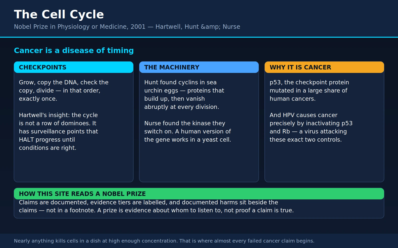
Table of Contents
- The Prize and the Three Men
- What a Cell Cycle Actually Is
- Hartwell's cdc Genes — and the Checkpoint Idea
- Nurse's cdc2: A Human Gene That Worked in a Yeast
- Hunt's Cyclins: The Protein That Kept Disappearing
- Why This Is Cancer's Mechanism
- Two Different Things Called "Checkpoints"
- The Drugs This Built
- Why Chemotherapy Makes Sense Here
- What This Does Not License
- What This Means for You Today
- Where Mainstream Medicine Agrees / What Remains Debated
- Key Research Papers
- Connections
- Featured Videos
1. The Prize and the Three Men
The Nobel Prize in Physiology or Medicine for 2001 went jointly to Leland H. Hartwell, R. Timothy (Tim) Hunt and Sir Paul M. Nurse, "for their discoveries of key regulators of the cell cycle." Between them, working on organisms nobody would mistake for a person — two kinds of yeast and the fertilised eggs of a sea urchin — they worked out the machinery that decides when a cell divides, and they showed that the same machinery runs in us.
That last part is the whole story. If the control system for cell division were a human peculiarity, you would have to study it in human cells, which grow slowly, cost a fortune, and cannot be crossed like peas. Because it turned out to be near-identical in a single-celled fungus, it could be taken apart with genetics in a matter of days per experiment. And because it is the system that fails in cancer, taking it apart in yeast turned out to be a way of taking apart cancer.
Leland Hartwell (born 1939)
An American geneticist who spent the productive part of his research career at the University of Washington in Seattle, working on budding yeast (Saccharomyces cerevisiae) — ordinary baker's and brewer's yeast. Beginning around 1970, Hartwell did something conceptually simple and, at the time, unfashionable: he collected mutant yeast cells that could not divide properly, and sorted them by where in the process they got stuck. He called the genes he found cdc genes, for "cell division cycle." He later added the idea that has arguably had the most clinical impact of anything in this story: the checkpoint. Hartwell went on to lead the Fred Hutchinson Cancer Research Center in Seattle for more than a decade, and spent much of his later career arguing for early detection and for molecular diagnostics.
Tim Hunt (born 1943)
A British biochemist trained at Cambridge, where he worked for years on how protein synthesis is controlled. He discovered cyclin almost by accident during a summer at the Marine Biological Laboratory in Woods Hole, Massachusetts, in the early 1980s, using sea urchin eggs because they are large, transparent, available by the bucket, and — crucially — because after fertilisation they all divide in step, so a whole population does the same thing at the same moment. He later moved to the Imperial Cancer Research Fund in London, which merged into Cancer Research UK, and ran a laboratory at Clare Hall.
Hunt's public life took a difficult turn in June 2015. Speaking at a lunch during the World Conference of Science Journalists in Seoul, he made remarks about women in laboratories — the widely reported version being that his "trouble with girls" was that "you fall in love with them, they fall in love with you, and when you criticise them they cry," followed by a suggestion about separate laboratories. Within days he resigned his honorary professorship at University College London after the university indicated he should, and he stepped down from a Royal Society awards committee and from the European Research Council's Scientific Council.
The record is genuinely contested on the detail. Hunt said the remarks were meant as a self-deprecating joke, apologised for the offence caused, and also said he had meant the part about having trouble with girls. An account from a European Commission official who was present reported that he had gone on, in the same speech, to say "Now, seriously" and to praise the contribution of women scientists — words absent from the first reports. Whether that changes the meaning of what came before is a question people answered very differently at the time and still do. This site's practice is to state what is on the record and stop there: the remarks were made, the resignations happened, the fuller transcript is disputed, and readers can weigh it themselves. It does not change a single result described on this page, and it is not a reason to leave the results out.
Paul Nurse (born 1949)
A British geneticist who worked on fission yeast (Schizosaccharomyces pombe) — a rod-shaped yeast that grows by getting longer and then splitting down the middle, which makes cell size unusually easy to measure. He identified the gene that controls entry into cell division, and then showed that the human version of that gene works inside a yeast cell. Nurse led the Imperial Cancer Research Fund and then Cancer Research UK, was president of Rockefeller University in New York from 2003 to 2011, served as president of the Royal Society from 2010 to 2015, and became the founding director of the Francis Crick Institute in London. He was knighted in 1999.
Nurse's own origin is worth recording, briefly, because he has chosen to talk about it publicly rather than keep it private. He grew up in London believing that his parents were his parents and that he had an older sister. While he was president of Rockefeller University, an application for United States permanent residency required documents his usual short-form birth certificate could not satisfy. When he obtained the full certificate, he found that the woman he had believed was his older sister was in fact his mother; she had been a teenager, unmarried, when he was born in Norfolk, and the family had raised him as her younger brother. He was in his fifties when he found out, and by then both women were dead. He has said, more than once and in public, that a man who had spent his life asking how cells know who they are had not known who he was.
Three careers, three organisms, one answer. What follows is that answer — and then the part that matters to a person reading this because of a diagnosis rather than because of the history.
2. What a Cell Cycle Actually Is
Every cell in your body that can divide does so by running a fixed sequence. Biologists call it the cell cycle, and it has four named parts.
- G1 — grow and decide. The cell gets bigger, makes proteins, and takes stock. Is there enough food? Is there a growth signal telling it to divide? Is it big enough? Is its DNA intact? G1 is the deliberation phase, and it is where a cell can step off the ride entirely into a resting state (called G0) and stay there for decades. Most of the cells in your brain, heart and muscles are sitting in G0 right now.
- S — copy the DNA. "S" is for synthesis. All three billion base pairs get duplicated, once and only once. At the end of S phase the cell holds two complete genomes.
- G2 — check the copy. A second growth-and-inspection gap. Was the copying finished? Was it accurate? Did anything break?
- M — divide. Mitosis. The duplicated chromosomes are lined up, pulled apart into two identical sets, and the cell pinches in two. This is the fastest phase — typically under an hour — and the most violent.
Written out like that it sounds like an assembly line, and the deep problem is easy to miss. The problem is order, and it is unforgiving.
The DNA must be copied before the cell divides, and copied exactly once. Copy it twice and you get a cell with four sets of chromosomes. Divide before copying finishes and you tear a chromosome in half, which is worse than losing it outright, because a broken chromosome end is chemically reactive and will fuse to whatever it finds. Divide with damaged DNA and the damage is now permanent in two daughter cells instead of temporary in one. A cell that gets the order wrong does not simply fail; it can produce descendants carrying rearranged, duplicated and deleted chromosomes — which is a recognisable description of what the inside of a tumour cell looks like.
So the question Hartwell, Hunt and Nurse were really answering is not "how does a cell divide?" It is "how does a cell make absolutely certain it does these things in the right order, exactly once, every time, for a lifetime?" Your body performs something on the order of tens of billions of cell divisions a day. The error rate has to be extraordinarily low or you would not survive childhood.
3. Hartwell's cdc Genes — and the Checkpoint Idea
The screen
Hartwell's method was blunt and beautiful. He mutated budding yeast at random and looked for temperature-sensitive mutants: cells that grow normally at 23 °C but fail at 36 °C. The trick is that you can keep such a strain alive indefinitely at the low temperature and then break it on command by warming the plate. If the broken gene is needed for cell division, the whole population stops — and, critically, it stops at the same place.
That last detail is what turned a mutant collection into a map. Under a microscope, budding yeast wears its cell-cycle stage on the outside: an unbudded cell is in G1; a small bud means early S; a large bud with a single nucleus means G2; two nuclei means mitosis is under way. Shift a mutant to the restrictive temperature and every cell in the flask piles up at whatever step that gene is required for, producing a flask full of identically-shaped cells. Hartwell could therefore say not just "this gene is needed for division" but "this gene is needed here."
He published the first batch in 1970 and the fuller picture in Science in 1974 (10.1126/science.183.4120.46). Ultimately the screen yielded roughly a hundred cdc genes, numbered in order of discovery — cdc1, cdc2, cdc28 and so on — and from their arrest points he assembled the sequence of dependent events that a yeast cell runs through. He named the commitment point in G1, after which the cell is going to divide whether or not conditions later worsen, START.
The idea that mattered more: checkpoints
The natural way to read a dependent sequence is as a chain of dominoes: step 1 physically causes step 2, which causes step 3. If DNA replication produces the signal that triggers mitosis, then a cell that has not finished replicating simply cannot enter mitosis, and order takes care of itself.
Hartwell and his student Ted Weinert showed this is not how it works, and the experiment is one of the cleanest in the field. In 1988 they studied a yeast gene called RAD9 (10.1126/science.3291120). Normal yeast, irradiated with X-rays, halts in G2 for a period roughly proportional to how much damage it took, repairs the breaks, and then carries on. Yeast lacking RAD9 does something startling: irradiated, it does not pause at all. It ploughs straight through mitosis, divides for a few more generations, and dies.
Then comes the part that settles the argument. Weinert and Hartwell took the rad9 mutants and forced them to stop in G2 artificially, using a drug that poisons the mitotic spindle. Held still by chemistry rather than by their own control system, those cells repaired their DNA perfectly well. The repair machinery had never been broken. What was missing was the pause.
So the cell does not stop because it is physically unable to continue. It stops because a surveillance system detects a problem and actively imposes a halt. Hartwell and Weinert named these surveillance systems checkpoints in a 1989 Science paper (10.1126/science.2683079) whose subtitle — "controls that ensure the order of cell cycle events" — is the whole idea in six words.
Why this is the sentence to remember
Your DNA is damaged constantly. Ultraviolet light, background radiation, the ordinary chemistry of oxygen, the copying errors of replication itself — a single cell sustains thousands of DNA lesions a day. Almost none of them become mutations. They do not because the checkpoint machinery notices the damage, freezes the cell cycle, and holds the cell still until the repair enzymes have finished.
Damage becomes mutation when the cell divides before the repair is done. That is the sentence. It reframes what a carcinogen is doing, what a tumour suppressor is protecting, and why a cell that has lost its checkpoints accumulates damage at a catastrophic rate even in an ordinary environment. It also explains the otherwise puzzling clinical observation that people who inherit a broken checkpoint gene — in conditions such as ataxia–telangiectasia or Li–Fraumeni syndrome — are not sick because their cells cannot repair DNA, but because their cells do not wait.
4. Nurse's cdc2: A Human Gene That Worked in a Yeast
Wee mutants
Nurse worked on fission yeast, which grows as a rod, extends at the ends, and divides across the middle at a fairly constant length. That makes size a readout you can measure with a ruler on a photograph, which is why his 1975 Nature paper was titled "Genetic control of cell size at cell division in yeast" (10.1038/256547a0).
Among his mutants were cells that divided when they were far too short — they were dubbed wee, after the Scots word, Nurse having done the work in Edinburgh. A wee mutant is the mirror image of a cdc mutant. A cdc mutant cannot enter mitosis and gets longer and longer; a wee mutant enters mitosis too eagerly and divides while still small. Having mutants that err in both directions is what let Nurse identify the rate-limiting step: the single control point where the decision to enter mitosis is actually made, rather than one of the many downstream steps that merely execute it.
That control point was a gene called cdc2. Nurse and Pierre Thuriaux showed in 1980 (10.1093/genetics/96.3.627) that cdc2 sits at the switch for entry into mitosis, with mutations in it capable of pushing division either way. It later emerged that fission yeast's cdc2 and budding yeast's CDC28, found independently in Hartwell's screen, are the same gene in two organisms — the first hint that this control system might be older and more general than anybody had assumed.
The experiment that ended the argument
In 1987, Melanie Lee and Paul Nurse ran an experiment that reads, even now, as slightly outrageous (10.1038/327031a0). They took a fission yeast strain carrying a broken cdc2 gene — a strain that cannot divide. They introduced into it a library of human DNA copies, essentially at random, one fragment per cell. Then they simply asked which yeast cells started dividing again.
Some did. The human fragment those cells had picked up encoded a protein whose sequence was strikingly similar to yeast Cdc2. It is the gene we now call CDK1, and it had substituted for its yeast counterpart well enough to rescue a fungus.
Consider what that requires. The human protein has to be made correctly by yeast machinery, fold correctly in a yeast cell, find and bind its yeast partner proteins, recognise its yeast targets, phosphorylate them at the right residues, and be switched on and off at the right moments by a yeast regulatory network it had never encountered. Yeasts and animals last shared an ancestor something on the order of a billion years ago. Nearly everything else about the two organisms has diverged beyond recognition. This did not.
The paper's own conclusion is stated with characteristic English understatement: the results "indicate that elements of the mechanism by which the cell cycle is controlled are likely to be conserved between yeast and humans." What it meant in practice was that every fact anybody could establish about the yeast cell cycle was now, provisionally, a fact about the human cell cycle — and that the fast, cheap, powerful genetics of yeast had been pointed directly at human cancer.
Cdc2/CDK1 turned out to be the founding member of a family. Humans have around twenty cyclin-dependent kinases. A kinase is an enzyme that attaches phosphate groups to other proteins, and attaching a phosphate is biology's standard way of flipping a switch: it changes a target protein's shape, and so its activity, in a way that another enzyme can later undo. CDKs are the switches that drive the cell cycle forward. But a CDK on its own does nothing. It needs a partner, and finding that partner is the third leg of this story.
5. Hunt's Cyclins: The Protein That Kept Disappearing
The observation
Hunt was not looking for the cell cycle. He was studying protein synthesis, and he was at Woods Hole because sea urchin eggs are a superb system for it: a fertilised egg contains a stockpile of maternal messenger RNA and begins translating it furiously the moment fertilisation occurs. He fed the eggs radioactive methionine so that newly-made proteins would be labelled, took samples at intervals, and ran them out on a gel to see which proteins were being made.
Most of the bands behaved sensibly, accumulating steadily. One did not. One band climbed steadily — and then, at the moment the eggs divided, vanished. Then it climbed again from nothing, and vanished again at the next division. And again. The eggs of Arbacia punctulata go through a run of rapid, synchronous cleavage divisions, so Hunt was watching the same disappearance happen over and over in the same tube.
The 1983 paper in Cell (10.1016/0092-8674(83)90420-8) reports the same behaviour in a second sea urchin species and in surf clam oocytes, and ends with one of the more consequential naming sentences in modern biology: "We propose to call these proteins the cyclins."
Why the pairing is the answer
Put Nurse's finding and Hunt's finding side by side and the control system falls out.
A cyclin-dependent kinase is a constant. Its level barely changes across the cell cycle. It is present in the cell all the time, and it is essentially inert on its own. A cyclin is a variable. Its level rises and falls dramatically, on a schedule. And a CDK becomes active only when a cyclin binds to it.
So the cell has built a clock out of two parts: a permanently-installed engine and a fuel supply that is manufactured and then deliberately destroyed on a timetable. Different cyclins peak at different phases — the D-type cyclins in G1, cyclin E at the G1-to-S transition, cyclin A through S phase, cyclin B before mitosis — and each partners a particular CDK to switch on the specific set of targets that phase requires. The rise of a cyclin turns a kinase on; the destruction of that cyclin turns it off again and lets the next one take over.
The destruction is the elegant part, and it is why Hunt's protein had to disappear rather than merely be inactivated. Making a protein takes time; degrading one can be effectively instantaneous. More importantly, degradation is irreversible in a way that inhibition is not. A cell exiting mitosis must not slide backwards into it. Destroying cyclin B guarantees the exit is one-way, which is precisely the property you need at a step that must happen once and not again.
The 2004 Chemistry connection
How is a protein destroyed on cue? In 1991, Michael Glotzer, Andrew Murray and Marc Kirschner showed that cyclin is tagged with ubiquitin — a small protein whose attachment marks another protein for demolition — and then chewed up by the cell's protein-shredding machinery (10.1038/349132a0). They found a short stretch near cyclin's front end that acts as the tag-me signal: fuse that stretch onto an unrelated protein and the unrelated protein now gets destroyed at mitosis too.
That ubiquitin system is itself Nobel-decorated: the 2004 Nobel Prize in Chemistry went to Aaron Ciechanover, Avram Hershko and Irwin Rose for the discovery of ubiquitin-mediated protein degradation. The cell-cycle clock, in other words, is built from two separately-honoured discoveries that turn out to be halves of the same mechanism — a kinase that is switched on by a partner protein, and a labelling system that destroys that partner on schedule.
It is also a mechanism with a drug attached: the proteasome inhibitor bortezomib, used in multiple myeloma, works by jamming the shredder that this pathway feeds.
6. Why This Is Cancer's Mechanism
Here is the connection this whole page exists to make.
Cancer is, at its root, cells dividing when they should not. Not cells that are poisoned, not cells that are inflamed, not cells that are acidic — cells that have escaped the control system governing whether and when to divide. Whatever else is true of a tumour, this is true of all of them. It follows that the cell-cycle control machinery is the place where cancer happens. Hartwell said so explicitly in a 1994 Science review with Michael Kastan titled, simply, "Cell cycle control and cancer" (10.1126/science.7997877).
p53, the guardian of the genome
The gene TP53, which encodes the p53 protein, is the checkpoint made flesh. When a cell's DNA is damaged, p53 accumulates and does one of two things depending on how bad the damage is. If it is repairable, p53 halts the cycle — principally by switching on a gene called CDKN1A, whose product p21 sits on cyclin–CDK complexes and blocks them — so the repair enzymes have time to work. If the damage is beyond repair, p53 orders the cell to kill itself.
In 1992, David Lane published a one-and-a-half-page commentary in Nature that gave the protein the name it has carried ever since: "p53, guardian of the genome" (10.1038/358015a0). Lose p53 and a cell keeps dividing through damage it should have paused for or died from. Mutations accumulate. Chromosomes rearrange. The cell becomes a mutation engine.
TP53 mutations are among the most frequent somatic events in human cancer. The IARC TP53 Database, the field's long-running catalogue, has compiled tens of thousands of somatic TP53 mutation records across essentially every tumour type (Bouaoun et al., Hum Mutat 2016;37(9):865-76). The often-quoted headline of "about half of all cancers" is a rough average that conceals enormous variation: TP53 is mutated in the great majority of high-grade serous ovarian cancers, in a large fraction of lung squamous and colorectal cancers, and in a small minority of some others. What is consistent is not the percentage — it is that when you sequence tumours at scale, TP53 keeps coming out at or near the top of the list.
Rb, the brake that CDKs release
The second great tumour suppressor in this story is the retinoblastoma protein (Rb), and it explains the G1 decision mechanically.
Rb's job is to hold down a set of transcription factors called E2F, which switch on the genes needed for DNA replication. As long as Rb has hold of E2F, the cell cannot enter S phase. When growth signals arrive, the cell makes cyclin D; cyclin D binds CDK4 and CDK6; that complex phosphorylates Rb; phosphorylated Rb lets go of E2F; E2F turns on the replication genes; the cell enters S phase and is committed.
Rb is a brake. Cyclin D–CDK4/6 is the foot that lifts off the brake. Remember that sentence — it is the entire rationale for a drug class in section 8.
The RB1 gene was the first human tumour suppressor gene identified, cloned in the mid-1980s from studies of retinoblastoma, a childhood eye cancer. Retinoblastoma is also where Alfred Knudson's two-hit hypothesis came from: a tumour suppressor has two copies, and both must be knocked out before the brake is gone. Children who inherit one broken copy need only one further hit, which is why the inherited form appears earlier and in both eyes.
The honest framing: it takes several failures, not one
It would be tidy to say "cancer is a p53 mutation." It is not true. Cells have overlapping, redundant restraints — the G1/S checkpoint, the intra-S checkpoint, the G2/M checkpoint, the spindle-assembly checkpoint, apoptosis, replicative senescence, immune surveillance — and knocking out any one of them generally produces a cell that is somewhat more dangerous, not a tumour.
When the Cancer Genome Atlas sequenced 3,281 tumours across twelve cancer types, it found 127 significantly mutated genes, and reported that most tumours carried mutations in only two to six of them — "indicating that the number of driver mutations required during oncogenesis is relatively small" (10.1038/nature12634). Small, but plural. Not one. That is why cancer is overwhelmingly a disease of later life, why it is rare in children except where an inherited first hit has already been supplied, and why the popular image of a single carcinogenic insult producing a tumour is misleading.
The virus that attacks exactly these two proteins
There is a case that ties this section together with uncommon neatness, and it belongs to another laureate on this site: Harald zur Hausen, who won the 2008 Nobel Prize for showing that human papillomavirus causes cervical cancer.
How does HPV cause a cancer? Through two small viral proteins. E6 promotes the destruction of p53 — shown by Scheffner and colleagues in 1990 (10.1016/0092-8674(90)90409-8), using, appropriately enough, the ubiquitin system. And E7 binds the retinoblastoma protein, prising it off E2F — shown by Dyson and colleagues in 1989 (10.1126/science.2537532).
The virus does this for its own reasons: it needs a cell in S phase to replicate its own genome, and it has evolved two proteins that force one. But the effect is that a high-risk HPV infection disables the two most important brakes in the human cell cycle simultaneously. A virus discovered by one line of research turns out to cause cancer by attacking, with precision, the exact two proteins identified by another. It is difficult to imagine better confirmation that the cell-cycle control system is where cancer lives — and it is why HPV vaccination is, mechanistically, one of the most direct cancer-prevention measures ever devised.
Two other laureate pages on this site sit alongside this one. Otto Warburg described the altered metabolism of tumour cells — a real and important phenomenon, and a downstream consequence of proliferation rather than its cause. Blackburn, Greider and Szostak worked out telomeres and telomerase, the counting mechanism that normally limits how many times a cell lineage can divide at all — another restraint that most cancers have to defeat.
7. Two Different Things Called "Checkpoints"
A short section, because this single word causes more confusion in cancer reading than almost any other.
Cell-cycle checkpoints are what this page has been about: internal surveillance systems inside a cell that halt its own division until DNA is repaired, replication is complete, and chromosomes are correctly attached. Hartwell and Weinert's word, 1989. The relevant proteins are p53, ATM, ATR, CHK1, CHK2, Rb.
Immune checkpoints are something entirely different: molecular brakes on T cells, not on cancer cells, which normally stop the immune system from attacking healthy tissue. Tumours exploit them to avoid being killed. Releasing those brakes with antibodies against CTLA-4 and PD-1 is the basis of checkpoint immunotherapy, and it earned James Allison and Tasuku Honjo the 2018 Nobel Prize.
Same word, unrelated machinery, opposite therapeutic logic. Cell-cycle checkpoint drugs generally aim to exploit a checkpoint the tumour has already lost. Immune checkpoint drugs aim to remove a brake the tumour is exploiting. If you read that a trial is testing "a checkpoint inhibitor," it is worth two seconds to establish which kind.
8. The Drugs This Built
Basic science pages often end with a vague gesture at "future therapies." This one does not have to. Two approved drug classes come directly out of the biology above, and they are used in oncology clinics today.
CDK4/6 inhibitors
Recall the mechanism from section 6: cyclin D binds CDK4 and CDK6, that complex phosphorylates Rb, phosphorylated Rb releases E2F, and the cell enters S phase. In hormone-receptor-positive breast cancer, oestrogen signalling drives cyclin D production, which is exactly how oestrogen drives proliferation. Block CDK4 and CDK6, and Rb stays unphosphorylated, and E2F stays suppressed, and the cell stalls in G1.
Three such drugs are approved: palbociclib (approved in the United States in 2015), ribociclib and abemaciclib (both 2017). All three are oral tablets. All three are used with endocrine therapy — an aromatase inhibitor or fulvestrant — in hormone-receptor-positive, HER2-negative advanced breast cancer. Abemaciclib and ribociclib also have approvals in earlier-stage disease.
Now the part that is routinely blurred, and should not be. There are two different survival questions, and these drugs answer them differently.
Progression-free survival (PFS) is the time from starting treatment until the cancer grows on a scan or the patient dies. Overall survival (OS) is the time from starting treatment until death from any cause. PFS is faster and cheaper to measure. OS is what most people mean when they ask whether a drug helps them live longer. A drug can improve the first without improving the second.
On PFS, the CDK4/6 inhibitors are unambiguous and impressive:
- PALOMA-2 (palbociclib + letrozole, first-line, 666 patients) reported median PFS of 24.8 months versus 14.5 months with letrozole alone — a hazard ratio of 0.58 (10.1056/NEJMoa1607303).
- MONARCH 3 (abemaciclib + aromatase inhibitor, first-line, 493 patients) reported a PFS hazard ratio of 0.54, with median PFS not yet reached in the abemaciclib arm against 14.7 months in the placebo arm at that analysis (10.1200/JCO.2017.75.6155).
On overall survival, the picture is genuinely mixed, and this is the honest summary:
- MONALEESA-2 (ribociclib + letrozole, first-line) did meet its overall survival endpoint. After a median follow-up of 6.6 years, median OS was 63.9 months versus 51.4 months, hazard ratio 0.76, two-sided P = 0.008 (10.1056/NEJMoa2114663).
- PALOMA-2 (palbociclib + letrozole, first-line) did not. At a median follow-up of 90.1 months, median OS was 53.9 months versus 51.2 months, hazard ratio 0.96, one-sided P = 0.34. The investigators themselves noted that an imbalance in patients with unknown survival status between the arms limited interpretation — but the honest reading is that overall survival was not significantly improved (10.1200/JCO.23.00137).
- MONARCH 3 (abemaciclib + aromatase inhibitor, first-line) narrowly missed. After a median 8.1 years, median OS was 66.8 versus 53.7 months — a 13.1-month difference — with a hazard ratio of 0.804, but P = 0.0664, above the significance threshold. The authors describe the result as clinically meaningful while stating plainly that statistical significance was not reached (10.1016/j.annonc.2024.04.013).
- PALOMA-3 (palbociclib + fulvestrant, after progression on endocrine therapy) found median OS of 34.9 versus 28.0 months, hazard ratio 0.81, P = 0.09 — not significant in the whole trial population, though the prespecified subgroup with prior endocrine sensitivity showed a 10-month difference (10.1056/NEJMoa1810527).
So: a large, consistent, reproducible delay in cancer growth across all three drugs; a clear overall-survival benefit demonstrated for ribociclib in the first-line setting; and results for palbociclib and abemaciclib that fall short of statistical significance, one of them by a hair. Media coverage compresses all of this into "CDK4/6 inhibitors help people live longer." That is defensible for ribociclib and, at best, unproven for the others. Anyone deciding among these drugs deserves to know which claim rests on which trial.
Toxicity. The main and predictable problem is neutropenia — a fall in the neutrophils that fight bacterial infection. This is not a surprise; it is the mechanism doing exactly what it says. Bone marrow is the body's most proliferative tissue, and a drug that arrests dividing cells in G1 arrests marrow precursors too. In PALOMA-2, grade 3 or 4 neutropenia occurred in 66.4% of patients on palbociclib against 1.4% on placebo. Crucially, though, febrile neutropenia — neutropenia with fever, the dangerous version — occurred in only 1.8%, and in none of the placebo group. The neutropenia from CDK4/6 inhibitors is generally reversible and managed with dose interruption or reduction; it behaves differently from chemotherapy-induced marrow suppression, which kills precursor cells outright rather than pausing them. Abemaciclib's dominant side effect is different again: diarrhoea, in about 80% of patients, mostly mild. Ribociclib requires ECG monitoring for QT prolongation and liver-function monitoring.
These are real, approved, effective drugs. They are also not cures. In advanced breast cancer they delay progression, sometimes substantially, and in at least one trial they extended life. They do not eliminate the disease. Any source telling you otherwise is not being careful with you.
PARP inhibitors and synthetic lethality
The second drug class comes from the checkpoint-and-repair side rather than the clock side, and it rests on an idea worth understanding because it is the template for a generation of cancer drugs.
Cells repair broken DNA by more than one route. Homologous recombination is the accurate route for double-strand breaks, and it needs the BRCA1 and BRCA2 proteins. A separate enzyme, PARP, is central to repairing single-strand breaks. A cell can survive losing either system on its own, because the other compensates. Lose both and it dies.
Now consider a woman with an inherited BRCA1 or BRCA2 mutation. Every cell in her body carries one broken copy; her tumour cells have lost the second. So her healthy cells still have working homologous recombination, and her cancer cells do not. Give a drug that blocks PARP, and the tumour cells — already down one repair system — lose the second and die, while the healthy cells, which retain homologous recombination, tolerate it. Two papers published back-to-back in Nature in 2005 demonstrated this (Farmer et al., 10.1038/nature03445; Bryant et al., 10.1038/nature03443).
The principle is called synthetic lethality: neither defect alone is fatal, but the combination is. It is the closest thing oncology has to a genuinely selective weapon, because the selectivity comes from a genetic difference between the tumour and the patient rather than from a difference in growth rate. Olaparib, the first PARP inhibitor, was approved in 2014; niraparib, rucaparib and talazoparib followed. They are used in BRCA-mutated and, more broadly, homologous-recombination-deficient ovarian, breast, prostate and pancreatic cancers. They are, like the CDK4/6 inhibitors, effective and not curative — resistance develops, sometimes through the tumour repairing its own BRCA gene.
9. Why Chemotherapy Makes Sense Here
Classical cytotoxic chemotherapy predates all of this science by decades. Nitrogen mustard was used against lymphoma in the 1940s; methotrexate, 5-fluorouracil and the rest arrived through empirical screening long before anyone could have drawn a cyclin. But the cell-cycle framework explains, retrospectively and completely, both why they work and why they hurt.
Why they work
Every classical chemotherapy drug attacks something that a dividing cell needs and a resting cell does not.
- Antimetabolites (methotrexate, 5-fluorouracil, gemcitabine, cytarabine) starve the cell of the nucleotide building blocks required for S phase. A cell in G0 is not copying DNA and does not care.
- Alkylating agents and platinums (cyclophosphamide, cisplatin, carboplatin) chemically damage and cross-link DNA. A cell that then tries to replicate through that damage stalls and dies; a cell that never replicates may repair it at leisure.
- Topoisomerase inhibitors (doxorubicin, etoposide, irinotecan) jam the enzymes that untangle DNA during replication, converting an ordinary replication fork into a double-strand break.
- Spindle poisons (paclitaxel, docetaxel, vincristine) target the microtubule apparatus that pulls chromosomes apart. Only a cell in mitosis has one.
The selectivity is therefore kinetic: the drugs harm proliferating cells preferentially, and tumours contain a higher fraction of proliferating cells than most normal tissues. It works because tumour cells divide more, not because tumour cells are recognised as foreign.
Why the side effects are what they are
This is where the mechanism becomes a prediction, and the prediction is correct. If a drug harms rapidly dividing cells, then the normal tissues that divide rapidly will be harmed too. Which are they?
- Bone marrow, which manufactures billions of blood cells daily. Hence neutropenia and infection risk, anaemia and fatigue, thrombocytopenia and bruising. Neutrophils live only hours to a couple of days in circulation, which is why the neutrophil count is the first to fall — the nadir typically arriving one to two weeks after a dose.
- The lining of the gut, which replaces itself every few days from crypt stem cells. Hence mouth ulcers, nausea, diarrhoea, appetite loss.
- Hair follicles, which are among the fastest-cycling tissues in the body. Hence alopecia — and hence the fact that hair grows back, because the follicle stem cells recover.
- The reproductive tissues, hence effects on fertility that ought to be discussed before treatment begins rather than after.
These are not mistakes, and they are not evidence that the treatment is poison rather than medicine. They are the same mechanism, arriving at the wrong address. A drug that kills dividing cells cannot know which dividing cells you wanted killed. The pattern of chemotherapy's toxicity is a direct, predictable readout of which of your tissues divide fastest — which is exactly what the cell-cycle framework says it should be. This is worth internalising, because it is the honest answer to a claim you will meet in the next section: that chemotherapy's side effects prove it is indiscriminate destruction. They prove it is insufficiently discriminating, which is a different and much more tractable problem.
The honest limits of that story
Two caveats keep this from being too neat.
First, not all chemotherapy toxicity is proliferation-related. Anthracycline damage to heart muscle, cisplatin damage to hearing and kidneys, platinum and taxane peripheral neuropathy — these hit cells that are not dividing at all, through separate mechanisms. Anyone who tells you chemotherapy side effects are entirely explained by rapid cell division is oversimplifying.
Second, the rapid-division story does not fully explain why chemotherapy works when it works. Some very slowly-growing tumours respond; some fast-growing ones do not. Modern understanding adds tumour-specific DNA-repair deficiencies, apoptotic thresholds, and immune effects to the picture.
What targeted therapy is trying to do is replace kinetic selectivity with molecular selectivity — to attack something the tumour has and your healthy tissue does not, rather than something the tumour does faster. PARP inhibitors in BRCA-mutated cancer are that idea in its purest form. Imatinib in chronic myeloid leukaemia, targeting a fusion protein that exists only in the malignant clone, is another. This is real progress, and it is incremental rather than total: most targeted agents still produce meaningful side effects, and most people with advanced cancer still receive cytotoxic chemotherapy at some point.
10. What This Does Not License
This is the most important section on the page, and it is written for someone who may be frightened, may have been given a bad prognosis, and may be reading late at night through a great many websites that sound confident.
Everything above is true. Cancer is a disease of the cell-division control system. From that true statement, an enormous amount of misleading advice has been built, and it is built by a single logical slip:
"Cancer is cells dividing too fast. Therefore anything that slows cell division treats cancer."
That inference is invalid, and it is worth seeing exactly why. Slowing cell division is not hard — almost anything does it at sufficient dose, including plain starvation, cold, and a great many ordinary chemicals. The difficulty in oncology has never been finding something that stops cells dividing. It has always been finding something that stops cancer cells dividing while leaving your bone marrow, gut lining and immune system functional enough that you survive the treatment. That is a therapeutic-window problem, not a growth-inhibition problem, and it is why "this compound inhibits cancer cell growth" is close to no information at all.
The petri-dish problem, which is where nearly every failed claim starts
Read enough alternative cancer material and you will notice a recurring shape: a compound was shown to inhibit growth of, or induce apoptosis in, a cancer cell line. Curcumin. EGCG from green tea. Resveratrol. Apigenin. Vitamin C at high dose. Various essential oils. Baking soda. Fenbendazole. Dozens more.
Those experiments were usually done honestly and usually reported accurately. The problem is what they mean, and the answer is: much less than it appears.
- Nearly anything kills cells in a dish at high enough concentration. This is not a slur on any particular compound; it is a property of cells in dishes. Cultured cancer cells are fragile, immortalised, adapted to plastic, and swimming in a medium with none of the protein binding, liver metabolism, kidney clearance or physiological buffering of a body.
- The concentrations are usually unreachable. The single most useful question to ask of any in-vitro cancer result is: what concentration, and can a human blood level get there? For many popular compounds the answer is no, not at any oral dose, often by one or two orders of magnitude. Curcumin is the standard example — it is poorly absorbed, rapidly conjugated, and the plasma levels achievable orally are far below what its cell-culture results require.
- Selectivity is rarely tested. A paper showing 60% killing of a cancer line frequently does not report what the same dose did to normal cells. When normal cells are tested, the difference is often modest — and a compound that kills tumour cells only slightly faster than it kills you is not a treatment.
- "Induces apoptosis" is a low bar. Apoptosis is a cell's built-in self-destruct programme, and a great many stresses trigger it. Inducing apoptosis in a dish tells you a compound is stressful to cells. It does not tell you it will shrink a tumour in a person, and it does not tell you it will spare healthy tissue — your gut lining runs on carefully regulated apoptosis too.
The gap between cell-culture activity and human benefit is where the overwhelming majority of cancer drug candidates die, including in the pharmaceutical industry, which spends billions on the transition and still fails most of the time. That is not a conspiracy; it is the difficulty of the problem.
Fasting, ketogenic diets, and "starving the cancer"
This one deserves care, because it is not simply nonsense, it is a real research area, and the honest answer is more interesting than either the enthusiasts' or the dismissers' version.
The underlying observation is real. Tumour cells often consume glucose heavily and ferment it even when oxygen is available — the phenomenon Otto Warburg described in the 1920s, and the basis of the FDG-PET scan, which finds tumours precisely by their glucose appetite. It is also true that fasting triggers autophagy, the cellular recycling programme whose genetics earned Yoshinori Ohsumi the 2016 Nobel Prize, and that fasting alters insulin and IGF-1 signalling in ways that plausibly interact with cancer cell growth.
From those real observations, three claims are commonly made. They are not equally supported.
Claim 1: "Sugar feeds cancer, so cutting carbohydrates starves it." This does not follow. Your body maintains blood glucose within a narrow range through gluconeogenesis regardless of what you eat — a person fasting for days still has blood glucose, because the liver manufactures it. You cannot lower blood glucose to a level that starves a tumour and remain conscious. Tumours also metabolise glutamine, fatty acids and ketone bodies. The ketogenic diet has genuine, established uses — refractory epilepsy, notably — and is under study in glioblastoma and elsewhere, but as a cancer treatment it remains investigational, and the trials done so far have been small and mixed.
Claim 2: "Fasting during chemotherapy improves outcomes." This is an active research area with genuine, small, early human trials — and it is not established therapy.
The largest randomised test to date is the Dutch DIRECT trial: 131 patients with HER2-negative stage II/III breast cancer randomised to a fasting-mimicking diet or their usual diet for three days before and during each cycle of neoadjuvant chemotherapy (10.1038/s41467-020-16138-3). What it found: no difference in toxicity between the groups, even though the fasting group had dexamethasone omitted; more frequent radiological response in the fasting group; and, in a per-protocol analysis — that is, restricted to the subset who actually completed the diet — a higher rate of near-complete pathological response. The fasting diet also reduced chemotherapy-induced DNA damage in circulating T cells. The authors' own conclusion is that the findings "encourage further exploration."
Read that carefully, because the structure matters. A per-protocol analysis is a weaker form of evidence than the randomised comparison, since the people who manage to complete a demanding diet during chemotherapy differ systematically from those who cannot. That is precisely where the tumour-response signal appeared. A smaller German cross-over trial of 34 women with breast and ovarian cancer found short-term fasting around chemotherapy was well tolerated and appeared to improve quality of life and fatigue, and concluded that "larger studies should prove the effect" (10.1186/s12885-018-4353-2).
That is a fair description of the field: promising, small, mostly in breast cancer, mostly measuring tolerability and short-term response rather than survival, and conducted under close supervision with dietitian input and defined protocols. It is a reasonable thing to ask your oncologist about, and a reasonable thing to enrol in a trial for. It is not a reason to decline treatment, and it is not established therapy.
Claim 3: "So I should fast, hard, on my own." This is where real harm happens, and it needs saying plainly.
Unsupervised fasting in someone who is already losing weight from cancer is dangerous. Cancer-associated weight loss is not ordinary dieting; it is a metabolic syndrome called cachexia in which the body breaks down skeletal muscle under inflammatory signalling, and it does not reverse simply by eating more. Low muscle mass is one of the more reliable predictors of poor chemotherapy tolerance, more dose reductions, more toxicity and worse survival. A person who fasts on top of cachexia loses more muscle, tolerates less treatment, and does worse. The DIRECT trial specifically excluded people with a BMI under 18.5 for exactly this reason. If you are losing weight without trying, the correct response is a referral to an oncology dietitian, not a fast.
Apoptosis-inducing supplement claims
A common formulation runs: "this compound selectively induces apoptosis in cancer cells while leaving healthy cells unharmed." Treat the word "selectively" as the load-bearing claim, and ask what was actually measured.
Selectivity is a dose-response comparison: how much is needed to harm a tumour cell versus a normal cell, expressed as a ratio. Where that comparison is done properly, the ratios for most popular natural compounds are modest — nothing like the ratios that would be required for a usable drug, and often within the range where the compound is simply toxic to everything. And any such claim still faces the concentration question above.
None of this means natural compounds are worthless or that the research is not worth doing. Some of oncology's most important drugs came from plants: paclitaxel from Pacific yew bark, vincristine and vinblastine from the Madagascar periwinkle, etoposide from mayapple, camptothecin derivatives from a Chinese tree. What made them medicines was not that they came from plants, and not that they killed cells in a dish. It was that somebody isolated the active molecule, established the dose that worked in a person without killing them, and ran the trials. That path is open to any compound. Very few survive it.
What a fair-minded reader should take from this
Interest in metabolism, diet and natural compounds in cancer is not foolish, and the people who hold it are not foolish. Some of it is being studied seriously right now by serious people. The failure mode is not curiosity — it is the leap from a laboratory observation to a treatment decision, and specifically the decision to delay or decline a treatment with demonstrated benefit in favour of one with a mechanism that sounds right.
Delay is the harm. Cancers that are curable at diagnosis frequently stop being curable a few months later, and the studies that have followed patients who chose alternative treatment instead of conventional treatment for curable cancers report substantially worse survival. If something in this space interests you, the productive move is to bring it to your oncology team as an addition to be checked for interactions — several popular supplements genuinely do interfere with chemotherapy metabolism or bleeding risk — rather than as a replacement.
11. What This Means for You Today
If you or someone close to you has a pathology report in hand, parts of it are direct descendants of the science on this page. Here is what those numbers actually measure.
Ki-67
Ki-67 is a protein present in cells that are in the cell cycle and absent in cells resting in G0. A pathologist stains the tumour for it and reports the percentage of tumour cell nuclei that light up. So a Ki-67 of 25% means roughly a quarter of the tumour cells sampled were actively cycling at the moment the tissue was fixed. It is a proliferation index — a snapshot of how much of the tumour is dividing.
Two honest caveats. First, reproducibility is a real problem: the same slide scored in two laboratories can give meaningfully different numbers, and scores from a core biopsy tend to run higher than from the matching surgical specimen. Second, and following from that, its clinical usefulness is narrower than its popularity suggests. The International Ki67 in Breast Cancer Working Group, reviewing the evidence, concluded that Ki-67 has clinical validity as a prognostic marker but that its clinical utility is limited — specifically, to estimating prognosis in anatomically favourable (T1–2, N0–1) oestrogen-receptor-positive, HER2-negative breast cancer, in order to identify patients who do not need adjuvant chemotherapy, using thresholds of 5% or less and 30% or more (10.1093/jnci/djaa201). A middling Ki-67 in that setting is genuinely uninformative, and a Ki-67 outside that setting should be weighed lightly. If your report shows a borderline value and it seems to be driving a major decision, that is a fair thing to ask about.
Mitotic index and grade
The mitotic count is simpler and older: a pathologist counts how many cells are caught in the act of mitosis in a defined area of tissue. It is one of the three components of the Nottingham grading system for breast cancer, alongside how well the tumour forms tubular structures and how abnormal the nuclei look. Each is scored 1–3 and the three are summed to give grade 1, 2 or 3.
Grade is not stage, and confusing them causes real distress.
- Grade describes how abnormal and how proliferative the cells look under the microscope. It is a property of the cells. Grade 1 is well differentiated and slow; grade 3 is poorly differentiated and fast.
- Stage describes how far the cancer has spread — size of the primary tumour, involvement of lymph nodes, presence of distant metastases. It is a property of the disease's extent in your body.
They are largely independent, and stage is generally the stronger driver of prognosis. A small, node-negative grade 3 tumour is usually a far better situation than a widely metastatic grade 1 tumour. Being told "grade 3" is frightening in a way that is often out of proportion to what it means for outcome once stage and receptor status are accounted for.
"Fast-growing" is not a synonym for "worse"
This deserves its own paragraph because the intuition is so strong and so unreliable.
Rapidly proliferating tumours are more sensitive to chemotherapy — a direct consequence of the mechanism in section 9. Some of the most curable cancers in adult medicine are among the fastest-growing: Burkitt lymphoma, whose Ki-67 approaches 100%, is curable in a large majority of patients with intensive chemotherapy; testicular germ cell tumours are highly proliferative and highly curable even when metastatic; acute promyelocytic leukaemia went from among the most rapidly lethal leukaemias to among the most curable. Meanwhile some indolent cancers are, with current treatment, not curable at all — follicular lymphoma grows slowly, responds to treatment, recurs, and is generally managed rather than cured over many years.
In breast cancer specifically, high Ki-67 is associated with worse prognosis if untreated and with better response to chemotherapy if treated. Both are true at once. This is why "how fast is it growing?" is a much less useful question than it feels, and why the questions that actually change management are: what type is it, what receptors does it carry, what stage is it, and what does the molecular testing say.
Reasonable questions to bring to an appointment
- What is my stage, and what is my grade — and which one is driving the treatment plan?
- Is this treatment intended to cure, to extend life, or to control symptoms? (Doctors often have a clear answer and often are not asked.)
- If a drug is being recommended on progression-free survival data, is there overall survival data, and what does it show?
- What should I do about the supplements I already take — and could any of them interact with this treatment?
- Am I losing weight, and should I see an oncology dietitian?
- Is there a clinical trial I am eligible for?
12. Where Mainstream Medicine Agrees / What Remains Debated
Settled — not seriously disputed by anyone working in the field
- The eukaryotic cell cycle is driven by cyclin-dependent kinases whose activity is controlled by the periodic synthesis and destruction of cyclins.
- This machinery is conserved from yeast to humans, to the extent that human genes can replace their yeast counterparts in a living yeast cell.
- Checkpoints are active surveillance systems that impose delay, not passive consequences of unfinished steps. The RAD9 experiment settled this.
- Cancer arises from failures of cell-cycle control and of the checkpoints that police it, and typically requires several such failures rather than one.
- TP53 is among the most frequently mutated genes in human cancer, and loss of Rb function is a recurring feature across many tumour types.
- High-risk HPV causes cancer by inactivating p53 and Rb through the viral E6 and E7 proteins.
- Classical chemotherapy's efficacy and its characteristic toxicities both follow from preferential harm to proliferating cells.
- CDK4/6 inhibitors substantially prolong progression-free survival in hormone-receptor-positive, HER2-negative advanced breast cancer.
Genuinely debated — where informed people disagree
- Whether the CDK4/6 inhibitors are interchangeable. Only ribociclib has clearly demonstrated an overall survival benefit in the first-line advanced setting; abemaciclib missed narrowly and palbociclib did not show one. Whether this reflects a real difference between the drugs, differences in trial design, follow-up and subsequent therapy, or chance, is actively argued. It matters for drug choice.
- How much Ki-67 should influence treatment. Its analytical reproducibility is poor enough that expert groups have restricted their recommended use of it, and some clinicians rely on it more than the evidence supports.
- Whether fasting or fasting-mimicking diets improve chemotherapy outcomes. The trials so far are small, mostly in breast cancer, and their most encouraging results come from per-protocol rather than intention-to-treat analyses. Larger trials are ongoing.
- The place of metabolic therapy generally. Cancer metabolism is a serious field with serious drugs emerging from it (IDH inhibitors, for instance). Whether dietary interventions can move the same levers usefully is unresolved, and the honest answer is "not yet demonstrated" rather than "impossible."
- How best to combine cell-cycle drugs with immunotherapy. CDK4/6 inhibition changes the immune environment of a tumour in ways that may be exploitable; the trials are ongoing and some combinations have proved unexpectedly toxic.
- Whether targeting other CDKs will work. Broad CDK inhibitors were tried for years and largely failed on toxicity, because the targets are essential in normal tissue. The success of CDK4/6 rested on those two kinases being unusually dispensable. Newer selective agents against CDK7, CDK9 and CDK12 are being tested, and the therapeutic-window question is open.
Not debated — positions no serious researcher holds
- That cancer can be reliably treated by slowing cell division with diet alone.
- That in-vitro growth inhibition or apoptosis induction, on its own, demonstrates a cancer treatment.
- That chemotherapy's side-effect profile shows it to be indiscriminate poison rather than an imperfectly selective drug class.
- That a single mutation is generally sufficient to produce a cancer.
13. Key Research Papers
- Hartwell LH, Culotti J, Pringle JR, Reid BJ. Genetic control of the cell division cycle in yeast. Science 1974;183(4120):46-51
- Nurse P. Genetic control of cell size at cell division in yeast. Nature 1975;256(5518):547-51
- Evans T, Rosenthal ET, Youngblom J, Distel D, Hunt T. Cyclin: a protein specified by maternal mRNA in sea urchin eggs that is destroyed at each cleavage division. Cell 1983;33(2):389-96
- Lee MG, Nurse P. Complementation used to clone a human homologue of the fission yeast cell cycle control gene cdc2. Nature 1987;327(6117):31-5
- Weinert TA, Hartwell LH. The RAD9 gene controls the cell cycle response to DNA damage in Saccharomyces cerevisiae. Science 1988;241(4863):317-22
- Hartwell LH, Weinert TA. Checkpoints: controls that ensure the order of cell cycle events. Science 1989;246(4930):629-34
- Glotzer M, Murray AW, Kirschner MW. Cyclin is degraded by the ubiquitin pathway. Nature 1991;349(6305):132-8
- Lane DP. Cancer. p53, guardian of the genome. Nature 1992;358(6381):15-6
- Hartwell LH, Kastan MB. Cell cycle control and cancer. Science 1994;266(5192):1821-8
- Kandoth C, McLellan MD, Vandin F, et al. Mutational landscape and significance across 12 major cancer types. Nature 2013;502(7471):333-339
- Finn RS, Martin M, Rugo HS, et al. Palbociclib and letrozole in advanced breast cancer (PALOMA-2). N Engl J Med 2016;375(20):1925-1936
- Hortobagyi GN, Stemmer SM, Burris HA, et al. Overall survival with ribociclib plus letrozole in advanced breast cancer (MONALEESA-2). N Engl J Med 2022;386(10):942-950
- Goetz MP, Toi M, Huober J, et al. Abemaciclib plus a nonsteroidal aromatase inhibitor as initial therapy for HR+, HER2- advanced breast cancer: final overall survival results of MONARCH 3. Ann Oncol 2024;35(8):718-727
- de Groot S, Lugtenberg RT, Cohen D, et al. Fasting mimicking diet as an adjunct to neoadjuvant chemotherapy for breast cancer in the multicentre randomized phase 2 DIRECT trial. Nat Commun 2020;11(1):3083
Live PubMed Searches
- Cell cycle checkpoint control
- Cyclin-dependent kinase discovery
- CDK4/6 inhibitor breast cancer survival
- p53 guardian of the genome
- Fasting chemotherapy trial
Connections
- All Notable Doctors
- Nobel Prize in Physiology or Medicine — every laureate from 1901 to 2025, including the 2001 award described here
- Otto Warburg — cancer metabolism and the Warburg effect: what "sugar feeds cancer" gets right and what it gets wrong
- Harald zur Hausen — HPV and cervical cancer; the virus whose E6 and E7 proteins disable p53 and Rb
- Blackburn, Greider & Szostak — telomeres and the replicative limit most cancers have to defeat
- Allison & Honjo — immune checkpoints: the other, entirely different thing called a checkpoint
- Barbara McClintock — jumping genes, broken chromosome ends, and genome instability
- Yoshinori Ohsumi — autophagy and fasting: what the biology does and does not support
- Shinya Yamanaka — reprogramming cell identity, and why the same genes that reset a cell can transform it
- Oncology — the full index of cancer topics on this site
- Cancer — the general overview page
- Breast Cancer — where the CDK4/6 inhibitors described in section 8 are actually used
- Retinoblastoma — the childhood eye cancer that gave us the first tumour suppressor gene and the two-hit hypothesis
- Cervical Cancer — the cancer caused by a virus that attacks p53 and Rb directly
- Genetics — inherited conditions, including the checkpoint-deficiency syndromes mentioned in section 3