Watson, Crick & Wilkins: The Double Helix — and Rosalind Franklin
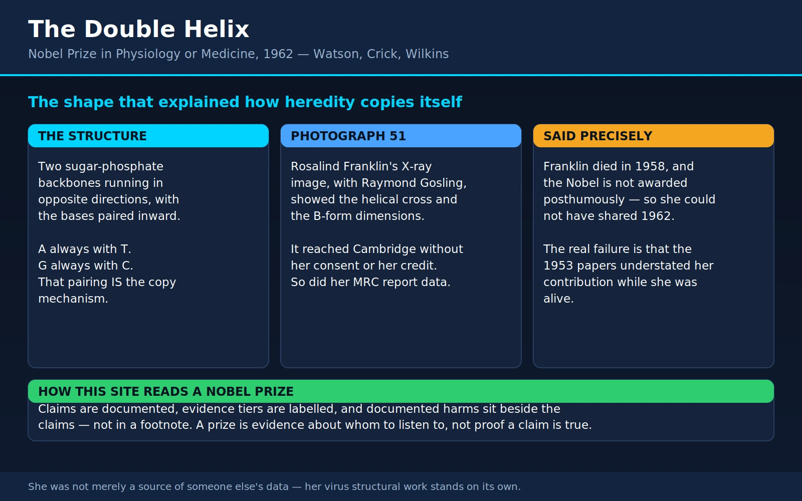
Table of Contents
- The Prize and the Four People
- What Was Actually Unknown in 1950
- Franklin's Photographs
- How Watson and Crick Saw the Data
- The Model, February 1953
- The Three Papers
- Pauling's Near-Miss
- What the Structure Explains
- What It Built, for You
- Franklin's Own Science, Beyond Photo 51
- Watson's Later Conduct, Stated Plainly
- Where Mainstream Science Agrees — and What the Record Complicates
- Key Research Papers
- Connections
- Featured Videos
1. The Prize and the Four People
The 1962 Nobel Prize in Physiology or Medicine went to three men — Francis Crick, James Watson and Maurice Wilkins — "for their discoveries concerning the molecular structure of nucleic acids and its significance for information transfer in living material." That is the most consequential structural discovery in the history of biology: the shape of DNA, and why its shape is the reason heredity works.
The fourth person was Rosalind Franklin, and almost everyone who hears this story hears it wrong in the same specific way. Franklin, born in London in 1920, was a physical chemist and X-ray crystallographer of unusual skill. From January 1951 to March 1953 she worked on DNA at King's College London, and the photographs she produced there — above all Photograph 51 — together with her measurements of the molecule's dimensions, are the experimental foundation the double helix rests on. She died of ovarian cancer on 16 April 1958, aged 37.
The Nobel Prize is not awarded posthumously. Franklin had been dead four years when the 1962 prize was announced. She therefore could not have shared it — not because a committee overlooked her, but because the statutes made her ineligible. (A second statute caps a prize at three recipients, which would have been a separate problem had she lived; it is not the reason she was left out.) This is the point garbled in almost every retelling, and getting it right matters, because the actual injustice is a different one and does not need embellishment.
The real question is whether Franklin would have been properly credited had she lived — and the honest answer is that the record from 1953, when she was very much alive, already understated her contribution. The paper announcing the double helix thanked the King's group in general terms; it did not say that its authors had seen her best photograph, or read her unpublished data, or that the model was built to fit measurements she had made. That was a choice made by living people, and it is the thing worth examining.
The principals: Crick (1916–2004), a physicist retrained in biology at Cambridge's Cavendish Laboratory, then 36 and without a doctorate; Watson (born 1928), an American phage biologist of 24; Wilkins (1916–2004), a New Zealand-born physicist who began the King's X-ray work on DNA before Franklin arrived; Franklin (1920–1958), fresh from four years in Paris; and Raymond Gosling (1926–2015), the student who exposed Photograph 51 under her direction and co-authored her 1953 paper.
2. What Was Actually Unknown in 1950
Today "DNA carries your genes" is something children learn before long division. In 1950 it was a recent and still-contested claim, and the molecule's shape was a complete blank. Most biochemists had assumed the hereditary material was protein, and the reasoning was not stupid: proteins are built from twenty amino acids and fold into endlessly varied shapes, while DNA has only four bases and, under the then-popular "tetranucleotide hypothesis," was thought to be a monotonous repeating chain — too boring to encode a nose or a blood type.
Two experiments dismantled that. In 1944, Oswald Avery, Colin MacLeod and Maclyn McCarty at the Rockefeller Institute ran down the "transforming principle" — the substance that converts harmless pneumococcus into virulent pneumococcus, an effect Frederick Griffith had reported in 1928. Destroying the proteins with proteases and the RNA with ribonuclease left the activity intact; destroying the DNA with deoxyribonuclease abolished it. The transforming principle was DNA — a result much of the field declined to believe.
In 1952, Alfred Hershey and Martha Chase closed the argument with an experiment of brutal simplicity. They grew bacteriophage — viruses that infect bacteria — in radioactive sulfur (which labels protein) or radioactive phosphorus (which labels DNA), let the phage infect E. coli, knocked the empty viral coats off in a kitchen blender, and spun the mixture down. The phosphorus went into the bacteria and appeared in the next generation of virus; the sulfur stayed outside in the discarded coats. Whatever carried the instructions was DNA.
Meanwhile Erwin Chargaff at Columbia had measured the base composition of DNA from different organisms and found two things. The proportions of the four bases varied from species to species — so DNA was not monotonous. And in every sample, adenine matched thymine and guanine matched cytosine. A = T. G = C. Chargaff reported it as an unexplained regularity, and it sat there for three years like a key with no lock.
So by 1950 the question had sharpened to a point. DNA carries heredity — what does it look like? That was not idle curiosity, because of a constraint any candidate must satisfy: a molecule that carries information must be able to be copied. Every time a cell divides the instructions have to be duplicated faithfully, and no proposed structure for DNA had ever suggested how. The structure was not decoration on the biology. The structure was the biology, if you could find one that copied itself.
3. Franklin's Photographs
Franklin came to King's in January 1951 from Paris, where she had spent four years with Jacques Mering learning to get diffraction data out of disordered, badly behaved materials — a harder craft than crystallography on a clean crystal, and exactly what DNA required. In X-ray fiber diffraction you draw a fiber, aim a narrow X-ray beam at it, and record the pattern the scattered rays make on film behind. You never see the molecule — only spots and arcs, from which, with mathematics and considerable judgement, its dimensions and symmetry can be reconstructed.
Her first major result was not a photograph but a discovery about the sample. With Raymond Gosling, she established that DNA fibers exist in two distinct forms depending on water content: a drier, crystalline "A" form, and a hydrated "B" form appearing above roughly 92 percent relative humidity. Earlier workers had been photographing uncontrolled mixtures of the two and getting uninterpretable patterns — two structures superimposed. Franklin's humidity-controlled setup produced each form cleanly, and without that nothing that followed was possible.
On 2 May 1952, Gosling exposed a B-form fiber for over sixty hours under Franklin's direction. The result, filed as Photograph 51, is the most famous X-ray image ever taken. Its pattern is a broad X, a cross of smeared spots radiating from the centre — and to a diffractionist that X is not suggestive but diagnostic. A helix scatters X-rays into precisely that cross, and the angle of its arms gives the ratio of pitch to diameter. Photograph 51 does not hint at a helix. It states one.
More than shape was readable in it. The layer-line spacing gives the pitch — about 34 Ångströms per turn; a heavy band on the meridian gives the spacing between stacked bases, about 3.4 Å, hence roughly ten bases per turn; the pattern's width gives a diameter near 20 Å. And a conspicuous absence — the missing fourth layer line — is the signature of two strands offset from each other along the axis. From the water content and the A form's symmetry she had also concluded that the phosphate backbone is on the outside, with the bases turned inward, which eliminated most competing models. She presented all of this at a King's colloquium in November 1951 — a talk Watson attended and, by his own admission, took poor notes at.
What she did not do was build a model, and this is the part most often told against her. Franklin was working through a Patterson function analysis of the A form — a rigorous, punishing method that extracts structure from the data itself rather than guessing a structure and checking the fit. It is the slower road and the safer one. Her notes from February 1952 record that the evidence suggested a helix; her documented skepticism concerned whether the crystalline A form was helical, not the B form, and on that narrow question she was being properly cautious about an ambiguous pattern. Refusing to publish a model your data does not yet compel is not timidity. It is the definition of good science. It is also, in a race, a way to lose.
4. How Watson and Crick Saw the Data
The popular version — "Watson stole Franklin's photograph" — is close enough to true to be repeated forever and wrong enough in its details to be dismissed by anyone who checks. Here is what the record supports. Watson and Crick were sixty miles away at the Cavendish and were not doing experiments on DNA at all: they were model builders, and their method, borrowed from Pauling, was to take other people's measurements, add the known bond lengths and angles of chemistry, and build metal models until something snapped into place that satisfied every constraint at once. That method is only as good as the measurements it is fed. They needed the King's data, and they got it twice, both times without Franklin's knowledge.
First, the photograph. On 30 January 1953 Watson travelled to King's, and in the course of the visit Wilkins showed him Photograph 51. Wilkins had the print legitimately: Franklin was leaving for Birkbeck, and Gosling — whose supervision was reverting to Wilkins — had passed it to him. Wilkins was not stealing it from a drawer. He was also not entitled to show it to a competitor at another laboratory, and he did not ask Franklin, whose photograph it was. Watson later wrote that his mouth fell open and his pulse began to race, because the X told him at once that the structure was a helix and roughly what size.
Second, the numbers. In December 1952 the King's group submitted a routine progress report to the Medical Research Council containing Franklin's B-form measurements and her analysis of the A form. It was not marked confidential and circulated to the MRC's biophysics committee — on which sat Max Perutz of the Cavendish, who passed it to Crick and Watson. What Crick saw there was arguably worth more than the photograph: Franklin had assigned the A form to the C2 monoclinic space group. Crick had spent years on that symmetry in hemoglobin and saw at once what it implied — a two-fold axis perpendicular to the molecule's length, meaning the two chains must run in opposite directions. Antiparallel: one of the two central features of the correct structure, straight out of Franklin's unpublished analysis. Perutz, later called to account, said the report bore no confidentiality marking and that he had been naive about etiquette — probably true, and beside the point.
So: neither act was theft in the legal sense. Wilkins owned a print he had been given; the MRC report was an unclassified internal document seen by a committee member. No lock was picked and no law was broken. And both were taken without her consent and used without her credit — a competitor at a rival institution obtained her best experimental result and her key unpublished analysis, built the winning model out of them, and thanked the King's group in general terms without saying what those results were or how central they had been.
Readers will weigh that differently depending on what they think scientific competition is for. What is not available is the comfortable position that nothing improper happened, or the equally comfortable one that Watson and Crick took what was not theirs and contributed nothing. Both are false.
5. The Model, February 1953
In the last week of February 1953 the pieces went together. The structure has two sugar-phosphate backbones running in opposite directions — antiparallel — twisting around a common axis, with the flat bases stacked inward like the treads of a spiral staircase. Ten base pairs per turn, 34 Å per turn, about 20 Å across: Franklin's numbers.
The unlock was chemical, and it came from a visitor. Jerry Donohue, an American structural chemist on sabbatical from Pauling's laboratory, shared their office. Watson had been trying to pair each base with another of the same kind, using the structures drawn in the textbooks. Donohue told him the textbooks were wrong: guanine and thymine were drawn in their enol tautomeric forms, when in reality the keto forms overwhelmingly predominate — and those present a different pattern of hydrogen-bond donors and acceptors. Watson redrew the bases as cardboard cutouts in the corrected forms, and the pairing fell out at once: adenine bonds to thymine (two hydrogen bonds), guanine to cytosine (three).
Two things happened at once. Chargaff's rules stopped being a curiosity and became an explanation: A equals T and G equals C in every organism because every A is bonded to a T on the other strand, and every G to a C. And — the feature that makes DNA capable of being a code at all — the two pairs are the same width. A purine always pairs with a pyrimidine, so an A–T rung and a G–C rung occupy the same span. The backbone therefore does not care what sequence it carries; any order of bases threads through the same regular helix without distorting it. That is why a molecule with four letters can hold unlimited information: the structure imposes no constraint on the message.
And each strand specifies the other completely: given one sequence, the partner is not merely compatible but determined. Watson and Crick put that in the paper's last paragraph, in what has become the most celebrated understatement in the scientific literature:
"It has not escaped our notice that the specific pairing we have postulated immediately suggests a possible copying mechanism for the genetic material."
Crick reportedly announced in the Eagle pub that they had found the secret of life; the Nature paper said it more quietly, and more effectively. Its diagram — the double ribbon everyone recognises — was drawn by Crick's wife, Odile Crick.
6. The Three Papers
Nature published the double helix on 25 April 1953, as three papers in sequence: Watson and Crick on pages 737–738 — the model, roughly 900 words and one figure; Wilkins, Stokes and Wilson on 738–740 — King's evidence that DNA is helical in intact biological material as well as in prepared fibers; and Franklin and Gosling on 740–741 — the B-form diffraction analysis, including Photograph 51.
The arrangement was brokered between the two laboratories and on its face looks generous: the King's groups got their own papers rather than an acknowledgment. In practice the ordering did something else. Model first, data second frames the sequence as hypothesis followed by supporting evidence, and a reader in 1953 — and every reader since — comes away believing the Cambridge model came first and the London photographs confirmed it.
The chronology was the other way around. Franklin's paper contains the data the model was built on, and her manuscript was substantially drafted before she saw the Watson–Crick structure. It is not a confirmation of their model; it is the independent presentation of the evidence that made their model possible. She revised it to add a line that her general ideas were "not inconsistent with the model proposed by Watson and Crick in the preceding communication" — a courteous sentence that has done more than any other to fix the wrong order in everyone's memory.
The Watson–Crick acknowledgment reads: "We have also been stimulated by a knowledge of the general nature of the unpublished experimental results and ideas of Dr M. H. F. Wilkins, Dr R. E. Franklin and their co-workers at King's College, London." Every word is technically accurate. It is also, as the historians Matthew Cobb and Nathaniel Comfort put it in 2023, misleading by omission — because "a knowledge of the general nature" is a remarkable way to describe having seen the photograph and read the numbers.
7. Pauling's Near-Miss
The reason February 1953 felt like a race was Linus Pauling, the most formidable structural chemist alive, who in 1951 had solved the alpha helix — the fundamental folding motif of proteins — by reasoning from the geometry of the peptide bond rather than from a diffraction pattern. In February 1953, weeks before the Cambridge model, he and Robert Corey published a proposed structure for DNA: a triple helix with the phosphate backbones on the inside and the bases pointing outward. It was wrong in a way that startled everyone who read it — to fit the phosphates into the crowded core he had left them un-ionised, so his proposed nucleic acid was not an acid. Watson and Crick, tipped off by Pauling's son Peter at the Cavendish, reckoned they had six weeks before he noticed. Why did the best structural chemist in the world get it inside out? He was working almost blind: his X-ray data on DNA were poor, and in 1952, in the thick of the McCarthy period, the US State Department refused to renew his passport over his anti-nuclear-testing activism, so he could not travel to London, where a Royal Society meeting would have put the King's diffraction work in front of him. The lesson is useful and not comfortable — brilliance without the right data loses to competence with it. Watson and Crick were not better chemists. They had Franklin's numbers and he did not.
8. What the Structure Explains
A correct structure is worth something only if it answers questions it was not designed to answer. This one did, repeatedly, for fifty years.
Replication. The mechanism they declined to spell out is the obvious one: unzip the strands, and each is a template for rebuilding its partner, because each base specifies exactly one opposite number. Every new molecule ends up with one old strand and one new — semi-conservative replication. In 1958 Matthew Meselson and Franklin Stahl confirmed that prediction with what is still called the most beautiful experiment in biology: they grew E. coli in heavy nitrogen (15N) until all its DNA was dense, switched to ordinary nitrogen, and spun the DNA in a caesium chloride density gradient at each generation. After one division every molecule was of exactly intermediate density; after two, half intermediate and half fully light — the pattern semi-conservative replication predicts, and the only pattern it predicts.
Information. If the backbone is indifferent to sequence, the sequence is free to mean something. By the mid-1960s the answer was in hand: bases are read in triplets, each specifying one amino acid, transcribed into RNA and translated into protein. The structure did not reveal the code, but it made a code conceivable.
Mutation. In a second Nature paper that May — "Genetical implications of the structure of deoxyribonucleic acid" — they noted that if a base briefly flipped into its rare tautomeric form during copying, it would pair with the wrong partner and the error would be inherited. A mutation is a change in sequence. Sickle cell disease, cystic fibrosis, Huntington's disease, the driver mutations in a tumour: all became specific misspellings in a specific text, findable and eventually readable.
And a lesson about method. "How does heredity work?" is a biological question, and its answer turned out to be a shape. Complementarity is not a metaphor but geometry — a hydrogen-bond donor sitting where an acceptor can reach it, a purine's bulk exactly offsetting a pyrimidine's slimness. This was the moment biology became, in substantial part, structural chemistry.
9. What It Built, for You
This is not distant history. Base pairing — A to T, G to C — is the operating principle behind a long list of things an ordinary person meets at a clinic.
- PCR tests. Every polymerase chain reaction test — COVID, influenza, strep, chlamydia, tuberculosis, hepatitis C viral load — uses a short primer complementary to a stretch of the target's genome, which finds that target and nothing else because base pairing is specific. That is the double helix used as a search engine.
- Newborn screening. The heel-prick card taken in a baby's first days is now substantially genetic; the conditions added most recently — spinal muscular atrophy, severe combined immunodeficiency — are caught by reading DNA directly, and finding them early is the difference between treatment and irreversible damage.
- Cancer risk testing. BRCA1 and BRCA2 testing tells a woman with a strong family history whether she carries a mutation raising her lifetime risk of breast cancer and ovarian cancer, turning anxiety into a prevention plan. Lynch syndrome testing, which reads the DNA mismatch-repair genes, identifies families needing colorectal cancer screening decades earlier than everyone else.
- Pharmacogenomics. Whether a drug helps you, does nothing, or poisons you can depend on which version of a metabolising enzyme you inherited. Clopidogrel needs CYP2C19 to activate it; DPYD variants make fluorouracil and capecitabine dangerously toxic, and TPMT and NUDT15 variants do the same for thiopurines. HLA-B*57:01 predicts a severe reaction to abacavir, and testing before prescribing has essentially abolished it.
- Ancestry and consumer DNA tests. Their genotyping chips read hundreds of thousands of single-letter variants by letting your DNA pair with immobilised probes. The population inferences drawn from that are far softer than the marketing implies, but the chemistry is straight base pairing.
- mRNA vaccine design. The SARS-CoV-2 spike sequence was published in January 2020 and vaccine sequences were designed within days — before anyone in the West had a physical sample of the virus. Turning that into a medicine took Katalin Karikó and Drew Weissman's discovery that modified nucleosides let synthetic RNA into a cell without triggering a destructive immune alarm. But the starting point was a text file.
- CRISPR. The gene-editing system approved in late 2023 for sickle cell disease and beta thalassemia is steered to its target by a short guide RNA that finds the right spot in three billion letters by base-pairing with it. The cutting enzyme is the easy part; the address system is the double helix.
None of this existed in 1962. All of it descends from a structure worked out on metal plates in a Cambridge office, from photographs taken in London.
10. Franklin's Own Science, Beyond Photo 51
There is a version of this story in which Franklin appears only as a source of data — the woman whose photograph was shown to the wrong person. That version is well-meant, and it is another way of erasing her, because it defines her by an episode in someone else's discovery. Her career stands on its own and would stand without DNA in it at all.
Coal and carbon (1942–1947). Her doctoral and wartime work, for the British Coal Utilisation Research Association, was on the physical structure of coals and carbons — their porosity, and how pore size determines which gas molecules can get in. It fed straight into gas-mask design, and produced a result of lasting importance: she showed that carbons divide into two classes, those that convert to graphite on heating and those that never do, and traced the difference to the arrangement of crystallites in the raw material. Those papers are still cited by people designing carbon fibers, battery electrodes and activated-carbon filters — fields with no idea they are reading a DNA scientist. In Paris (1947–1950), with Jacques Mering, she learned the diffraction technique behind Photograph 51.
Viruses, at Birkbeck (1953–1958). Franklin left King's in March 1953 for J. D. Bernal's laboratory at Birkbeck College and turned to tobacco mosaic virus, then the standard model organism of structural virology. In five years, with a small group and precarious funding, she established its architecture: a helical stack of identical protein subunits forming a rod that is hollow along its central axis — contradicting the assumption of a solid core — with the viral RNA embedded within the protein at a fixed radius rather than free in the middle, following the same helical path. That was the first complete description of how any virus is assembled. She built a five-foot model of it for the 1958 Brussels World's Fair; it went on display the day after she died.
Her group had by then started on poliovirus — far harder, because polio is spherical and its crystals are hazardous to handle. After her death Aaron Klug and John Finch carried it on and published the polio structure the following year. Klug, who joined her group in 1954 and became her closest collaborator, won the 1982 Nobel Prize in Chemistry for determining the structures of nucleic acid–protein complexes — work that grew out of the Birkbeck laboratory Franklin built. He also spent much of his life setting the DNA record straight, from her notebooks.
She was 37 when she died in April 1958, having worked between operations almost to the end. Whether her heavy occupational X-ray exposure contributed cannot be established; she also came from an Ashkenazi Jewish family, a population with an elevated frequency of inherited BRCA1 and BRCA2 mutations — plausible, entirely unproven. The irony needs no decoration: the genetic testing that identifies exactly that risk today exists because of the structure her photographs revealed.
11. Watson's Later Conduct, Stated Plainly
This site does not launder records, so this belongs on the page. James Watson led the Cold Spring Harbor Laboratory for decades and turned it into one of the world's leading centres of molecular biology, and was the first director of the NIH's Human Genome Project. He has also, repeatedly and publicly, asserted that Black people are innately less intelligent than white people.
In 2007 he told a British newspaper that he was "inherently gloomy about the prospect of Africa" because Western social policy assumes "their intelligence is the same as ours — whereas all the testing says not really." Cold Spring Harbor suspended him and he resigned as chancellor. In a 2019 documentary he said he had not changed his views, attributing the average difference in measured IQ between Black and white Americans to genetics. Cold Spring Harbor then revoked his remaining honorary titles — Chancellor Emeritus, Oliver R. Grace Professor Emeritus, Honorary Trustee — describing the statements as reprehensible and unsupported by science.
The claim has no scientific support. Human genetic variation does not partition into the categories invoked: most of it lies within any so-called racial group rather than between groups, and the differences that do track ancestry do not map onto the trait in question. Measured IQ gaps track schooling, nutrition, lead exposure, poverty and test familiarity, and have narrowed within living memory — not how a fixed genetic difference behaves. Sequencing the human genome, a project Watson himself launched, is a principal reason we know the claim is wrong.
His 1968 memoir The Double Helix is part of the same record. Its portrait of "Rosy" is condescending in a way that reads worse every decade — remarks on her clothes and manner, a depiction of her as an obstacle who did not understand her own results. Crick and Wilkins both objected, and Harvard University Press dropped the book before another publisher took it. Watson's epilogue conceding that his impressions had often been wrong is genuine, and it does not undo the several hundred preceding pages that shaped how two generations pictured her.
None of this unmakes 1953. The double helix is correct; the model was real intellectual work; the discovery reorganised biology. A person can do something extraordinary and later say things that are false and harmful, and the honest response is to record both.
12. Where Mainstream Science Agrees — and What the Record Complicates
Not in dispute
- The structure is right, confirmed by every technique invented since — crystallography, NMR, cryo-electron microscopy, sequencing itself.
- Watson and Crick built it. Having the right data is not the same as recognising what it means: the antiparallel arrangement, the keto-form pairing, and the leap from Chargaff's ratios to a copying mechanism were genuine acts of insight.
- Franklin's data was essential, and no serious historian argues otherwise. The pitch, the rise per base, the diameter, the outside phosphates and the C2 symmetry that implied antiparallel chains came from King's, and mostly from Franklin.
What the record complicates
- The 1953 acknowledgment was misleading by omission. "A knowledge of the general nature of the unpublished experimental results" does not describe having seen the photograph and read the report. This is the substantive charge, and it holds.
- The Nobel question is not the interesting one. Franklin was ineligible in 1962 because she was dead. Arguing about the prize avoids the harder question of how she was treated in 1953, when it could still have been put right.
- "Franklin didn't realise what she had" is false, and Cobb and Comfort's 2023 Nature reassessment is the best correction of it. Drawing on an unpublished draft news article by the journalist Joan Bruce and on contemporary correspondence, they show that Franklin grasped the significance of her results and was working, in effect, within a two-laboratory effort on one problem. She was a peer in a race, treated as a resource.
- The "stolen photograph" version is too simple, and framing it as a heist invites easy rebuttal — letting the real failure, consent and credit, slip away in the argument.
- Wilkins is flattened in most tellings. He started the King's DNA work, produced the first good oriented fibers, and shared a real Nobel for real contributions — and he showed a colleague's photograph to a competitor. The conflict was partly institutional: the department head's ambiguous instructions had left Franklin and Wilkins each believing the DNA project was theirs, poisoning a collaboration that might have solved the structure at King's.
The version worth telling is not a morality play with a villain and a martyr. It is a story about how discovery happens: distributed across laboratories, dependent on other people's measurements, decided partly by temperament, and written up afterwards by the winners, whose account of who contributed what is the one that gets remembered. The double helix is a triumph; the way credit for it was assigned is a caution. Both are permanent parts of the record.
13. Key Research Papers
- Watson JD, Crick FHC. Molecular structure of nucleic acids; a structure for deoxyribose nucleic acid. Nature 1953;171(4356):737-8
- Franklin RE, Gosling RG. Molecular configuration in sodium thymonucleate. Nature 1953;171(4356):740-1
- Wilkins MHF, Stokes AR, Wilson HR. Molecular structure of deoxypentose nucleic acids. Nature 1953;171(4356):738-40
- Avery OT, MacLeod CM, McCarty M. Studies on the chemical nature of the substance inducing transformation of pneumococcal types. J Exp Med 1944;79(2):137-58
- Hershey AD, Chase M. Independent functions of viral protein and nucleic acid in growth of bacteriophage. J Gen Physiol 1952;36(1):39-56
- Meselson M, Stahl FW. The replication of DNA in Escherichia coli. Proc Natl Acad Sci U S A 1958;44(7):671-82
- Klug A. Rosalind Franklin and the discovery of the structure of DNA. Nature 1968;219(5156):808-10
- Cobb M, Comfort N. What Rosalind Franklin truly contributed to the discovery of DNA's structure. Nature 2023;616(7958):657-60
- Franklin RE, Holmes KC. The helical arrangement of the protein subunits in tobacco mosaic virus. Biochim Biophys Acta 1956;21(2):405-6
Live PubMed Searches
- DNA double helix structure history
- Rosalind Franklin photograph 51
- Semiconservative DNA replication
- DNA structure discovery credit
- Tobacco mosaic virus structure Franklin
Connections
- All Notable Doctors
- Nobel Prize in Physiology or Medicine — the full roll, and how the prize's rules work
- Prizes That Aged Badly — its credit-disputes section covers the Franklin case
- Linus Pauling — the rival racing them, whose triple helix was not even an acid
- Katalin Karikó & Drew Weissman — mRNA vaccines designed from a published sequence
- Blackburn, Greider & Szostak — telomeres, the repeated DNA caps at the ends of chromosomes
- Shinya Yamanaka — four genes that reset an adult cell: same genome, different readout
- Genetics — inherited conditions, and what testing can tell you
- Ovarian Cancer — the disease that killed Franklin at 37; risk and BRCA management
- Breast Cancer — where BRCA testing changes screening and prevention decisions
- Colorectal Cancer — Lynch syndrome and why family history moves the screening age
- Cancer — how sequence changes become disease