Calcium – Essential Mineral for Human Health
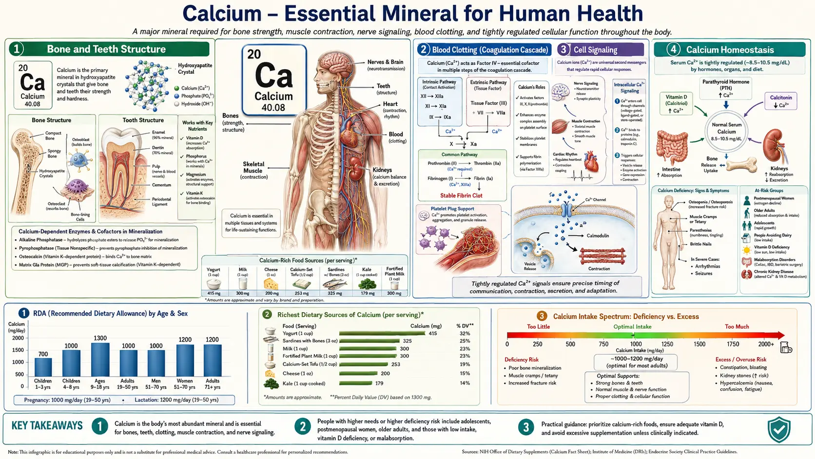
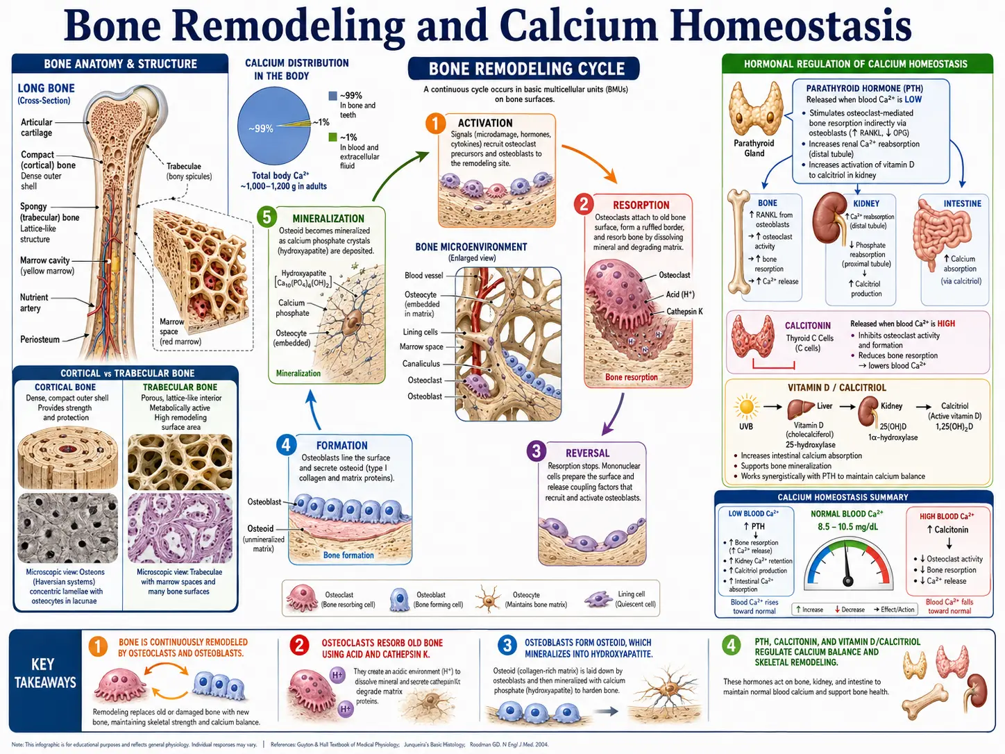
Calcium is the most abundant mineral in the human body; a 1990 clinical reference puts the adult total at about 1,200 grams (Goldstein 1990). The NIH Office of Dietary Supplements (ODS) states that almost all of it (98%) is stored in the bones and that more than 99% is in the form of hydroxyapatite in bones and teeth, where it provides structural integrity. The small remainder, under 1%, circulates in the blood, resides in extracellular fluid, and is distributed within cells, where it performs a remarkably diverse array of physiological functions. Maintaining precise calcium concentrations in these compartments is critical to survival, and the body employs a sophisticated hormonal regulatory system to achieve this balance.
Interactive Visualization Bone Remodeling & Calcium Balance Watch osteoclasts tunnel and osteoblasts rebuild — then drop vitamin D and see PTH quietly strip calcium out of your skeleton. Launch → Interactive Visualization Magnesium — the mineral that makes ATP work Bare ATP is useless and enzymes reject it. Watch magnesium clamp the phosphates and switch it on, plug the NMDA channel, and let a cramping muscle finally let go. Launch → Interactive Visualization Calcium & PTH — defend the blood calcium level Watch four tiny parathyroid glands pull calcium from bone, kidney and gut with PTH and vitamin D — then drop your vitamin D and see PTH climb and quietly rob the skeleton. Launch → Interactive Visualization Fracture Healing — rebuild a broken bone Follow a break through its four stages — blood clot, soft cartilage callus, hard bony bridge, then the long remodel back to normal — and see what smoking, movement or missing calcium do to it. Launch → Interactive Visualization Kidney Stones — grow one, then prevent it Watch minerals in concentrated urine crystallize and clump into a stone — then feel it drop into the ureter and block it, and learn why water and dietary calcium actually prevent them. Launch → Interactive Visualization How Cavities Form (and How to Stop Them) Feed the plaque bacteria sugar and watch the acid dissolve enamel below the surface — then brush with fluoride and watch the tooth re-harden before a cavity breaks through. Launch →Table of Contents
- Bone and Teeth Structure
- Muscle Contraction
- Nerve Transmission
- Blood Clotting (Coagulation Cascade)
- Enzyme Activation
- Hormone Secretion
- Cell Signaling
- Cardiovascular Function
- Acid-Base Balance
- Calcium Homeostasis
- Intake, Food Sources and Supplements
- Deficiency and Excess
- Key Research Papers
- Connections
- Featured Videos
Bone and Teeth Structure
Calcium serves as the principal structural component of the skeletal system and dentition. Within bone and tooth enamel, calcium combines with phosphate to form crystalline hydroxyapatite (Ca10(PO4)6(OH)2), a mineral lattice that confers rigidity and compressive strength.
- Hydroxyapatite – Bone is roughly 70% inorganic mineral by weight, mainly this calcium-phosphate crystal, and about 30% organic matrix, mainly type I collagen (Venkatesan 2014). The hydroxyapatite lattice is deposited within a collagen matrix, creating a composite material that is both hard and somewhat flexible, allowing bones to resist fracture under mechanical stress.
- Bone as a calcium reservoir – Beyond its structural role, the skeleton functions as a dynamic reservoir from which calcium can be mobilized into the bloodstream when dietary intake is insufficient. This process is tightly regulated to prevent excessive bone resorption.
- Dental enamel – Tooth enamel is the hardest and most highly mineralized tissue in the body, with nearly 96% inorganic content (mainly hydroxyapatite) and about 4% organic material and water (Pushpalatha 2023). Enamel mineralizes while the tooth develops; unlike bone, teeth are not continuously remodeled afterwards (ODS).
- Dentin and cementum – Beneath enamel, dentin is about 65 to 70% mineral by weight, depending on the source (Pushpalatha 2023 gives 65% inorganic content; Grawish 2022 gives 70% hydroxyapatite, 20% organic matrix and 10% water). Cementum, which covers the tooth root, is also a calcium-phosphate mineralized tissue.
Muscle Contraction
Calcium ions (Ca2+) are indispensable for the contraction of all three types of muscle tissue: skeletal, cardiac, and smooth muscle. The mechanism by which calcium triggers contraction is known as excitation-contraction coupling.
- Skeletal muscle – When a motor neuron fires an action potential, calcium is released from the sarcoplasmic reticulum into the cytoplasm. Calcium binds to troponin C on the thin filament, causing a conformational change that moves tropomyosin and exposes myosin-binding sites on actin. Cross-bridge cycling then produces force and shortening.
- Cardiac muscle – Calcium enters cardiac myocytes through L-type voltage-gated calcium channels during the plateau phase of the action potential. This extracellular calcium triggers a much larger release from the sarcoplasmic reticulum via ryanodine receptors, a process called calcium-induced calcium release (CICR).
- Smooth muscle – In smooth muscle, calcium binds to calmodulin rather than troponin. The calcium-calmodulin complex activates myosin light-chain kinase (MLCK), which phosphorylates the myosin regulatory light chain and permits cross-bridge cycling.
Nerve Transmission
Calcium plays a pivotal role in synaptic transmission, the process by which neurons communicate with one another and with effector cells such as muscle fibers and glandular cells.
- Neurotransmitter release – When an action potential arrives at the presynaptic terminal, voltage-gated calcium channels open, allowing Ca2+ to flow into the terminal. The rise in intracellular calcium triggers the fusion of synaptic vesicles with the presynaptic membrane, releasing neurotransmitters such as acetylcholine, norepinephrine, and glutamate into the synaptic cleft.
- Synaptotagmin – Calcium binds to synaptotagmin, a sensor protein on synaptic vesicles, which then interacts with SNARE proteins to catalyze membrane fusion and exocytosis.
- Neural excitability – Extracellular calcium concentration influences the threshold for neuronal firing. Low calcium levels (hypocalcemia) increase neural excitability, which can manifest as tingling, numbness, and in severe cases, tetany or seizures.
Blood Clotting (Coagulation Cascade)
Calcium is designated as Factor IV in the coagulation cascade and is required at multiple steps in the clotting process. Without adequate ionized calcium, blood cannot coagulate properly.
- Gamma-carboxyglutamate (Gla) domains – Several clotting factors (II, VII, IX, and X) contain Gla domains that require calcium ions to bind to phospholipid surfaces on activated platelets. This membrane binding is essential for the assembly of the tenase and prothrombinase complexes.
- Intrinsic and extrinsic pathways – Calcium participates in both the intrinsic pathway (involving factors XII, XI, IX, and VIII) and the extrinsic pathway (involving tissue factor and factor VII). It is also required for the common pathway, where prothrombin is converted to thrombin.
- Fibrin stabilization – Factor XIII, a transglutaminase activated by thrombin in the presence of calcium, cross-links fibrin monomers to form a stable, insoluble clot.
- Clinical relevance – Chelating agents such as EDTA and citrate prevent coagulation in blood collection tubes by binding calcium, underscoring its essential role in the clotting cascade.
Enzyme Activation
Calcium ions serve as cofactors or regulators for numerous enzymes throughout the body, influencing metabolic pathways in virtually every organ system.
- Protein kinase C (PKC) – Calcium, along with diacylglycerol (DAG), activates PKC, a family of enzymes that phosphorylate serine and threonine residues on target proteins. PKC is involved in cell proliferation, differentiation, and immune responses.
- Phospholipase A2 – This enzyme, which releases arachidonic acid from membrane phospholipids (a precursor for prostaglandins and leukotrienes), requires calcium for its activity.
- Calpains – These calcium-dependent cysteine proteases participate in cytoskeletal remodeling, cell migration, apoptosis, and signal transduction.
- Calcineurin – A calcium- and calmodulin-dependent serine/threonine phosphatase that plays a critical role in T-cell activation and immune function. It is the target of immunosuppressive drugs cyclosporine and tacrolimus.
Hormone Secretion
Calcium regulates the release of hormones from endocrine glands through mechanisms similar to neurotransmitter exocytosis.
- Insulin secretion – In pancreatic beta cells, glucose metabolism raises intracellular ATP, which closes ATP-sensitive potassium channels, depolarizes the cell membrane, and opens voltage-gated calcium channels. The resulting calcium influx triggers insulin granule exocytosis.
- Parathyroid hormone (PTH) – The calcium-sensing receptor (CaSR) on parathyroid cells detects extracellular calcium levels. When calcium falls, CaSR signaling decreases, permitting increased PTH secretion. When calcium is high, CaSR activation suppresses PTH release.
- Aldosterone – Angiotensin II stimulates aldosterone secretion from the adrenal zona glomerulosa partly through calcium signaling pathways.
- Catecholamines – Calcium influx into adrenal chromaffin cells is required for the exocytotic release of epinephrine and norepinephrine.
Cell Signaling
Calcium functions as one of the most versatile and ubiquitous second messengers in cellular signaling. The free calcium concentration in the cytoplasm of a resting cell is at the 10−7 M level (about 100 nanomolar), while outside the cell it is at the 10−3 M level (about 1 millimolar), a 10,000-fold gradient across the cell membrane (Sobue 2024). When a cell is stimulated, calcium entering through membrane channels or released from internal stores raises the cytoplasmic level to 10−6 to 10−5 M, and that transient rise is the signal (Sobue 2024).
- IP3-mediated calcium release – Many G-protein-coupled receptors activate phospholipase C, which cleaves PIP2 into IP3 and DAG. IP3 binds to receptors on the endoplasmic reticulum, releasing stored calcium into the cytoplasm.
- Calmodulin – This ubiquitous calcium-binding protein undergoes a conformational change upon binding four Ca2+ ions, enabling it to activate a wide array of downstream targets, including kinases, phosphatases, and ion channels.
- CaMKII (calcium/calmodulin-dependent protein kinase II) – A multifunctional kinase activated by calcium-calmodulin that plays key roles in synaptic plasticity, learning, memory, and cardiac function.
- Store-operated calcium entry (SOCE) – When intracellular calcium stores are depleted, the ER sensor STIM1 activates Orai1 channels in the plasma membrane, allowing sustained calcium entry from the extracellular space.
- Calcium oscillations – Many cell types exhibit rhythmic fluctuations in cytoplasmic calcium concentration. The frequency and amplitude of these oscillations encode information that determines specific cellular responses such as gene transcription, secretion, or contraction.
Cardiovascular Function
Beyond its role in cardiac muscle contraction, calcium influences multiple aspects of cardiovascular physiology.
- Cardiac pacemaker activity – Calcium currents contribute to the spontaneous depolarization of sinoatrial node cells, helping to establish and maintain heart rhythm. The "calcium clock" mechanism, involving rhythmic calcium release from the sarcoplasmic reticulum, works in concert with the membrane voltage clock.
- Vascular smooth muscle tone – Calcium influx through L-type calcium channels in vascular smooth muscle cells is a primary determinant of vessel tone and blood pressure. Calcium channel blockers (e.g., amlodipine, nifedipine) lower blood pressure by reducing this influx.
- Endothelial function – Calcium signaling in endothelial cells stimulates nitric oxide synthase (eNOS), promoting nitric oxide production and vasodilation.
- Cardiac action potential – The plateau phase (phase 2) of the ventricular action potential depends on inward calcium current through L-type channels, which sustains depolarization and allows sufficient time for ventricular contraction before repolarization.
Acid-Base Balance
Calcium metabolism is closely linked to the body's acid-base status, with changes in blood pH directly affecting calcium availability and function.
- pH and protein binding – About 40% of serum calcium is bound to proteins, predominantly albumin; roughly 50% is free (ionized) and about 9% is complexed with small anions such as phosphate, bicarbonate and citrate (Sahota, StatPearls). Other sources give slightly different splits; these are compared on the serum calcium test page. Alkalosis increases albumin's affinity for calcium, reducing the ionized (physiologically active) fraction and potentially causing symptoms of hypocalcemia even when total calcium is normal. Acidosis has the opposite effect, increasing ionized calcium.
- Bone buffering – Chronic metabolic acidosis stimulates calcium release from bone as the skeleton's alkaline mineral salts help buffer excess acid. Prolonged acidosis can therefore contribute to bone demineralization.
- Renal handling – The kidneys adjust calcium reabsorption in response to acid-base status. Acidosis tends to increase urinary calcium excretion, while alkalosis promotes calcium retention.
Calcium Homeostasis
The body maintains serum ionized calcium within a narrow range — MedlinePlus gives an example adult range of 1.20 to 1.40 mmol/L (4.8 to 5.6 mg/dL), and each laboratory prints its own — through a tightly coordinated hormonal system involving three primary regulators: parathyroid hormone, calcitonin, and active vitamin D (calcitriol).
Parathyroid Hormone (PTH)
- Stimulus – PTH is secreted by the four parathyroid glands in response to a fall in serum ionized calcium, detected by the calcium-sensing receptor (CaSR).
- Bone effects – PTH stimulates osteoclast-mediated bone resorption, releasing calcium and phosphate from the skeleton into the bloodstream.
- Renal effects – PTH increases calcium reabsorption in the distal convoluted tubule and thick ascending limb of Henle, reduces phosphate reabsorption in the proximal tubule (promoting phosphaturia), and stimulates the enzyme 1-alpha-hydroxylase, which converts 25-hydroxyvitamin D to its active form, 1,25-dihydroxyvitamin D (calcitriol).
- Net effect – PTH raises serum calcium and lowers serum phosphate.
Calcitonin
- Stimulus – Calcitonin is released by the parafollicular C cells of the thyroid gland in response to elevated serum calcium.
- Mechanism – Calcitonin inhibits osteoclast activity, reducing bone resorption and thereby lowering serum calcium. It also promotes renal calcium excretion.
- Physiological significance – The role of calcitonin in adult human calcium homeostasis is considered modest compared to PTH and vitamin D. However, it may be more important during periods of calcium stress such as pregnancy, lactation, and skeletal growth.
Vitamin D (Calcitriol)
- Activation pathway – Vitamin D3 (cholecalciferol) is synthesized in the skin upon UVB exposure or obtained from the diet. It is hydroxylated in the liver to 25-hydroxyvitamin D (calcidiol) and then in the kidney to the active hormone 1,25-dihydroxyvitamin D (calcitriol) by 1-alpha-hydroxylase, an enzyme stimulated by PTH.
- Intestinal absorption – Calcitriol's most important action is to increase intestinal absorption of calcium (and phosphate). It induces the expression of calbindin, a calcium-binding protein in enterocytes, and upregulates calcium transport channels (TRPV6) and basolateral calcium pumps.
- Bone effects – Calcitriol supports bone mineralization by maintaining adequate serum calcium and phosphate concentrations. At high levels, it can also stimulate osteoclast differentiation and bone resorption through the RANK/RANKL pathway.
- Renal effects – Calcitriol enhances calcium reabsorption in the distal nephron, working in concert with PTH.
Integrated Homeostatic Response
When serum calcium falls, PTH secretion increases within seconds to minutes. PTH rapidly mobilizes calcium from bone, conserves calcium at the kidney, and stimulates calcitriol production. Calcitriol then enhances intestinal calcium absorption over hours to days. When serum calcium rises above the set point, PTH secretion is suppressed, calcitonin secretion increases modestly, renal calcium excretion rises, and the system returns to equilibrium. This multi-organ, multi-hormone feedback loop ensures that calcium levels remain within the physiological range necessary for cellular function and survival.
Intake, Food Sources and Supplements
The U.S. reference values below were set by the Food and Nutrition Board of the National Academies and are reproduced in the NIH Office of Dietary Supplements (ODS) calcium fact sheet for health professionals (updated June 22, 2026). ODS defines the RDA as the average daily intake sufficient to meet the requirements of nearly all (97%–98%) healthy individuals, and the UL (Tolerable Upper Intake Level) as the maximum daily intake unlikely to cause adverse health effects. For infants the first figure is an Adequate Intake (AI), set when the evidence is insufficient to develop an RDA.
Recommended Intakes and Upper Limits (ODS)
- 0–6 months – AI 200 mg/day; UL 1,000 mg/day
- 7–12 months – AI 260 mg/day; UL 1,500 mg/day
- 1–3 years – RDA 700 mg/day; UL 2,500 mg/day
- 4–8 years – RDA 1,000 mg/day; UL 2,500 mg/day
- 9–13 years – RDA 1,300 mg/day; UL 3,000 mg/day
- 14–18 years (including pregnancy and lactation) – RDA 1,300 mg/day; UL 3,000 mg/day
- 19–50 years (including pregnancy and lactation) – RDA 1,000 mg/day; UL 2,500 mg/day
- 51–70 years – RDA 1,000 mg/day for men and 1,200 mg/day for women; UL 2,000 mg/day
- Over 70 years – RDA 1,200 mg/day; UL 2,000 mg/day
ODS notes that the ULs rest on observational evidence from the Women's Health Initiative (WHI) linking supplemental calcium with kidney stones, and that two later systematic reviews (one of 10 studies in more than 8,000 adults with osteoporosis, one of 11 trials in 51,419 adults aged 50 and older) found no such association.
Food Sources
ODS describes milk, yogurt and cheese as rich natural sources and states that about 72% of calcium intake in the United States comes from dairy products and foods with added dairy ingredients; nondairy sources include canned fish eaten with the bones and certain vegetables. Calcium per serving, from the ODS food table unless noted:
- Sardines, canned in oil, with bones, 3 ounces – 325 mg
- Milk, whole (3.25% milk fat), 1 cup – 276 mg
- Salmon, pink, canned, solids with bones, 3 ounces – 181 mg
- Turnip greens, fresh, boiled, ½ cup – 99 mg
- Kale, fresh, cooked, 1 cup – 94 mg
- Chia seeds, 1 tablespoon – 76 mg
- Chinese cabbage (bok choy), raw, shredded, 1 cup – 74 mg
- Natto (fermented soybeans) – 217 mg per 100 g (USDA FoodData Central, FDC ID 172443)
Food-by-food detail is on the calcium food sources page.
How Much Is Absorbed
- Intake and age – ODS states that absorption from food is about 45% at intakes of 200 mg/day but only 15% at intakes above 2,000 mg/day, and that net absorption is as high as 60% in infants and young children and about 25% in adults, declining further with age.
- Supplement forms – The two most common forms are calcium carbonate (40% calcium by weight) and calcium citrate (21% calcium). According to ODS, carbonate dissolves less well in people with low stomach acid unless it is taken with a meal, citrate depends less on stomach acid and can be absorbed without food, and absorption of supplements is generally greater with food either way.
- Amount per dose – ODS states that absorption from supplements is highest at doses of 500 mg or less; for example, about 36% of a 300 mg dose and 28% of a 1,000 mg dose is absorbed.
- Medication interaction – ODS reports that calcium carbonate can interfere with the absorption of levothyroxine, and that the drug's FDA-approved label instructs patients to separate the two by 4 hours.
Supplements and Cardiovascular Risk: A Disputed Signal
Whether calcium supplements raise heart-attack risk has been argued since at least 2010, and the published analyses point in both directions.
- Bolland 2010 (BMJ) – A meta-analysis of 15 placebo-controlled trials of calcium supplements given without vitamin D (500 mg/day or more). In the five trials with patient-level data (8,151 participants), 143 people allocated to calcium had a myocardial infarction against 111 on placebo (hazard ratio 1.31, 95% CI 1.02–1.67); the trial-level analysis gave a pooled relative risk of 1.27 (1.01–1.59). Increases in stroke, the composite end point and death were not statistically significant.
- Bolland 2011 (BMJ) – A reanalysis of the WHI calcium and vitamin D trial found that in the 16,718 women not taking their own calcium supplements at randomisation, hazard ratios for cardiovascular events ranged from 1.13 to 1.22; in a meta-analysis combining those women with eight trials of calcium supplements (28,072 participants in all), calcium with or without vitamin D was associated with a relative risk of 1.24 (1.07–1.45) for myocardial infarction and 1.15 (1.03–1.27) for myocardial infarction or stroke.
- Lewis 2015 (J Bone Miner Res) – A meta-analysis of 18 trials (63,563 participants) that counted coronary events in elderly women only when verified by clinical review, hospital record or death certificate (five trials contributed such events) found a relative risk of 1.02 (0.96–1.09) for coronary heart disease, 1.08 (0.92–1.26) for myocardial infarction and 0.96 (0.91–1.02) for all-cause mortality.
- Chung 2016 (Ann Intern Med) – A systematic review of 4 randomized trials and 27 observational studies concluded that calcium intake within the tolerable upper intake levels (2,000 to 2,500 mg/day) is not associated with cardiovascular risk in generally healthy adults. The accompanying National Osteoporosis Foundation and American Society for Preventive Cardiology guideline (Kopecky 2016) graded the evidence of no relationship as moderate quality.
- Myung 2021 (Nutrients) – A meta-analysis of 13 double-blind, placebo-controlled trials found calcium supplements associated with a 15% higher risk of cardiovascular disease (RR 1.15, 1.06–1.25) and 16% higher risk of coronary heart disease (RR 1.16, 1.05–1.28) in healthy postmenopausal women.
- WHI long-term follow-up (Thomson 2024) – A post hoc analysis after a median 22.3 years found cardiovascular mortality 6% higher with calcium plus vitamin D than with placebo (HR 1.06, 1.01–1.12), cancer mortality 7% lower (HR 0.93, 0.87–0.99), and no effect on all-cause mortality (HR 1.00, 0.97–1.03).
Fractures and Bone Density
- WHI calcium plus vitamin D trial (Jackson 2006) – 36,282 postmenopausal women aged 50 to 79 were randomly assigned to daily calcium carbonate with 400 IU of vitamin D3, or placebo, and followed for an average of 7.0 years. (The 2006 report gives the dose as 1,000 mg of elemental calcium; the 2024 follow-up report describes it as 1,000 mg of calcium carbonate, 400 mg elemental.) Hip bone density was 1.06% higher in the supplement group, but the hazard ratios were 0.88 (0.72–1.08) for hip fracture, 0.90 (0.74–1.10) for clinical spine fracture and 0.96 (0.91–1.02) for total fractures, none statistically significant. Kidney stones increased (HR 1.17, 1.02–1.34). When follow-up was censored at the point women stopped taking the study pills, the hip-fracture hazard ratio was 0.71 (0.52–0.97).
- Bolland 2015 (BMJ) – In this systematic review, most cohort studies found no association between dietary calcium, milk or dairy intake and fracture. Across 26 trials, calcium supplements were associated with fewer total fractures (RR 0.89, 0.81–0.96) and vertebral fractures (0.86, 0.74–1.00) but not hip (0.95, 0.76–1.18) or forearm fractures (0.96, 0.85–1.09); in the four trials at lowest risk of bias (44,505 participants) there was no effect at any site. The authors concluded that the evidence that supplements prevent fractures is weak and inconsistent.
- Tai 2015 (BMJ) – A meta-analysis of 59 trials found that more calcium from food or supplements raised bone mineral density by about 0.6–1.8%, with no further gain after the first year, which the authors judged unlikely to produce a clinically significant reduction in fractures.
- U.S. Preventive Services Task Force – As summarised by ODS, the Task Force concluded with moderate certainty that daily doses below 1,000 mg calcium and 400 IU vitamin D do not prevent fractures in postmenopausal women, and that evidence on larger doses, and on men and premenopausal women, is inadequate.
Kidney Stones: Food Calcium Versus Supplements
- Curhan 1993 (NEJM) – In 45,619 men aged 40 to 75 followed for four years (505 stones), those in the highest fifth of dietary calcium intake had a lower risk of symptomatic kidney stones than those in the lowest (relative risk 0.56, 0.43–0.73, after age adjustment; 0.66, 0.49–0.90, after adjustment for other risk factors).
- Curhan 1997 (Ann Intern Med) – In 91,731 women in the Nurses' Health Study followed for 12 years (864 stones), the highest fifth of dietary calcium intake carried a relative risk of 0.65 (0.50–0.83), while women taking calcium supplements had a relative risk of 1.20 (1.02–1.41) compared with non-users. In 67% of supplement users the calcium was not taken with a meal or was taken with low-oxalate meals; the authors suggested that calcium eaten with food binds dietary oxalate and lowers its absorption.
- WHI trial – Calcium plus vitamin D increased kidney stones (HR 1.17, Jackson 2006); the later systematic reviews summarised by ODS above did not find the association.
Decisions about supplements belong with a clinician.
Deficiency and Excess
Too little or too much calcium both cause problems. Low blood calcium can trigger tingling, muscle cramps, and in severe cases tetany or seizures, while high blood calcium can cause fatigue, kidney stones, confusion, and abnormal heart rhythms. For a full, patient-friendly guide to the symptoms, causes, and treatment of each, see Hypocalcemia (Low Calcium) and Hypercalcemia (High Calcium).
Key Research Papers
- Goldstein DA. Serum calcium. In: Walker HK, Hall WD, Hurst JW, editors. Clinical Methods: The History, Physical, and Laboratory Examinations. 3rd edition. Boston: Butterworths; 1990. Chapter 143. — PubMed PMID: 21250094
- Sahota RJ, Leslie SW, Sharma S. Physiology, Calcium. In: StatPearls. Treasure Island (FL): StatPearls Publishing; 2026 (updated 2026 Sep 14). — PubMed PMID: 29489276
- Venkatesan J, Nithya R, Sudha PN, Kim SK. Role of alginate in bone tissue engineering. Advances in Food and Nutrition Research. 2014;73:45–57. — PubMed PMID: 25300542
- Pushpalatha C, Gayathri VS, Sowmya SV, Augustine D, Alamoudi A, Zidane B, et al. Nanohydroxyapatite in dentistry: a comprehensive review. The Saudi Dental Journal. 2023;35(6):741–752. — PubMed PMID: 37817794
- Grawish ME, Grawish LM, Grawish HM, Grawish MM, Holiel AA, Sultan N, et al. Demineralized dentin matrix for dental and alveolar bone tissues regeneration: an innovative scope review. Tissue Engineering and Regenerative Medicine. 2022;19(4):687–701. — PubMed PMID: 35429315
- Sobue K. Calmodulin: a highly conserved and ubiquitous Ca2+ sensor. Proceedings of the Japan Academy, Series B. 2024;100(7):368–386. — PubMed PMID: 39085063
- Bolland MJ, Avenell A, Baron JA, Grey A, MacLennan GS, Gamble GD, Reid IR. Effect of calcium supplements on risk of myocardial infarction and cardiovascular events: meta-analysis. BMJ. 2010;341:c3691. — PubMed PMID: 20671013
- Bolland MJ, Grey A, Avenell A, Gamble GD, Reid IR. Calcium supplements with or without vitamin D and risk of cardiovascular events: reanalysis of the Women's Health Initiative limited access dataset and meta-analysis. BMJ. 2011;342:d2040. — PubMed PMID: 21505219
- Lewis JR, Radavelli-Bagatini S, Rejnmark L, Chen JS, Simpson JM, Lappe JM, et al. The effects of calcium supplementation on verified coronary heart disease hospitalization and death in postmenopausal women: a collaborative meta-analysis of randomized controlled trials. Journal of Bone and Mineral Research. 2015;30(1):165–175. — PubMed PMID: 25042841
- Chung M, Tang AM, Fu Z, Wang DD, Newberry SJ. Calcium intake and cardiovascular disease risk: an updated systematic review and meta-analysis. Annals of Internal Medicine. 2016;165(12):856–866. — PubMed PMID: 27776363
- Kopecky SL, Bauer DC, Gulati M, Nieves JW, Singer AJ, Toth PP, et al. Lack of evidence linking calcium with or without vitamin D supplementation to cardiovascular disease in generally healthy adults: a clinical guideline from the National Osteoporosis Foundation and the American Society for Preventive Cardiology. Annals of Internal Medicine. 2016;165(12):867–868. — PubMed PMID: 27776362
- Myung SK, Kim HB, Lee YJ, Choi YJ, Oh SW. Calcium supplements and risk of cardiovascular disease: a meta-analysis of clinical trials. Nutrients. 2021;13(2):368. — PubMed PMID: 33530332
- Thomson CA, Aragaki AK, Prentice RL, Stefanick ML, Manson JE, Wactawski-Wende J, et al. Long-term effect of randomization to calcium and vitamin D supplementation on health in older women: postintervention follow-up of a randomized clinical trial. Annals of Internal Medicine. 2024;177(4):428–438. — PubMed PMID: 38467003
- Jackson RD, LaCroix AZ, Gass M, Wallace RB, Robbins J, Lewis CE, et al.; Women's Health Initiative Investigators. Calcium plus vitamin D supplementation and the risk of fractures. New England Journal of Medicine. 2006;354(7):669–683. — PubMed PMID: 16481635
- Bolland MJ, Leung W, Tai V, Bastin S, Gamble GD, Grey A, Reid IR. Calcium intake and risk of fracture: systematic review. BMJ. 2015;351:h4580. — PubMed PMID: 26420387
- Tai V, Leung W, Grey A, Reid IR, Bolland MJ. Calcium intake and bone mineral density: systematic review and meta-analysis. BMJ. 2015;351:h4183. — PubMed PMID: 26420598
- Curhan GC, Willett WC, Rimm EB, Stampfer MJ. A prospective study of dietary calcium and other nutrients and the risk of symptomatic kidney stones. New England Journal of Medicine. 1993;328(12):833–838. — PubMed PMID: 8441427
- Curhan GC, Willett WC, Speizer FE, Spiegelman D, Stampfer MJ. Comparison of dietary calcium with supplemental calcium and other nutrients as factors affecting the risk for kidney stones in women. Annals of Internal Medicine. 1997;126(7):497–504. — PubMed PMID: 9092314
- NIH Office of Dietary Supplements. Calcium: Fact Sheet for Health Professionals (updated June 22, 2026). — ods.od.nih.gov
- MedlinePlus Medical Encyclopedia. Calcium – ionized (normal results). — medlineplus.gov
- U.S. Department of Agriculture, FoodData Central. Natto (SR Legacy), FDC ID 172443. — fdc.nal.usda.gov
Connections
- Fluoride Chelation — calcium binds fluoride in the gut and reduces how much is absorbed
- All Minerals
- How Cavities Form — interactive animation
- How Kidney Stones Form (and How to Prevent Them) — interactive animation
- How a Broken Bone Heals — interactive animation
- Calcium Control: PTH, Vitamin D & Your Blood Level — interactive animation
- Magnesium: The Mineral That Makes ATP Work — interactive animation
- Bone Remodeling & Calcium Balance — interactive animation
- Hypocalcemia (Low Calcium) — symptoms, causes, and recovery from low blood calcium
- Hypercalcemia (High Calcium) — symptoms, causes, and risks of high blood calcium
- Calcium Benefits Deep Dive
- Calcium for Bone Health
- Calcium for Muscle Function
- Calcium for Cardiovascular Health
- Calcium for Nerve Transmission
- Vitamin K2 (Menaquinone) — the vitamin that activates calcium-binding proteins in bone (osteocalcin) and artery walls (matrix Gla protein)
- Calcium Chloride — the IV emergency salt, and why the "acidify the body" theory is a myth
- Calcium and Muscle Function — excitation-contraction coupling, cramps, tetany, and the calcium-magnesium balance for cardiac and skeletal muscle.
- Osteoporosis
- Vitamin D3
- Vitamin K
- Magnesium
- Potassium
- Phosphorus
- Boron
- Silicon
- Manganese and Bone Formation
- Collagen
- Milk
- Cramp Prevention
- Hypertension
- Kidney Disease
- Vitamin C
- Celiac Disease
- Taurine for Retinal and Eye Health — taurine's role in calcium handling inside photoreceptors.
- Glutamate Neurotransmission — calcium influx through NMDA receptors in learning and excitotoxicity.
- Chanca Piedra Kidney-Stone Protocol
- Valerian for Muscle Relaxation
- DiGeorge Syndrome (22q11.2 Deletion Syndrome)
- Bananas
- Swiss Chard
- Primary Hyperoxaluria — a rare genetic stone disease in which adequate dietary calcium is protective, not harmful.
- Calcium Pyrophosphate Deposition Disease (CPPD, Pseudogout) — serum calcium and PTH are core to its metabolic workup, since hyperparathyroid hypercalcemia is the leading secondary cause.
- Williams Syndrome — idiopathic infantile hypercalcemia affects about 15% of babies with the 7q11.23 deletion, so supplemental calcium and vitamin D are restricted.
- Rhabdomyolysis — calcium is driven into dying muscle causing early hypocalcemia, then released back for a rebound hypercalcemia during recovery.
- Aspartate Neurotransmission and the NMDA Receptor — calcium influx through NMDA receptors in memory formation and excitotoxicity.
- Magnesium: The Silent Epidemic and How to Replenish — Morley Robbins' argument that standalone calcium supplements can functionally lower magnesium status.
- Vitamin K2 for Bone Health — how osteocalcin carboxylation locks calcium into bone.
- Vitamin D3 for Autoimmune Disease — why high-dose D3 protocols demand calcium restriction and serum-calcium monitoring.
- Quercetin for Allergy and Histamine — how blocking store-operated calcium entry stabilises mast cells.
- D-Serine and the NMDA Receptor — why no calcium enters the synapse until the D-Serine co-agonist site is occupied.
- Renal Tubular Acidosis — acid retention pulls calcium out of the skeleton, causing hypercalciuria, nephrocalcinosis, and bone loss.
- Discussed in depth on these pages: Stinging Nettle Nutrition, Iron and Nettle as Food · Rothman, Schekman and Südhof: The Cell's Delivery Service, and Wh · Cashews - Beneficial Foods · Katz, von Euler and Axelrod: How Nerves Talk, and How Antidepressants · What Every Camp Says About Removing Fluoride
- Conditions on this site that reference this page: Natto, Cheonggukjang, and Fermented Foods Made with · Bacillus subtilis · Arrhythmia · Atrial Fibrillation · Long QT Syndrome · Valvular Heart Disease · Periodontitis (Gum Disease) · Dentistry & Oral Health