Calcium and Muscle Function: Excitation-Contraction Coupling, Cramps, and the Mineral Balance
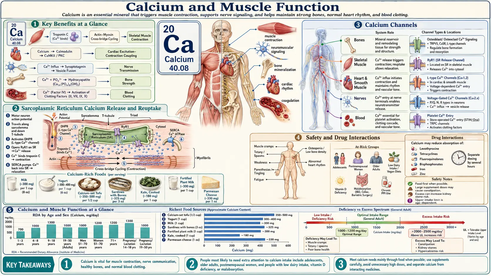
Calcium is the switch that turns muscle contraction on and off. Muscle contracts when calcium in the cell fluid (cytosol) rises, either by entering from outside the cell or by release from stores inside it (Kuo & Ehrlich, 2015, PMID 25646377), and relaxes when that calcium is pumped away again. In heart muscle cells, pumps, exchangers and channels hold the resting cytosolic calcium level at about 100 nM, more than 10,000 times lower than outside the cell (Schaub et al., 2006, PMID 16765984). This page describes that machinery in skeletal, cardiac and smooth muscle; what the clinical literature reports about low and high blood calcium (cramps, tetany, heart-rhythm changes); and what trials have found about calcium, magnesium and other treatments for muscle cramps.
Table of Contents
- Calcium as a Universal Signaling Ion
- Skeletal Muscle Excitation-Contraction Coupling
- Cardiac Muscle and Calcium-Induced Calcium Release
- Smooth Muscle and Vasoconstriction
- Deficiency Symptoms: Cramps, Tetany, Arrhythmia
- The Calcium-Magnesium Balance
- Vitamin D, Vitamin K2, and Potassium Interactions
- Dietary Calcium vs Supplement Forms
- Evidence for Cramp Prevention
- Hypercalcemia and Excess Risks
- Summary of the Findings
- Key Research Papers
- Connections
- Featured Videos
Calcium as a Universal Signaling Ion
Inside muscle cells calcium works as a signal rather than as building material. A 2000 review describes cells as carrying a calcium-signalling “toolkit” whose parts are mixed and matched to control processes as different as fertilization, learning and memory, contraction and secretion — all within the limit that calcium is highly toxic, so that a signal exceeding its normal spatial and temporal bounds can kill the cell through necrosis or apoptosis (Berridge, Lipp & Bootman, 2000, PMID 11413485).
- Resting cytosolic [Ca2+] — about 100 nM in heart muscle cells, more than 10,000-fold lower than outside the cell; the sarcoplasmic reticulum (SR) holds the bulk of the calcium used for each heartbeat (Schaub et al., 2006, PMID 16765984).
- Pumping calcium back into the store — the SR calcium ATPase (SERCA) moves calcium from the cytosol into the SR; its activity is modulated by phospholamban and sarcolipin in cardiac and skeletal muscle (Periasamy & Kalyanasundaram, 2007, PMID 17286271).
- Pumping calcium out of the cell — in heart muscle, reuptake into the SR during relaxation is backed up by calcium efflux across the cell membrane through the Na+/Ca2+ exchanger and the membrane calcium pump (Fabiato, 1983, PMID 6346892).
- Blood calcium — held within a narrow range. Roughly 40% of serum calcium is free (ionized) and biologically active, about 45% is bound to plasma proteins (mainly albumin) and about 15% to small anions; one clinical reference gives normal ranges of 8.5–10.5 mg/dL (2.12–2.62 mmol/L) for total calcium and 1.16–1.31 mmol/L for ionized calcium (Anastasopoulou & Goyal, StatPearls, PMID 28613662).
Skeletal Muscle Excitation-Contraction Coupling
In skeletal muscle the link between the electrical signal on the membrane and calcium release from the SR is a physical one. In 1987 Rios and Brum showed that low concentrations of a dihydropyridine drug inhibited the voltage sensor’s “charge movement” and SR calcium release in parallel, and proposed that the voltage sensor in the transverse-tubule (T-tubule) membrane is the dihydropyridine receptor (DHPR); they noted that fewer than 5% of muscle dihydropyridine receptors work as functional calcium channels (Rios & Brum, 1987, PMID 2434854). A 2014 review describes “an overwhelming body of work” supporting a physical interaction between the skeletal-muscle DHPR (an L-type calcium channel) and the skeletal ryanodine receptor (RyR1), the SR calcium-release channel, while noting that the exact contact residues on RyR1 have not been identified and that the DHPR beta subunit may take part (Rebbeck et al., 2014, PMID 24374102). An older detailed review of the field is Melzer, Herrmann-Frank & Lüttgau (1995), PMID 7742348.
The sequence, as these sources describe it:
- An action potential spreads along the muscle membrane and into the T-tubules, where voltage-sensing DHPRs sit (Rios & Brum, 1987, PMID 2434854).
- The voltage change is passed to RyR1, which opens and releases calcium from the SR into the cytosol (Rebbeck et al., 2014, PMID 24374102).
- Calcium binds troponin C (TnC). In a 1975 binding study, full activation of the muscle’s ATPase required calcium binding to the two calcium-specific sites on troponin, not to the two high-affinity calcium–magnesium sites (Potter & Gergely, 1975, PMID 124731); a later review places these regulatory sites at the NH2-terminal end of TnC (Gordon, Homsher & Regnier, 2000, PMID 10747208).
- Calcium binding strengthens the TnC–troponin I interaction, loosens troponin I from actin and lets tropomyosin move across the actin surface, exposing myosin-binding sites that tropomyosin had blocked (Gordon et al., 2000, PMID 10747208).
- Myosin cross-bridges bind strongly to actin, produce force and release the products of ATP hydrolysis; strongly bound cross-bridges also help switch on neighbouring actin units (Gordon et al., 2000, PMID 10747208).
- SERCA pumps calcium back into the SR, cytosolic calcium falls and the muscle relaxes (Periasamy & Kalyanasundaram, 2007, PMID 17286271).
Cardiac Muscle and Calcium-Induced Calcium Release
Heart muscle uses a calcium trigger instead. A 2005 review summarizes cardiac excitation-contraction coupling this way:
- The action potential lets calcium flow into the heart muscle cell through L-type calcium channels in the cell membrane.
- That entering calcium triggers the release of additional calcium from the SR through type 2 ryanodine receptors (RyR2) — calcium-induced calcium release (CICR).
- Catecholamines released during emotional or physical stress boost CICR through protein kinase A phosphorylation of RyR2 (Kontula et al., 2005, PMID 15913575).
Fabiato’s 1983 experiments on skinned heart cells supported the CICR hypothesis: calcium entering across the cell membrane did not activate the contractile filaments directly but through calcium release from the SR, and the release was not all-or-none. He also noted that the hypothesis could be challenged because of uncertainties in the skinned-cell methods (Fabiato, 1983, PMID 6346892). During relaxation SERCA returns calcium to the SR; its activity is modulated by phospholamban and sarcolipin (Periasamy & Kalyanasundaram, 2007, PMID 17286271). Reviews of the subject include Bers (2002), Nature, “Cardiac excitation-contraction coupling,” PMID 11805843 (doi:10.1038/415198a) and Bers (2008), Annual Review of Physiology, PMID 17988210, which covers calcium’s roles in heart-cell electrophysiology and arrhythmias, contraction, energy supply and cell death.
Disordered calcium handling is tied to some arrhythmias and to heart failure. In catecholaminergic polymorphic ventricular tachycardia (CPVT), an inherited disorder of effort-induced ventricular arrhythmias in a structurally normal heart, mutations in the RyR2 gene cause the autosomal dominant form and mutations in calsequestrin 2 (CASQ2) other forms, and inappropriate “leakiness” of RyR2 channels is thought to play a role (Kontula et al., 2005, PMID 15913575). SERCA expression and activity are reported to be lower in aging and in heart failure (Periasamy & Kalyanasundaram, 2007, PMID 17286271).
Smooth Muscle and Vasoconstriction
Smooth muscle, which lines the walls of various organs and tubes in the body and is not under voluntary control, switches contraction on differently. Contraction starts when calcium binds calmodulin and the calcium–calmodulin complex stimulates phosphorylation of the myosin light chain. The RhoA/Rho kinase pathway “sensitizes” the contractile proteins to calcium by inhibiting myosin phosphatase, the enzyme that removes that phosphate, so force is maintained. Relaxation begins when calcium is removed from the cytosol and myosin phosphatase is stimulated (Webb, 2003, PMID 14627618). Smooth and striated muscle contract in response to depolarization or activation of G-protein-coupled receptors, with the calcium coming from outside the cell or from internal stores (Kuo & Ehrlich, 2015, PMID 25646377).
Drugs that block L-type calcium channels are in wide clinical use. A clinical reference lists their indications as including hypertension, coronary spasm, angina, supraventricular arrhythmias, hypertrophic cardiomyopathy and pulmonary hypertension; it divides them into dihydropyridines and non-dihydropyridines (verapamil, diltiazem) and notes that this drug class is one of the main contributors to drug-related deaths (McKeever, Patel & Hamilton, StatPearls, PMID 29494080).
Deficiency Symptoms: Cramps, Tetany, Arrhythmia
Clinical references describe the effects of low blood calcium (hypocalcemia) as ranging from mild paresthesias (tingling) to life-threatening tetany, seizures and cardiac arrhythmias (Anastasopoulou & Goyal, StatPearls, PMID 28613662). A 1979 review noted much individual variability, with the speed at which hypocalcemia develops determining whether symptoms appear (Juan, 1979, PMID 226022).
- Paresthesias and carpopedal spasm — perioral tingling and carpopedal spasm, with bone pain, were the presenting complaints in a case of hypocalcemia after bariatric surgery (Ghazi & Amirbaigloo, 2017, PMID 29263738).
- Chvostek’s sign — first described in 1876 as a clue to latent tetany and elicited by tapping (percussion) at the angle of the jaw; clinicians have questioned how strongly it is tied to latent tetany, particularly in children (Hasan, Absamara & Ahmed, 2014, PMID 25088339). It is absent in about one-third of patients with hypocalcemia (Schnur et al., 2021, PMID 33692059).
- Trousseau’s sign — carpopedal spasm brought on by inflating a blood-pressure cuff to 20 mm Hg above systolic pressure for 3 minutes (Schnur et al., 2021, PMID 33692059).
- Tetany, seizures, reduced or absent deep tendon reflexes and mental changes — listed among the signs and symptoms in the 1979 review (Juan, 1979, PMID 226022).
- Prolonged QT interval and weakened heart pumping — a 2014 systematic review of 47 published cases found that lower corrected calcium went with a longer corrected QT interval and a lower left-ventricular ejection fraction, and described the cardiac dysfunction as reversible; the authors judged the evidence very limited (Newman et al., 2014, PMID 23355181).
Common causes listed in clinical references include disorders of the parathyroid glands, vitamin D deficiency, chronic kidney disease and disturbed magnesium balance (Anastasopoulou & Goyal, StatPearls, PMID 28613662); a 2020 review names postsurgical hypoparathyroidism as the most frequent cause (Pepe et al., 2020, PMID 32367335). Because the skeleton is a large calcium reserve, the Linus Pauling Institute notes that a low blood calcium level usually points to abnormal parathyroid function rather than low intake. Most muscle cramps are a different matter: the Cochrane magnesium review describes typical skeletal muscle cramps — seen with pregnancy, older age, exercise or motor neuron disease — as usually having no obvious underlying pathology, which is why they are termed idiopathic (Garrison et al., 2020, PMID 32956536). A general clinical review of hypocalcemia is Cooper & Gittoes (2008), BMJ, PMID 18535072.
The Calcium-Magnesium Balance
Calcium and magnesium interact at several points in muscle. In the 1975 troponin binding study, magnesium competed with calcium for the two high-affinity calcium–magnesium sites on troponin C, while the two calcium-specific sites that switch on contraction did not bind magnesium (Potter & Gergely, 1975, PMID 124731). In the heart, Mg2+ is one of the ions that interact with the RyR2 channel complex (Kontula et al., 2005, PMID 15913575).
- Low magnesium can cause low calcium. The Linus Pauling Institute states that magnesium deficiency can impair parathyroid hormone (PTH) secretion and lower the responsiveness of bone cells to PTH, so that magnesium is required to correct hypocalcemia in people with low serum magnesium. A 2025 literature review describes this “paradoxical hypoparathyroidism” and reports that hypocalcemia from magnesium deficiency cannot be corrected adequately by calcium, vitamin D or both (Albert et al., 2025, PMID 40740723).
- Magnesium in muscle and heart. A 2015 review describes magnesium as playing an important physiological role particularly in the brain, heart and skeletal muscles, and lists diuretics, epidermal growth factor receptor inhibitors, calcineurin inhibitors and proton pump inhibitors among drugs that can lower blood magnesium (de Baaij, Hoenderop & Bindels (2015), Physiol Rev, PMID 25540137 (doi:10.1152/physrev.00012.2014)).
Trial evidence on magnesium supplements for cramps is covered under Evidence for Cramp Prevention.
Vitamin D, Vitamin K2, and Potassium Interactions
- Vitamin D. The Linus Pauling Institute describes how PTH stimulates the kidneys to convert vitamin D into its active form, 1,25-dihydroxyvitamin D (calcitriol), which decreases urinary calcium loss and stimulates intestinal calcium absorption by promoting the synthesis of calcium-binding proteins. Vitamin D receptors are also present in muscle: a 2001 study of biopsies from 20 women found strong staining for the receptor in the nuclei of human skeletal muscle cells (Bischoff et al., 2001, PMID 11352397). A widely cited review of vitamin D deficiency is Holick (2007), N Engl J Med, PMID 17634462.
- Vitamin K2 (menaquinone-7, MK-7). In a three-year double-blind trial in 244 healthy postmenopausal women, 180 µg/day of MK-7 halved circulating inactive (desphospho-uncarboxylated) matrix Gla-protein compared with placebo and improved measures of arterial stiffness, especially in women whose arteries were stiffer at the start (Knapen et al., 2015, PMID 25694037). In the same group of women, MK-7 slowed the age-related decline in bone mineral density at the lumbar spine and femoral neck, but not at the total hip (Knapen et al., 2013, PMID 23525894). The research raises a concern for people on vitamin K antagonist anticoagulants: in 15 healthy volunteers stabilized on acenocoumarol, MK-7 doses as low as 10–20 µg/day lowered the INR to a clinically relevant degree in some participants, and the authors concluded that MK-7 supplements need to be avoided during such therapy (Theuwissen et al., 2013, PMID 23530987).
- Potassium. A clinical reference lists muscle weakness, fatigue, cramping, palpitations and constipation among the symptoms of low blood potassium (hypokalemia), with fatal arrhythmias or respiratory muscle paralysis possible when it is severe; causes include poor nutrition, endocrine disorders, diarrhea and medications (Castro & Sharma, StatPearls, PMID 29494072).
Dietary Calcium vs Supplement Forms
The Recommended Dietary Allowances set by the Institute of Medicine in 2011, as listed by the Linus Pauling Institute, are 1,000 mg/day for adults aged 19–50 and men aged 51–70, and 1,200 mg/day for women over 50 and everyone over 70. The Linus Pauling Institute notes that most experts recommend obtaining as much calcium as possible from food, because calcium in food comes with other nutrients that help the body use it.
Calcium per 100 g from USDA FoodData Central (SR Legacy), with absorption figures where an isotope study measured them:
- Whole milk and whole-milk yogurt — 113 mg (whole milk) and 121 mg (plain whole-milk yogurt). In two isotope studies at Creighton University, 27.6% and 32.1% of the calcium in milk was absorbed (Heaney, Weaver & Recker, 1988, PMID 3354496; Heaney & Weaver, 1990, PMID 2321572).
- Sardines and canned salmon with bones — 382 mg for Atlantic sardines canned in oil (drained solids with bone) and 213 mg for canned pink salmon (solids with bone and liquid).
- Fermented soy — 217 mg for natto; 111 mg for tempeh.
- Low-oxalate leafy greens — 150 mg for cooked kale, 141 mg for collards, 137 mg for turnip greens, 118 mg for mustard greens and 93 mg for bok choy (all boiled and drained). In 11 women, 40.9% of the calcium in kale was absorbed, against 32.1% from milk (Heaney & Weaver, 1990, PMID 2321572), and the Linus Pauling Institute describes the calcium in kale-family greens, bok choy among them, as being as bioavailable as the calcium in milk.
- Spinach — 136 mg cooked, but in a randomized cross-over study of 13 adults only 5.1% of spinach calcium was absorbed, against 27.6% from milk; the spinach tested held slightly more oxalate than needed to bind all of its calcium (Heaney, Weaver & Recker, 1988, PMID 3354496). The Linus Pauling Institute lists spinach and rhubarb as high in oxalate, the most potent inhibitor of calcium absorption.
- Almonds and sesame seeds — 269 mg for almonds and 975 mg for whole dried sesame seeds.
- Calcium-fortified drinks — a 2007 review reports that absorption from calcium-fortified beverages varies and in general is not equal to that of milk (Straub, 2007, PMID 17507729).
Supplement forms, as described by the Linus Pauling Institute and a 2007 review of supplement forms (Straub, 2007, PMID 17507729):
- Calcium carbonate — described by both as the most economical form, best absorbed with a meal. In a 1985 study of 11 fasting patients with achlorhydria (no stomach acid), mean absorption was 4.2% from calcium carbonate against 45.2% from a pH-adjusted calcium citrate; taken with a normal breakfast, the carbonate was absorbed normally (Recker, 1985, PMID 4000241). The Linus Pauling Institute notes that H2 blockers and proton-pump inhibitors may decrease absorption of calcium carbonate.
- Calcium citrate and calcium citrate malate — can be taken with or without food; both sources describe citrate as the preferred form for people with achlorhydria or taking H2 blockers or proton-pump inhibitors. In a 1988 study of 21 adults, the rise in urinary calcium (a marker of absorption) was higher after calcium citrate than after calcium carbonate at every dose tested (Harvey, Zobitz & Pak, 1988, PMID 3213620).
- Calcium lactate and calcium gluconate — less concentrated forms, which the 2007 review describes as not practical oral supplements (Straub, 2007, PMID 17507729).
- Calcium hydroxyapatite — the 2007 review describes research on it as a calcium source as limited (Straub, 2007, PMID 17507729).
On dose size, the same 1988 study found that absorption rose steeply up to a 0.5 g load, with only slight further increases at 1.0 and 2.0 g (Harvey, Zobitz & Pak, 1988, PMID 3213620); the Linus Pauling Institute advises taking no more than 500 mg of elemental calcium at one time to maximize absorption.
Evidence for Cramp Prevention
Leg cramps in pregnancy. The 2020 Cochrane review included eight small trials (576 women). Results for oral magnesium were inconsistent; the evidence on calcium was very low to low certainty (one small study suggested slightly fewer cramps); one small trial suggested vitamin B supplements may reduce cramp frequency and intensity; and vitamin D, or calcium with vitamin D, made little or no difference in one trial of 84 women. The authors concluded that it is unclear whether any of the interventions is effective (Luo et al., 2020, PMID 33275278).
Magnesium for cramps. The 2020 Cochrane review of 11 trials (735 people) concluded that magnesium is unlikely to provide clinically meaningful cramp prevention for older adults with idiopathic cramps, found conflicting results in pregnancy, and found no randomized trials of magnesium for exercise-associated cramps; oral magnesium was associated mostly with gastrointestinal adverse events such as diarrhea, in 11% to 37% of participants against 10% to 14% in control groups (Garrison et al., 2020, PMID 32956536).
Quinine. The 2015 Cochrane review of 23 trials (1,586 participants) found low-quality evidence that quinine (200–500 mg daily) reduced cramp number over two weeks by 28% and cramp days by 20%, with moderate-quality evidence for a 10% reduction in intensity. Minor adverse events, mainly gastrointestinal, were more common than on placebo; serious adverse events can rarely be fatal, and the review notes that in some countries prescription of quinine is severely restricted (El-Tawil et al., 2015, PMID 25842375).
Exercise-associated muscle cramps. A 2022 evidence-based review concluded that these cramps arise from a combination of intrinsic and extrinsic factors rather than a single cause; it describes gentle static stretching as the continuing treatment for an acute cramp and judged individualized prevention more likely to work than generalized advice such as drinking more fluids (Miller et al., 2022, PMID 34185846). In a 2010 laboratory study of dehydrated men, electrically induced cramps lasted 49 seconds less after drinking pickle juice than after water; the drink barely changed blood composition within 5 minutes, and the authors suspected a reflex starting in the mouth and throat that inhibits the motor neurons of the cramping muscle (Miller et al., 2010, PMID 19997012).
Hypercalcemia and Excess Risks
The Linus Pauling Institute gives malignancy and primary hyperparathyroidism as the most common causes of high blood calcium (hypercalcemia). Mild hypercalcemia may cause no symptoms, or loss of appetite, nausea, vomiting, constipation, abdominal pain, fatigue, frequent urination and hypertension; more severe hypercalcemia may cause confusion, delirium and coma, and can be fatal if untreated. On the heart, a 2018 case report describes a man with myeloma-related hypercalcemia whose ECG showed a short QT interval and who died suddenly a few hours after admission; the authors describe marked QT shortening from hypercalcemia as an underappreciated warning sign of arrhythmia risk (Liu et al., 2018, PMID 30497751).
- Calcium-alkali (milk-alkali) syndrome — the Linus Pauling Institute states that hypercalcemia has not been associated with calcium occurring naturally in food, but has been reported with large amounts of calcium supplements taken with absorbable alkali, at supplement levels from 1.5 to 16.5 g/day for 2 days to 30 years; those now at greater risk include postmenopausal and pregnant women, transplant recipients, people with bulimia and people on dialysis.
- Thiazide diuretics — the same source notes that calcium supplements taken with thiazide diuretics raise the risk of hypercalcemia because the kidneys reabsorb more calcium.
- Vitamin D toxicity — a 2018 review describes it as rare; overdose from very high-dose preparations is marked by serum 25-hydroxyvitamin D above 150 ng/mL (375 nmol/L), and excess production of active vitamin D in granulomatous disorders and some lymphomas can also cause it (Marcinowska-Suchowierska et al., 2018, PMID 30294301).
Calcium supplements and heart attack risk have been debated. A 2010 meta-analysis of placebo-controlled trials of calcium supplements given without vitamin D found more myocardial infarctions with calcium (patient-level data: hazard ratio 1.31, 95% CI 1.02–1.67; trial-level data: relative risk 1.27, 1.01–1.59), with non-significant increases in stroke and death (Bolland et al. (2010), BMJ, PMID 20671013 (doi:10.1136/bmj.c3691)). The Linus Pauling Institute reports that a later meta-analysis of 18 randomized trials in 63,563 postmenopausal women found no evidence of increased coronary artery disease or all-cause mortality with calcium supplementation of at least 500 mg/day for at least a year.
Summary of the Findings
- Intake values. The Institute of Medicine’s 2011 RDAs are 1,000–1,200 mg/day for adults, depending on age and sex (as listed by the Linus Pauling Institute).
- Food sources. In USDA data, sardines and canned salmon eaten with their bones, natto, almonds, sesame seeds and cooked low-oxalate greens list substantial calcium; in isotope studies 40.9% of kale calcium was absorbed against 32.1% from milk, and 5.1% of spinach calcium against 27.6% from milk.
- Supplement forms. Calcium carbonate absorption depends on stomach acid or a meal; calcium citrate was absorbed without food in people with achlorhydria. Absorption per dose rose little above about 500 mg at a time.
- Vitamin D and vitamin K2. Active vitamin D drives intestinal calcium absorption. One three-year trial of MK-7 reported less arterial stiffening and slower bone loss in postmenopausal women; MK-7 interfered with vitamin K antagonist anticoagulants even at low doses.
- Magnesium. Low blood magnesium impairs PTH secretion and can cause hypocalcemia that calcium and vitamin D alone do not correct; magnesium supplements were unlikely to prevent idiopathic cramps in older adults. See Magnesium.
- Potassium. Weakness and cramping are listed among the symptoms of low potassium.
- Cramps. Most cramps have no obvious underlying cause. In the Cochrane reviews, the evidence for calcium, magnesium and vitamin D in preventing cramps was unclear or negative; quinine reduced cramps modestly in trials, with rare but serious adverse events; pickle juice shortened electrically induced cramps in one laboratory study.
- Heart risk. One meta-analysis linked calcium supplements without vitamin D to more heart attacks; a later meta-analysis in postmenopausal women did not find increased coronary disease or mortality.
Decisions about calcium, magnesium and vitamin supplements belong with a clinician.
Key Research Papers
- Rios E, Brum G (1987). Involvement of dihydropyridine receptors in excitation-contraction coupling in skeletal muscle. Nature 325(6106):717-720. — PubMed PMID: 2434854
- Melzer W, Herrmann-Frank A, Lüttgau HC (1995). The role of Ca2+ ions in excitation-contraction coupling of skeletal muscle fibres. Biochimica et Biophysica Acta 1241(1):59-116. — PubMed PMID: 7742348
- Rebbeck RT, Karunasekara Y, Board PG, Beard NA, Casarotto MG, Dulhunty AF (2014). Skeletal muscle excitation-contraction coupling: who are the dancing partners? International Journal of Biochemistry & Cell Biology 48:28-38. — PubMed PMID: 24374102
- Bers DM (2002). Cardiac excitation-contraction coupling. Nature 415(6868):198-205. doi:10.1038/415198a. — PubMed PMID: 11805843
- Webb RC (2003). Smooth muscle contraction and relaxation. Advances in Physiology Education 27(1-4):201-206. — PubMed PMID: 14627618
- Cooper MS, Gittoes NJ (2008). Diagnosis and management of hypocalcaemia. BMJ 336(7656):1298-1302. — PubMed PMID: 18535072
- Potter JD, Gergely J (1975). The calcium and magnesium binding sites on troponin and their role in the regulation of myofibrillar adenosine triphosphatase. Journal of Biological Chemistry 250(12):4628-4633. — PubMed PMID: 124731
- de Baaij JHF, Hoenderop JGJ, Bindels RJM (2015). Magnesium in man: implications for health and disease. Physiological Reviews 95(1):1-46. doi:10.1152/physrev.00012.2014. — PubMed PMID: 25540137
- Holick MF (2007). Vitamin D deficiency. New England Journal of Medicine 357(3):266-281. — PubMed PMID: 17634462
- Knapen MH, Braam LA, Drummen NE, Bekers O, Hoeks AP, Vermeer C (2015). Menaquinone-7 supplementation improves arterial stiffness in healthy postmenopausal women. A double-blind randomised clinical trial. Thrombosis and Haemostasis 113(5):1135-1144. — PubMed PMID: 25694037
- Luo L, Zhou K, Zhang J, Xu L, Yin W (2020). Interventions for leg cramps in pregnancy. Cochrane Database of Systematic Reviews 12:CD010655. — PubMed PMID: 33275278
- Miller KC, McDermott BP, Yeargin SW, Fiol A, Schwellnus MP (2022). An evidence-based review of the pathophysiology, treatment, and prevention of exercise-associated muscle cramps. Journal of Athletic Training 57(1):5-15. — PubMed PMID: 34185846
- El-Tawil S, Al Musa T, Valli H, Lunn MP, Brassington R, El-Tawil T, Weber M (2015). Quinine for muscle cramps. Cochrane Database of Systematic Reviews 2015(4):CD005044. — PubMed PMID: 25842375
- Bolland MJ, Avenell A, Baron JA, Grey A, MacLennan GS, Gamble GD, Reid IR (2010). Effect of calcium supplements on risk of myocardial infarction and cardiovascular events: meta-analysis. BMJ 341:c3691. doi:10.1136/bmj.c3691. — PubMed PMID: 20671013
- Schaub MC, Hefti MA, Zaugg M (2006). Integration of calcium with the signaling network in cardiac myocytes. Journal of Molecular and Cellular Cardiology 41(2):183-214. — PubMed PMID: 16765984
- Berridge MJ, Lipp P, Bootman MD (2000). The versatility and universality of calcium signalling. Nature Reviews Molecular Cell Biology 1(1):11-21. — PubMed PMID: 11413485
- Gordon AM, Homsher E, Regnier M (2000). Regulation of contraction in striated muscle. Physiological Reviews 80(2):853-924. — PubMed PMID: 10747208
- Periasamy M, Kalyanasundaram A (2007). SERCA pump isoforms: their role in calcium transport and disease. Muscle & Nerve 35(4):430-442. — PubMed PMID: 17286271
- Fabiato A (1983). Calcium-induced release of calcium from the cardiac sarcoplasmic reticulum. American Journal of Physiology 245(1):C1-C14. — PubMed PMID: 6346892
- Bers DM (2008). Calcium cycling and signaling in cardiac myocytes. Annual Review of Physiology 70:23-49. — PubMed PMID: 17988210
- Kontula K, Laitinen PJ, Lehtonen A, Toivonen L, Viitasalo M, Swan H (2005). Catecholaminergic polymorphic ventricular tachycardia: recent mechanistic insights. Cardiovascular Research 67(3):379-387. — PubMed PMID: 15913575
- Kuo IY, Ehrlich BE (2015). Signaling in muscle contraction. Cold Spring Harbor Perspectives in Biology 7(2):a006023. — PubMed PMID: 25646377
- McKeever RG, Patel P, Hamilton RJ. Calcium Channel Blockers. In: StatPearls (2024). — PubMed PMID: 29494080
- Anastasopoulou C, Goyal A. Hypocalcemia. In: StatPearls (2026). — PubMed PMID: 28613662
- Juan D (1979). Hypocalcemia. Differential diagnosis and mechanisms. Archives of Internal Medicine 139(10):1166-1171. — PubMed PMID: 226022
- Ghazi AA, Amirbaigloo A (2017). Hypocalcemia and osteomalacia after bariatric surgery. Clinical Cases in Mineral and Bone Metabolism 14(2):227-229. — PubMed PMID: 29263738
- Hasan ZU, Absamara R, Ahmed M (2014). Chvostek’s sign in paediatric practice. Current Pediatric Reviews 10(3):194-197. — PubMed PMID: 25088339
- Schnur J, Sinawe H, Yoham AL, Casadesus D (2021). Trousseau’s sign and QT prolongation in hypocalcaemia. BMJ Case Reports 14(3):e240260. — PubMed PMID: 33692059
- Newman DB, Fidahussein SS, Kashiwagi DT, Kennel KA, Kashani KB, Wang Z, Altayar O, Murad MH (2014). Reversible cardiac dysfunction associated with hypocalcemia: a systematic review and meta-analysis of individual patient data. Heart Failure Reviews 19(2):199-205. — PubMed PMID: 23355181
- Pepe J, Colangelo L, Biamonte F, Sonato C, Danese VC, Cecchetti V, et al. (2020). Diagnosis and management of hypocalcemia. Endocrine 69(3):485-495. — PubMed PMID: 32367335
- Albert A, Hinkel UP, Bohlender T, Stieger P, Braun-Dullaeus RC, Albert C (2025). Paradoxical inadequate parathyroid hormone secretion secondary to severe hypomagnesemia: a review of the literature. Kidney Medicine 7(8):101046. — PubMed PMID: 40740723
- Garrison SR, Korownyk CS, Kolber MR, Allan GM, Musini VM, Sekhon RK, Dugré N (2020). Magnesium for skeletal muscle cramps. Cochrane Database of Systematic Reviews 9:CD009402. — PubMed PMID: 32956536
- Miller KC, Mack GW, Knight KL, Hopkins JT, Draper DO, Fields PJ, Hunter I (2010). Reflex inhibition of electrically induced muscle cramps in hypohydrated humans. Medicine & Science in Sports & Exercise 42(5):953-961. — PubMed PMID: 19997012
- Bischoff HA, Borchers M, Gudat F, Duermueller U, Theiler R, Stähelin HB, Dick W (2001). In situ detection of 1,25-dihydroxyvitamin D3 receptor in human skeletal muscle tissue. Histochemical Journal 33(1):19-24. — PubMed PMID: 11352397
- Knapen MH, Drummen NE, Smit E, Vermeer C, Theuwissen E (2013). Three-year low-dose menaquinone-7 supplementation helps decrease bone loss in healthy postmenopausal women. Osteoporosis International 24(9):2499-2507. — PubMed PMID: 23525894
- Theuwissen E, Teunissen KJ, Spronk HM, Hamulyák K, Ten Cate H, Shearer MJ, Vermeer C, Schurgers LJ (2013). Effect of low-dose supplements of menaquinone-7 (vitamin K2) on the stability of oral anticoagulant treatment: dose-response relationship in healthy volunteers. Journal of Thrombosis and Haemostasis 11(6):1085-1092. — PubMed PMID: 23530987
- Castro D, Sharma S. Hypokalemia. In: StatPearls (2025). — PubMed PMID: 29494072
- Heaney RP, Weaver CM, Recker RR (1988). Calcium absorbability from spinach. American Journal of Clinical Nutrition 47(4):707-709. — PubMed PMID: 3354496
- Heaney RP, Weaver CM (1990). Calcium absorption from kale. American Journal of Clinical Nutrition 51(4):656-657. — PubMed PMID: 2321572
- Recker RR (1985). Calcium absorption and achlorhydria. New England Journal of Medicine 313(2):70-73. — PubMed PMID: 4000241
- Harvey JA, Zobitz MM, Pak CY (1988). Dose dependency of calcium absorption: a comparison of calcium carbonate and calcium citrate. Journal of Bone and Mineral Research 3(3):253-258. — PubMed PMID: 3213620
- Straub DA (2007). Calcium supplementation in clinical practice: a review of forms, doses, and indications. Nutrition in Clinical Practice 22(3):286-296. — PubMed PMID: 17507729
- Liu Y, Pérez-Riera AR, Barbosa-Barros R, Daminello-Raimundo R, de Abreu LC, Nikus K, Baranchuk A (2018). Severe hypercalcemia from multiple myeloma as an acquired cause of short QT. Journal of Electrocardiology 51(6):939-940. — PubMed PMID: 30497751
- Marcinowska-Suchowierska E, Kupisz-Urbańska M, Łukaszkiewicz J, Płudowski P, Jones G (2018). Vitamin D toxicity — a clinical perspective. Frontiers in Endocrinology 9:550. — PubMed PMID: 30294301
- Linus Pauling Institute — Calcium, Micronutrient Information Center (RDAs, absorption, supplement forms, toxicity)
- U.S. Department of Agriculture, Agricultural Research Service — FoodData Central, SR Legacy (calcium values per 100 g). — fdc.nal.usda.gov
Connections
- All Minerals
- Calcium Overview
- Calcium for Bone Health
- Calcium for Cardiovascular Health
- Calcium for Muscle Function (Benefits sub-article)
- Calcium for Nerve Transmission
- Magnesium
- Potassium
- Phosphorus
- Vitamin D3
- Vitamin K (K2/MK-7)
- Cramp Prevention
- Hypertension and Calcium Channel Blockers
- Osteoporosis
- Milk
- Collagen