Arrhythmia
Interactive Visualization The Heart & Circulation — watch a blood cell make the loop Trace a blood cell through all four chambers and both circuits, with a live ECG, chamber pressures, and an exercise mode. Launch → Interactive Visualization Potassium & the Heartbeat — bend the ECG with one slider Slide your blood potassium up and down and watch the ECG morph in real time — peaked T waves and a widening QRS as it climbs, U waves and a stretched QT as it falls. Launch → Interactive Visualization The Heart's Electrical System — trace the spark on the ECG Follow the spark from the SA-node pacemaker across the atria, through the AV-node pause, and down the fast fibres to fire the ventricles — read it live on the ECG, then break it into heart block or fibrillation. Launch →
Table of Contents
What is Arrhythmia?
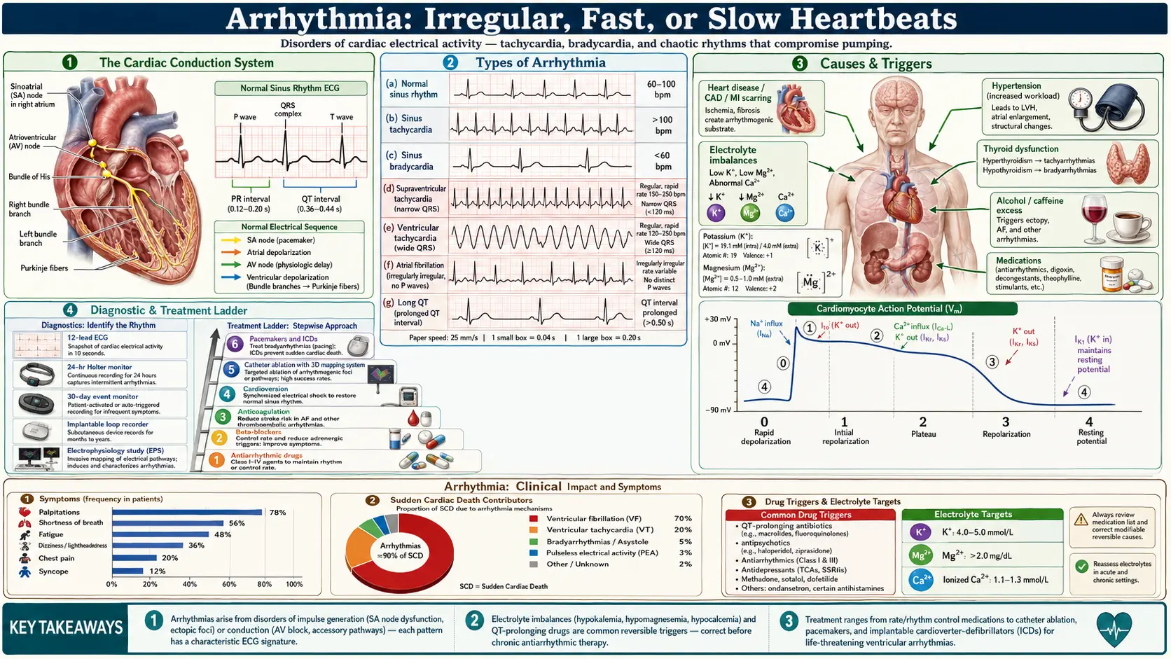
Arrhythmia is a condition characterized by an irregular heartbeat, which can be too fast (tachycardia), too slow (bradycardia), or erratic. It can affect the heart's ability to pump blood effectively.
The wiring, in plain language
Your heart is a pump with its own electrical system, and the electrics are what go wrong in an arrhythmia — not the muscle, at least not at first. Think of it as a relay race run on a fixed track. The starting gun is the sinoatrial (SA) node, a patch of self-firing cells in the top-right chamber that ticks 60–100 times a minute at rest. The signal spreads across both upper chambers (the atria), squeezing them so they top up the lower chambers. It then hits the atrioventricular (AV) node, which does something unusual: it deliberately slows down, holding the signal for about a fifth of a second. That pause is the whole point — it gives the ventricles time to finish filling before they are told to squeeze. From there the signal drops down fast fibres into the ventricles, which contract and push blood to the lungs and the body.
On an ECG that whole sequence is the familiar squiggle: the small P wave is the atria firing, the flat stretch after it is the AV node's deliberate pause (the PR interval, normally 120–200 milliseconds), the tall spike is the ventricles firing (the QRS complex), and the bump after it is the ventricles resetting (the T wave). Almost every arrhythmia can be located on that picture — a doctor reading your ECG is asking which part of the relay went wrong.
Three ways the relay breaks
Nearly all arrhythmias come down to one of three faults, and knowing which one you have explains why a particular treatment is being offered:
- A rogue starter. A cell somewhere other than the SA node starts firing on its own and jumps the gun. This is what an extra beat — a premature atrial or ventricular contraction — actually is. If a whole cluster of such cells fires fast enough it can take over the rhythm entirely.
- A signal chasing its own tail (re-entry). If the electrical wave finds a loop it can run around — typically around a patch of scar, an extra strand of conducting tissue, or a ring of tissue at the top of the heart — it circles endlessly, firing the heart every time round. This is the mechanism behind atrial flutter, most supraventricular tachycardias, Wolff–Parkinson–White, and most ventricular tachycardia that follows a heart attack. It is also why ablation works so well for these: burn or freeze one point on the loop and the circuit cannot complete.
- An unstable reset (triggered activity). While a heart cell is recovering it can twitch and fire an extra time. Anything that stretches out the recovery phase — low potassium, low magnesium, certain antibiotics and antipsychotics, or an inherited channel fault — makes this more likely. This is the mechanism behind torsades de pointes, the dangerous rhythm that long QT syndrome predisposes to.
Atrial fibrillation sits slightly apart: it is driven by rapid firing from sleeves of muscle around the pulmonary veins, which then breaks down into chaotic, disorganized activity across both atria. That specific anatomy is why the standard ablation for it is pulmonary vein isolation — ring-fencing those veins electrically.
The Main Types — and Which Ones Matter
"Arrhythmia" is an umbrella term covering rhythms that range from completely harmless to immediately lethal. Lumping them together is the single biggest source of unnecessary fear. The practical question is always which chamber and how well is the heart otherwise working.
Extra beats (usually benign)
- Premature atrial contractions (PACs) and premature ventricular contractions (PVCs) — single early beats. Essentially everyone has some. They are felt as a "skip", a thud, or a flip in the chest, and the sensation is usually the next beat, which is stronger because the heart had extra time to fill.
- In a structurally normal heart these are generally not dangerous. What changes the calculation is burden — the percentage of all beats that are PVCs, measured on a monitor. In a study of 174 patients referred for PVC ablation, a burden above 24% best separated those with reduced pumping function from those without, and the lowest burden that produced a reversible cardiomyopathy was 10% [11]. A burden of a few per cent, with a normal echocardiogram, is a very different situation from a burden of 30%.
- Crucially, PVC-induced cardiomyopathy is usually reversible once the PVCs are suppressed or ablated [10]. This is one of the few kinds of heart failure that can genuinely be cured.
Fast rhythms from the upper chambers
- Atrial fibrillation — the most common sustained arrhythmia. Irregularly irregular; the main danger is stroke, not the rhythm itself.
- Atrial flutter — a single organized loop, usually in the right atrium. Often produces a metronomic 150 beats per minute. Carries the same stroke risk as fibrillation but is far more curable by ablation.
- Supraventricular tachycardia (SVT) — sudden-onset, sudden-offset racing, often 150–250 beats per minute, classically in otherwise healthy young people. Frightening, rarely dangerous, and often permanently curable with an ablation.
- Wolff–Parkinson–White — an extra conducting strand present from birth. Mostly benign, but it is the one supraventricular pattern that can occasionally degenerate into a lethal rhythm, so it is taken seriously.
Fast rhythms from the lower chambers (the ones that matter most)
- Ventricular tachycardia — a fast rhythm arising below the AV node. In a scarred heart it is a medical emergency; in a structurally normal heart certain forms are far more benign.
- Ventricular fibrillation — disorganized quivering with no effective output. This is cardiac arrest. It is the rhythm a defibrillator exists to treat, and the reason sudden cardiac death is survivable only when a shock arrives within minutes.
- Long QT syndrome and Brugada syndrome — inherited faults in the heart's ion channels that leave a normal-looking heart electrically vulnerable. They are rare but they are why fainting during exercise, or a family history of unexplained young death, is never dismissed.
Slow rhythms
- Sinus bradycardia — a slow but normal rhythm. Trained endurance athletes frequently run 40–50 beats per minute at rest and this is a sign of fitness, not disease. Slow only matters when it causes symptoms.
- Sick sinus syndrome — an ageing SA node that alternates between too slow and too fast (the "tachy–brady" pattern). Awkward to treat with drugs alone, because slowing the fast phase worsens the slow phase; this is a common reason for a pacemaker.
- Heart block — the AV node's pause becomes a stumble or a complete blockade. First-degree is usually harmless. Third-degree (complete) block means the atria and ventricles have stopped talking altogether, and needs a pacemaker.
Causes and Risk Factors
- Heart disease: Coronary artery disease or a heart attack can damage heart tissue and lead to arrhythmias. Scar tissue does not conduct, so signals must detour around it — and a detour is exactly the loop that re-entry needs.
- High blood pressure: Increases the risk by causing the heart to work harder. Years of hypertension thicken and stiffen the left ventricle, which raises pressure in the left atrium, which stretches it — and a stretched atrium is the classic substrate for atrial fibrillation.
- Electrolyte imbalances: Affect the heart's electrical impulses. Low potassium and low magnesium are the two that come up most often in practice, and both are correctable (see below).
- Medications: Some drugs, including those for treating heart conditions, can trigger arrhythmias. Beyond cardiac drugs, the common culprits are certain antibiotics (azithromycin, levofloxacin), antifungals, some antipsychotics and antidepressants, and anti-nausea drugs such as ondansetron — mostly by stretching out the QT interval. Diuretics cause trouble indirectly, by flushing out potassium and magnesium.
- Alcohol or caffeine: Excessive intake can lead to irregular heartbeats. The evidence for these two is genuinely different in size and direction — see Electrolytes, Diet and Lifestyle.
- Thyroid disease: An overactive thyroid is a classic reversible cause of atrial fibrillation, which is why a TSH test is standard in anyone newly diagnosed. See Thyroid Disorders.
- Obstructive sleep apnea: Repeated overnight oxygen dips and pressure swings remodel the atria. Untreated sleep apnea roughly doubles the odds of atrial fibrillation coming back after an ablation, and treating it with CPAP substantially reduces that recurrence [13]. If you have atrial fibrillation and you snore, this is one of the highest-yield things to chase.
- Obesity, deconditioning and heavy endurance training: Both ends of the spectrum raise atrial fibrillation risk — the first through atrial stretch and inflammation, the second through years of high-volume endurance load. Moderate activity sits in the protective middle.
- Age and genetics: Risk climbs steeply after 60. A first-degree relative with unexplained sudden death under 40, or with a pacemaker or defibrillator young, is worth telling your doctor about.
Symptoms of Arrhythmia
Common signs and symptoms include:
- Palpitations (fluttering or pounding in the chest)
- Shortness of breath
- Fatigue
- Dizziness or lightheadedness
- Chest pain
- Fainting (syncope) in severe cases
Why two people with the same rhythm feel completely different things
This is the part that confuses patients most, and it is worth saying plainly: how bad an arrhythmia feels has very little to do with how dangerous it is. A run of harmless extra beats can be terrifying. A dangerous rhythm can be silent. Several things drive the difference:
- How aware you are of your own heartbeat. Some people simply perceive cardiac sensation more strongly. The same PVC burden produces incapacitating symptoms in one person and nothing at all in another.
- Whether the atria are still contributing. In a normal beat the atria add roughly a fifth of the ventricle's filling. Lose that — as you do in atrial fibrillation — and a young flexible heart barely notices, while a stiff, thickened heart such as in HFpEF can tip into breathlessness almost immediately.
- How fast, and for how long. A brief burst at 180 is a fright; sustained weeks at 130 quietly weakens the pumping muscle (tachycardia-induced cardiomyopathy) and often shows up as fatigue and swelling rather than palpitations.
- Whether it is silent. A large share of atrial fibrillation produces no symptoms at all and is found incidentally — on a blood-pressure cuff that reports an irregular beat, a routine ECG, or a wearable. Silent atrial fibrillation carries the same stroke risk as the symptomatic kind, which is the entire argument for taking a watch notification seriously rather than dismissing it.
Patterns that help your doctor more than the word "palpitations" on its own: whether it starts and stops abruptly (suggests SVT) or fades in and out (more typical of sinus tachycardia or anxiety); whether the beat is regular-but-fast or completely irregular; what you were doing when it began; and how long it lasted. A dated note on your phone with those four items is more useful than any description given weeks later in a clinic.
Diagnosis and Treatment
Diagnostic Tests
To diagnose arrhythmia, doctors may use:
- Electrocardiogram (ECG): A test that records the heart's electrical activity.
- Holter monitor: A portable ECG device worn for a day or more to track heart activity.
- Event monitor: Similar to a Holter monitor but used for longer periods.
- Electrophysiological study: To map electrical pathways in the heart.
Matching the monitor to how often it happens
The central problem in diagnosing an arrhythmia is catching it in the act. A standard 12-lead ECG records about ten seconds; if your episodes come once a fortnight, the odds of that ten seconds landing on one are essentially zero. A normal ECG therefore does not mean nothing is wrong — it means nothing was happening during those ten seconds. Pick the monitor by how often symptoms occur:
- Daily symptoms → a 24- to 48-hour Holter is enough.
- Weekly symptoms → a 7- to 14-day adhesive patch monitor, worn under clothes and posted back.
- Monthly symptoms → a 30-day event or loop recorder that you activate when you feel something.
- Rare but serious events (unexplained fainting, suspected but unproven atrial fibrillation after a stroke) → an implantable loop recorder, a device the size of a paperclip placed under the skin that watches for up to about three years.
Supporting tests: an echocardiogram (ultrasound) to answer the question that changes everything — is the heart muscle structurally normal, and what is the ejection fraction; blood tests for thyroid function, potassium, magnesium, kidney function and blood count; and, where exercise brings symptoms on, a stress test. An electrophysiology study, where catheters are threaded to the heart to provoke and map the rhythm, is generally reserved for when an ablation is already being contemplated.
Consumer wearables: useful, and commonly misread
Smartwatch alerts have become one of the most common routes into a cardiology clinic. The evidence is worth knowing precisely. In the Apple Heart Study, 419,297 participants were monitored for a median of 117 days; only 0.52% ever received an irregular-pulse notification. Among those who returned an ECG patch afterwards, atrial fibrillation was confirmed in 34%, and when a further notification fired while the patch was recording, the two agreed 84% of the time [4].
Read that both ways. A notification is a real signal that deserves a proper evaluation — but roughly two thirds of people who chased one did not have atrial fibrillation documented on the patch. The correct response is neither panic nor dismissal: save the watch's single-lead ECG strip, note the date and time, and take it to a clinician. That saved strip is often the single most useful thing you can bring.
Treatment Options
- Medications: Antiarrhythmic drugs, beta-blockers, and anticoagulants to manage the condition.
- Cardioversion: A procedure using electrical shocks to restore a normal heart rhythm.
- Catheter ablation: A minimally invasive procedure to destroy the heart tissue causing the arrhythmia.
- Implantable devices: Pacemakers or implantable cardioverter-defibrillators (ICDs) for severe cases.
What each treatment is actually for
Treatment answers three separate questions, and it helps enormously to keep them apart:
- Should the rate be slowed? Beta-blockers (metoprolol, bisoprolol, atenolol, carvedilol) and the rate-limiting calcium channel blockers (diltiazem, verapamil) both slow conduction through the AV node, taking a heart running at 140 down toward 80. They are old, generic, and among the cheapest drugs in any pharmacy. Typical side effects are tiredness, cold hands, and a blunted heart-rate response to exercise; verapamil and diltiazem must be avoided in significantly reduced pumping function.
- Should the rhythm be restored? This is rhythm control — cardioversion, antiarrhythmic drugs, or ablation. Antiarrhythmics divide sharply by heart structure. Flecainide and propafenone work well but only in structurally normal hearts; after a heart attack this class increased mortality in the CAST trial, and that finding still governs prescribing today. Sotalol and dofetilide require QT monitoring. Amiodarone is the most effective and the most toxic — long-term use demands periodic thyroid, liver and lung monitoring, so it tends to be reserved for older patients or where nothing else works.
- Should the stroke risk be treated? For atrial fibrillation and flutter this is decided separately from the rhythm, using the CHA2DS2-VASc score, which adds points for heart failure, hypertension, age, diabetes, prior stroke, vascular disease and sex. It is worth knowing two things about it. First, restoring rhythm does not by itself remove the need for anticoagulation. Second, aspirin is not adequate stroke prevention in atrial fibrillation — a belief that persists among patients decades after the guidelines moved on. The direct oral anticoagulants (apixaban, rivaroxaban, edoxaban, dabigatran) have largely replaced warfarin because they need no routine blood monitoring and cause less bleeding into the brain.
Ablation: what the trials actually found
Catheter ablation is now genuinely first-line for several rhythms, but the evidence differs by situation and the honest summary is more nuanced than the marketing.
- SVT, atrial flutter, WPW — ablation is frequently curative, with high success rates, and is often preferable to a lifetime of drugs in a young person.
- Atrial fibrillation, symptom relief — in the CABANA trial (2,204 patients), ablation did not significantly reduce the primary composite of death, disabling stroke, serious bleeding or cardiac arrest by intention-to-treat, though it clearly reduced recurrence and improved quality of life [8]. Read plainly: ablation for atrial fibrillation is chiefly a quality-of-life operation in the average patient.
- Atrial fibrillation with reduced ejection fraction — here it is different. In CASTLE-AF, ablation in patients with heart failure and an implanted defibrillator reduced the composite of death from any cause or hospitalization for worsening heart failure [7]. If you have both atrial fibrillation and a weak heart, ablation is a stronger proposition than the CABANA headline suggests.
- Treating early rather than late — EAST-AFNET 4 randomized 2,789 patients diagnosed within the previous year and found that early rhythm control (drugs or ablation) lowered cardiovascular death, stroke and hospitalization compared with usual care [6]. Time from diagnosis matters; "let's wait and see" is not a free option.
Practical points to ask about before an ablation: how many of this specific procedure the operator does per year; whether a repeat procedure is likely (for atrial fibrillation, a second procedure is common); the risk of the specific complications that matter (tamponade, phrenic nerve injury, vascular access problems); and how long you will stay on anticoagulation afterwards — which, again, is decided by your stroke score, not by whether the ablation worked.
Electrolytes, Diet and Lifestyle
This is where most of the internet's advice about arrhythmia lives, and where the evidence is most uneven. Below, each item is labelled with how strong the support actually is — including the popular ones that do not hold up.
Magnesium — moderate evidence, worth attending to
In the Framingham Offspring cohort, 3,530 people free of atrial fibrillation and cardiovascular disease were followed for up to 20 years. Those in the lowest quartile of serum magnesium (at or below 1.77 mg/dL) were about 50% more likely to develop atrial fibrillation than those in the upper quartiles (adjusted hazard ratio 1.52) [9]. That is an association, not proof that supplementing prevents it — but hypomagnesemia is common, cheap to check, and cheap to fix.
In the acute setting the evidence is more direct: intravenous magnesium added to standard therapy improves rate control in rapid atrial fibrillation in the emergency department [12]. That is a hospital treatment, not something to attempt at home, but it tells you the mineral is doing real electrophysiological work.
Practically: ask for a serum magnesium alongside your potassium — it is frequently left off. Note that serum magnesium is an imperfect measure, since most of the body's supply sits inside cells and in bone, so a result at the bottom of the "normal" range in someone on a diuretic or a proton-pump inhibitor is worth taking seriously. Food first: pumpkin seeds, almonds, cashews, spinach and chard, black beans, avocado, dark chocolate, and brown rice are the reliable everyday sources. Where a supplement is used, glycinate and citrate are absorbed better and are far gentler on the gut than magnesium oxide, which is mostly a laxative. See Magnesium.
Potassium — strong evidence, but individualized
Potassium sets the resting voltage of every heart cell. When it falls, the recovery phase stretches out and the heart becomes vulnerable to exactly the triggered activity described above — this is why low potassium and long QT are such a dangerous pair. The most common cause in practice is a diuretic, sometimes combined with vomiting or diarrhoea.
Practically: food sources are the safest route — potatoes with the skin, beans and lentils, spinach and other leafy greens, avocado, bananas, oranges, salmon, and plain yogurt. Do not start potassium supplements on your own if you have kidney disease or take an ACE inhibitor, an ARB, or spironolactone: those drugs retain potassium, and high potassium causes its own dangerous arrhythmias. This is a case where the mineral must be measured, not guessed. See Potassium.
Alcohol — strong evidence, and one of the biggest levers you control
This is the most useful randomized trial in the whole lifestyle literature for arrhythmia. Voskoboinik and colleagues took 140 regular drinkers (10 or more standard drinks a week) who had atrial fibrillation and randomized them to abstain or carry on. Over six months, atrial fibrillation recurred in 53% of the abstinence group versus 73% of controls, and the total time spent in atrial fibrillation fell from a median of 1.2% to 0.5% [2].
Those are large effects for a free intervention, and they are worth putting beside the numbers for ablation. If you drink and you have atrial fibrillation, cutting alcohol is not a footnote to treatment — it is one of the main treatments.
Caffeine — weaker than its reputation
Caffeine is the first thing most people are told to give up, and the evidence does not really support a blanket ban. The CRAVE trial randomized 100 adults, day by day, to drink caffeinated coffee or avoid caffeine, while continuously monitoring their hearts. Coffee days produced no significant increase in premature atrial contractions (58 vs 53 per day). Premature ventricular contractions did rise meaningfully (154 vs 102 per day), and people slept about 36 minutes less — while walking roughly 1,000 more steps [3].
So: if your problem is atrial, moderate coffee is probably not the villain. If your problem is frequent PVCs, or if lost sleep is itself one of your triggers, cutting back is reasonable. Energy drinks are a separate matter — the caffeine doses are much higher and often combined with other stimulants.
Fish oil — popular, and pointing the wrong way for this condition
This one deserves to be stated bluntly, because it contradicts what most people believe. A meta-analysis of seven cardiovascular outcome trials covering 81,210 patients found that marine omega-3 supplementation increased the risk of atrial fibrillation (hazard ratio 1.25). The effect was dose-dependent: 1.49 in trials using more than 1 g/day, 1.12 at 1 g/day or less, with risk rising a further 11% per additional gram [5].
This does not make eating fish harmful — the trials tested concentrated supplements, often at doses far above what food provides. But if you have atrial fibrillation and you are taking high-dose fish oil capsules for general heart health, that is worth raising with your doctor rather than assuming it is helping. See Omega-3 Fatty Acids.
Sleep, weight and blood pressure — the unglamorous, high-yield trio
Treating obstructive sleep apnea reduces atrial fibrillation recurrence after ablation [13]; sustained weight loss and good blood-pressure control both reduce atrial fibrillation burden. None of these is exciting, and collectively they outperform every supplement on this page.
Red Flags: When to Get Help Now
Call emergency services immediately if palpitations come with any of the following:
- Fainting, or nearly fainting — especially during exertion. Fainting while exercising is the single most concerning cardiac symptom there is and should never be attributed to heat or dehydration without evaluation.
- Chest pain or pressure, particularly if it spreads to the arm, jaw or back.
- Severe breathlessness or an inability to speak a full sentence.
- Confusion, one-sided weakness, facial droop or slurred speech — these are stroke signs, and atrial fibrillation is a common cause. See Stroke.
- A racing heart that will not stop after 20–30 minutes of rest, or a sustained rate above roughly 150 at rest.
Arrange an urgent, not emergency, appointment for: a first episode of an irregular pulse; palpitations that are new, more frequent, or lasting longer than before; palpitations plus ankle swelling or unusual fatigue; a repeated smartwatch atrial fibrillation notification; or a family history of sudden unexplained death under 40.
Interactions and traps worth knowing
- Grapefruit raises blood levels of several antiarrhythmics and calcium channel blockers. Check with a pharmacist rather than assuming.
- St John's wort speeds the breakdown of many drugs and can drop levels of anticoagulants and antiarrhythmics into ineffectiveness — a genuinely dangerous herbal interaction, not a theoretical one.
- Licorice (real licorice root, not the candy flavouring) lowers potassium and raises blood pressure with regular use.
- NSAIDs such as ibuprofen and naproxen raise bleeding risk on an anticoagulant and can raise blood pressure.
- Decongestants containing pseudoephedrine or phenylephrine are common, avoidable triggers.
- Stacking QT-prolonging drugs. Any single one may be fine; two or three together, on a background of low potassium, is how torsades happens. Keep one complete medication list, over-the-counter items and supplements included, and show it to a pharmacist whenever anything new is added.
Prognosis
The prognosis for arrhythmia depends on the type and underlying cause. Many arrhythmias are manageable with treatment, allowing individuals to lead healthy lives.
To put shape on that: extra beats in a structurally normal heart carry an essentially normal life expectancy. SVT, atrial flutter and Wolff–Parkinson–White are frequently cured outright by a single ablation. Atrial fibrillation is usually a long-term condition rather than a curable one, but with anticoagulation appropriate to your stroke score the great majority of its danger is removed — and treating it early appears to work better than treating it late [6]. Ventricular arrhythmias in a scarred or weakened heart are the serious end of the spectrum, and their prognosis is driven mainly by the underlying muscle disease rather than the rhythm itself.
Two situations deserve emphasis because they are the hopeful ones. PVC-induced cardiomyopathy and tachycardia-induced cardiomyopathy both describe a heart weakened purely by the rhythm, and in both the pumping function commonly recovers once the rhythm is controlled [10]. Being told your ejection fraction is low is not, in these cases, a permanent verdict.
Key Research Papers
The following PubMed topic searches return current peer-reviewed literature relevant to this condition. Each link opens a live PubMed query.
Key Research Papers
Every citation below was verified against its PubMed record before publication — author list, journal, year and title all checked against the source.
- Joglar JA, Chung MK, Armbruster AL, et al. 2023 ACC/AHA/ACCP/HRS Guideline for the Diagnosis and Management of Atrial Fibrillation: A Report of the American College of Cardiology/American Heart Association Joint Committee on Clinical Practice Guidelines. Circulation. 2024;149(1):e1-e156 — PubMed PMID: 38033089 · doi:10.1161/CIR.0000000000001193
- Voskoboinik A, Kalman JM, De Silva A, et al. Alcohol Abstinence in Drinkers with Atrial Fibrillation. N Engl J Med. 2020;382(1):20-28 — PubMed PMID: 31893513 · doi:10.1056/NEJMoa1817591
- Marcus GM, Rosenthal DG, Nah G, et al. Acute Effects of Coffee Consumption on Health among Ambulatory Adults. N Engl J Med. 2023;388(12):1092-1100 — PubMed PMID: 36947466 · doi:10.1056/NEJMoa2204737
- Perez MV, Mahaffey KW, Hedlin H, et al. Large-Scale Assessment of a Smartwatch to Identify Atrial Fibrillation. N Engl J Med. 2019;381(20):1909-1917 — PubMed PMID: 31722151 · doi:10.1056/NEJMoa1901183
- Gencer B, Djousse L, Al-Ramady OT, et al. Effect of Long-Term Marine ɷ-3 Fatty Acids Supplementation on the Risk of Atrial Fibrillation in Randomized Controlled Trials of Cardiovascular Outcomes: A Systematic Review and Meta-Analysis. Circulation. 2021;144(25):1981-1990 — PubMed PMID: 34612056 · doi:10.1161/CIRCULATIONAHA.121.055654
- Kirchhof P, Camm AJ, Goette A, et al. Early Rhythm-Control Therapy in Patients with Atrial Fibrillation. N Engl J Med. 2020;383(14):1305-1316 — PubMed PMID: 32865375 · doi:10.1056/NEJMoa2019422
- Marrouche NF, Brachmann J, Andresen D, et al. Catheter Ablation for Atrial Fibrillation with Heart Failure. N Engl J Med. 2018;378(5):417-427 — PubMed PMID: 29385358 · doi:10.1056/NEJMoa1707855
- Packer DL, Mark DB, Robb RA, et al. Effect of Catheter Ablation vs Antiarrhythmic Drug Therapy on Mortality, Stroke, Bleeding, and Cardiac Arrest Among Patients With Atrial Fibrillation: The CABANA Randomized Clinical Trial. JAMA. 2019;321(13):1261-1274 — PubMed PMID: 30874766 · doi:10.1001/jama.2019.0693
- Khan AM, Lubitz SA, Sullivan LM, et al. Low serum magnesium and the development of atrial fibrillation in the community: the Framingham Heart Study. Circulation. 2013;127(1):33-8 — PubMed PMID: 23172839 · doi:10.1161/CIRCULATIONAHA.111.082511
- Latchamsetty R, Bogun F. Premature Ventricular Complex-Induced Cardiomyopathy. JACC Clin Electrophysiol. 2019;5(5):537-550 — PubMed PMID: 31122375 · doi:10.1016/j.jacep.2019.03.013
- Baman TS, Lange DC, Ilg KJ, et al. Relationship between burden of premature ventricular complexes and left ventricular function. Heart Rhythm. 2010;7(7):865-9 — PubMed PMID: 20348027 · doi:10.1016/j.hrthm.2010.03.036
- Ramesh T, Lee PYK, Mitta M, et al. Intravenous magnesium in the management of rapid atrial fibrillation: A systematic review and meta-analysis. J Cardiol. 2021;78(5):375-381 — PubMed PMID: 34162502 · doi:10.1016/j.jjcc.2021.06.001
- Congrete S, Bintvihok M, Thongprayoon C, et al. Effect of obstructive sleep apnea and its treatment of atrial fibrillation recurrence after radiofrequency catheter ablation: A meta-analysis. J Evid Based Med. 2018;11(3):145-151 — PubMed PMID: 30091301 · doi:10.1111/jebm.12313
PubMed Topic Searches
Each link opens a live PubMed query returning current peer-reviewed literature on that sub-topic.
- PubMed: Cardiac arrhythmia diagnosis
- PubMed: Supraventricular tachycardia
- PubMed: Ventricular tachycardia
- PubMed: Bradyarrhythmia pacemaker
- PubMed: Arrhythmia pathophysiology
- PubMed: Antiarrhythmic drugs
- PubMed: Catheter ablation arrhythmia
- PubMed: Implantable cardioverter defibrillator
- PubMed: Sudden cardiac death arrhythmia
- PubMed: Long QT syndrome
- PubMed: Arrhythmia electrocardiogram
- PubMed: Arrhythmia management guidelines
Connections
- Cardiology
- The Heart’s Electrical System & the ECG — interactive animation
- Potassium & the Heartbeat — interactive animation
- The Heart & Circulation — interactive animation
- Atrial Fibrillation
- Heart Failure
- Cardiomyopathy
- Valvular Heart Disease
- Heart Palpitations
- Lightheadedness
- Chest Pain
- Shortness of Breath
- Magnesium
- Potassium
- Calcium
- Omega-3 Fatty Acids
- Stress Management
- Coronary Artery Disease
- POTS
- Lead Poisoning
- Thyroid Disorders
- Obstructive Sleep Apnea
- Cryosurgery and Cryoablation (cardiac cryoablation)
- Palpitations: Causes and Workup — the symptom-side approach, from benign ectopy to ambulatory monitoring.
- Herbs covered on this site that discuss this condition: Motherwort · Vietnamese Balm (Elsholtzia ciliata) · Horehound · Licorice (Glycyrrhiza glabra) · Sida cordifolia (Bala)
- Atrial Flutter — a single organized re-entry loop; same stroke risk as fibrillation, far more curable by ablation
- Supraventricular Tachycardia — abrupt-onset racing from above the AV node, often permanently curable
- Wolff-Parkinson-White Syndrome — an extra conducting strand present from birth
- Ventricular Tachycardia — the fast rhythm that arises below the AV node
- Ventricular Fibrillation — disorganized quivering with no output — this is cardiac arrest
- Long QT Syndrome — a stretched recovery phase that predisposes to torsades de pointes
- Brugada Syndrome — an inherited sodium-channel fault in a structurally normal heart
- Heart Block — when the AV node's deliberate pause becomes a stumble or a blockade
- Sudden Cardiac Death — why a shock within minutes is the only thing that works
- HFpEF — a stiff ventricle that depends on the atrial kick, and suffers most when it is lost