Rhabdomyolysis
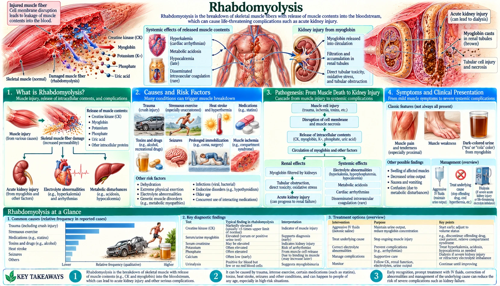
Table of Contents
- How Rhabdomyolysis Works
- Causes and Risk Factors
- Pathogenesis: From Muscle Death to Kidney Injury
- Symptoms and Clinical Presentation
- Diagnosis and Laboratory Findings
- Acute Kidney Injury Risk Stratification
- Treatment and Fluid Resuscitation
- Compartment Syndrome and Fasciotomy
- Genetic and Metabolic Forms
- Key Research Papers
- Connections
- Featured Videos
How Rhabdomyolysis Works
Rhabdomyolysis is a potentially life-threatening syndrome caused by the breakdown (lysis) of skeletal muscle cells, releasing their intracellular contents — most critically myoglobin, creatine kinase (CK), potassium, phosphate, and uric acid — into the bloodstream. The word comes from Greek: rhabdo (rod-shaped), myo (muscle), and lysis (breakdown), reflecting the rod-shaped appearance of skeletal muscle fibers.
The hallmark complication is acute kidney injury (AKI): myoglobin released from destroyed muscle cells is freely filtered by the glomerulus, but at high concentrations it precipitates in the renal tubules, causing direct tubular toxicity through oxidative injury and physical obstruction. Rhabdomyolysis accounts for 5–7% of all cases of acute kidney injury in the United States and is responsible for approximately 26,000 hospitalizations per year. How often the kidneys fail badly enough to need dialysis depends on how sick the group studied was. Among 2,371 hospital patients whose creatine kinase was above 5,000 U/L, 8.0% needed dialysis or another form of renal replacement therapy and 14.1% died in hospital (McMahon et al., 2013). In a 10-year series of 232 intensive-care patients with confirmed rhabdomyolysis, 45 (19%) developed acute kidney injury and 29 (12.5%) needed dialysis, and death was far more common in those who did (62% versus 18%) (Simpson et al., 2016).
The classic diagnostic triad is muscle pain and weakness + dark tea-colored urine (myoglobinuria) + markedly elevated serum creatine kinase. However, fewer than half of patients with confirmed rhabdomyolysis present with all three components simultaneously — particularly in exertional or drug-induced cases where the presentation may be subtle.
Causes and Risk Factors
Rhabdomyolysis has a remarkably diverse array of causes, unified by the common mechanism of skeletal muscle cell membrane disruption. Understanding the cause is essential because it dictates not only acute treatment but prevention of recurrence.
Traumatic Causes
- Crush injury: The archetypal cause — building collapse, industrial accidents, natural disasters (earthquakes). "Crush syndrome" describes the systemic consequences of releasing myoglobin, potassium, and inflammatory mediators from large volumes of compressed muscle into the circulation after pressure is released. First described systematically after the London Blitz (1941).
- Compartment syndrome: Pressure buildup within a closed fascial compartment exceeds perfusion pressure, causing muscle ischemia and cell death. Most common in the lower leg after fractures, vascular injuries, or prolonged compression. Pressure above 30 mmHg is typically treated surgically.
- Prolonged immobilization: Patients found down for extended periods (comatose patients, post-surgical positioning, drug overdose with inability to move) develop pressure-related muscle ischemia. A classic scenario is the elderly patient found on the floor many hours after a fall.
- Severe burns: Both direct thermal muscle injury and massive fluid shifts contribute.
- Lightning strike and electrical injury: Electrical current directly disrupts muscle cell membranes; can cause massive rhabdomyolysis even with minimal visible external burns.
Exertional Causes
- Extreme exercise in unconditioned individuals: Military recruits, first-time gym participants, marathon runners. Historically described as "march hemoglobinuria" in soldiers. The 2020 COVID-19 lockdown was associated with a spike in exertional rhabdomyolysis cases from sudden exercise resumption after deconditioning.
- Heat stroke: Hyperthermia directly disrupts muscle cell membranes and adenosine triphosphate (ATP) production; creatine kinase elevation is nearly universal in exertional heat stroke.
- Exercise + sickle cell trait: Sickle cell trait markedly increases exertional rhabdomyolysis risk during intense activity — an important consideration in athletic settings.
Drugs and Toxins
- Statins: The most clinically important pharmacological cause. All statins can cause myopathy and rhabdomyolysis, but the risk is highest with lipophilic statins and with drug interactions that raise statin plasma levels (fibrate co-administration, cytochrome P450 3A4 (CYP3A4) inhibitors — cyclosporine, azole antifungals, macrolides, amiodarone, diltiazem). Cerivastatin was withdrawn from the market in 2001 after rhabdomyolysis deaths. Risk is increased in hypothyroidism, vitamin D deficiency, and genetic polymorphisms in SLCO1B1 (hepatic statin transporter).
- Fibrates: Independently myotoxic; combination with statins synergistically increases risk.
- Cocaine and amphetamines: Vasoconstriction → muscle ischemia + direct myotoxicity + hyperthermia. Common in emergency department rhabdomyolysis presentations.
- Alcohol: Both acute (binge drinking, prolonged immobilization) and chronic (alcoholic myopathy, hypomagnesemia, hypokalemia, hypophosphatemia reduce muscle membrane stability).
- Heroin and opioids: Primarily via prolonged unconsciousness and pressure-related muscle ischemia.
- Neuroleptic malignant syndrome (NMS): Dopamine receptor blockade by antipsychotics (haloperidol, clozapine, risperidone) → hyperthermia + muscle rigidity + rhabdomyolysis. The creatine kinase can exceed 100,000 international units (IU)/L in severe NMS.
- Colchicine toxicity: Affects microtubule function in muscle cells; can cause severe rhabdomyolysis in overdose or when combined with cytochrome P450 3A4 inhibitors (e.g., clarithromycin).
Electrolyte and Metabolic Causes
- Hypokalemia: Severe potassium deficiency (<2.5 mEq/L) impairs muscle cell membrane repolarization. Classically seen in patients with hyperaldosteronism, severe diuretic use, or prolonged vomiting/diarrhea. Rhabdomyolysis from hypokalemia is often exertional — muscles can't cope with exertion in the potassium-depleted state.
- Hypophosphatemia: adenosine triphosphate synthesis requires phosphate; severe hypophosphatemia (<1.0 mg/dL) in refeeding syndrome or alcoholism depletes muscle adenosine triphosphate, causing membrane instability and cell death.
- Hyponatremia and hypernatremia: Osmotic shifts damage muscle cell membranes.
- Hypothyroidism: Thyroid hormone is essential for normal muscle metabolism; untreated hypothyroidism causes baseline creatine kinase elevation and markedly increases statin-induced myopathy risk.
Infections
- Viral: Influenza A and B are the most common viral causes; Coxsackievirus, Epstein-Barr virus (EBV), cytomegalovirus (CMV), and human immunodeficiency virus (HIV) can all cause myositis with rhabdomyolysis. SARS-CoV-2 has been associated with rhabdomyolysis, sometimes as a presenting feature.
- Bacterial: Group A Streptococcus toxic shock syndrome, Legionella pneumophila (Legionella myositis), Staphylococcal toxic shock. Rhabdomyolysis from bacterial infection carries a worse prognosis because of the combined organ insult from sepsis plus myoglobin-mediated acute kidney injury.
Inflammatory and Autoimmune Causes
Inflammatory myopathies — polymyositis, dermatomyositis, immune-mediated necrotizing myopathy (IMNM, sometimes statin-triggered) — cause chronic myositis that can flare into acute rhabdomyolysis. IMNM associated with anti-SRP or anti-HMGCR antibodies can present as acute rhabdomyolysis and requires immunosuppression, not simply statin discontinuation.
Malignant Hyperthermia
A rare but potentially lethal pharmacogenetic disorder triggered by volatile anesthetic agents (halothane, sevoflurane, desflurane) or succinylcholine in genetically susceptible individuals. Mutations in the ryanodine receptor gene (RYR1) or dihydropyridine receptor cause uncontrolled sarcoplasmic reticulum calcium release in skeletal muscle → massive hypermetabolism + rigidity + hyperthermia + rhabdomyolysis. Treated emergently with dantrolene.
Pathogenesis: From Muscle Death to Kidney Injury
Regardless of the triggering cause, muscle cell death in rhabdomyolysis follows a common pathway centered on loss of cellular energy and uncontrolled calcium influx into the muscle cell.
Step 1: Muscle Cell Adenosine Triphosphate Depletion and Calcium Overload
Normal skeletal muscle cells tightly regulate intracellular calcium at concentrations approximately 10,000-fold lower than extracellular calcium. This gradient is maintained by adenosine triphosphate-dependent calcium pumps (SERCA on the sarcoplasmic reticulum; plasma membrane Ca-ATPase). When adenosine triphosphate production fails — from ischemia, toxins, infections, or metabolic depletion — these pumps fail. Sodium and calcium pour into the cell through non-selective cation channels. Intracellular calcium activates destructive enzymes: phospholipases (degrade membrane phospholipids), proteases (including calpain, which degrades cytoskeletal proteins), and mitochondrial permeability transition pores (causing irreversible mitochondrial damage and further adenosine triphosphate depletion). This creates a self-amplifying cycle that ends in cell death.
Step 2: Intracellular Contents Released into Circulation
Disruption of the muscle cell plasma membrane releases the cell's contents into the interstitium and then the bloodstream:
- Myoglobin: The oxygen-carrying protein of muscle cells (analogous to hemoglobin in red blood cells); brown-red pigment; responsible for the dark urine color in myoglobinuria.
- Creatine kinase (CK): The predominant isoenzyme released is CK-MM (skeletal muscle isoform); the serum level reflects the extent of muscle destruction and is the primary diagnostic marker.
- Potassium: The primary intracellular cation; massive potassium release can cause life-threatening hyperkalemia, cardiac arrhythmias, and cardiac arrest.
- Phosphate: Intracellular phosphate release → hyperphosphatemia → complexes with calcium → hypocalcemia. Calcium-phosphate precipitates also deposit in damaged muscle tissue, worsening local injury.
- Uric acid: From breakdown of purines in damaged cells → hyperuricemia → renal tubular uric acid crystal precipitation (contributes to acute kidney injury alongside myoglobin).
- Lactate dehydrogenase (LDH), aldolase, aspartate aminotransferase (AST): Released from necrotic muscle cells, causing elevated liver enzyme panels — this is commonly misinterpreted as hepatic injury in patients with rhabdomyolysis.
Step 3: Myoglobin-Induced Acute Kidney Injury
Myoglobin causes acute kidney injury through three complementary mechanisms:
- Direct tubular toxicity: Myoglobin is filtered at the glomerulus and reabsorbed in the proximal tubule. When present in high concentrations, the heme group of myoglobin undergoes oxidation to generate reactive oxygen species (ROS), particularly in acidic urine, directly damaging proximal and distal tubular epithelial cells. The Fe3+ (ferrihemate) form is the most nephrotoxic — it generates lipid peroxidation of tubular cell membranes.
- Tubular obstruction: Myoglobin precipitates with Tamm-Horsfall protein (uromodulin) in the distal tubule and collecting duct, forming dense brown casts that physically obstruct urine flow. Acidic urine and concentrated urine dramatically accelerate precipitation — explaining why alkaline urine and high urine output protect the kidney.
- Renal vasoconstriction: Myoglobin scavenges nitric oxide in the renal vasculature, causing afferent arteriolar vasoconstriction and reducing glomerular filtration. Volume depletion (common in crush and exertional cases) synergistically worsens this effect.
Symptoms and Clinical Presentation
The clinical presentation of rhabdomyolysis spans a wide spectrum — from an asymptomatic incidental finding of elevated creatine kinase on routine labs to a catastrophic emergency with acute kidney injury, cardiac arrhythmias, and disseminated intravascular coagulation (DIC).
Classic Triad (Present Together in <50% of Cases)
- Myalgia: Muscle pain, tenderness, weakness, and swelling — particularly in the proximal muscles (thighs, calves, lower back, shoulders) in exertional or toxic causes. Severe muscle swelling may signal evolving compartment syndrome.
- Myoglobinuria (dark urine): Tea-colored, cola-colored, or brown-red urine from myoglobin. Present in only about 50% of cases — myoglobin has a short half-life (2–3 hours) and may have already cleared by the time of presentation. Important diagnostic key: the urine dipstick tests strongly positive for "blood" (heme), but microscopy shows few or no red blood cells — this discordance (dipstick-positive, RBC-microscopy negative) is pathognomonic for myoglobinuria and distinguishes it from hematuria.
- Elevated serum creatine kinase: The most reliable diagnostic marker. A creatine kinase above 1,000 international units/L is generally considered diagnostic; most clinically significant cases have creatine kinase levels of 5,000–100,000 international units/L or higher. Peak creatine kinase occurs approximately 24–72 hours after the muscle injury event.
Signs of Severe or Complicated Rhabdomyolysis
- Oliguria/anuria: Early sign of myoglobin-mediated acute kidney injury; urine output less than 0.5 mL/kg/h.
- Weakness or paralysis: Severe electrolyte abnormalities (hyperkalemia, hypocalcemia) or extensive muscle necrosis may cause profound weakness.
- Cardiac arrhythmias: Hyperkalemia from massive potassium release can cause peaked T-waves, wide QRS, and ventricular fibrillation; electrocardiogram (ECG) monitoring is essential.
- Tetany or seizures: From hypocalcemia (calcium shifts into damaged muscle tissue) + hyperphosphatemia.
- Altered consciousness: From uremia (in acute kidney injury), severe electrolyte disturbance, or the underlying trigger (e.g., drug intoxication, heat stroke).
Delayed Hypercalcemia in Recovery
A clinically important but counterintuitive phenomenon: after the acute phase, calcium that deposited in necrotic muscle tissue during the injury phase is remobilized as damaged muscle is cleared, causing rebound hypercalcemia in the recovery phase (days 1–3 after the acute event). This is why calcium is held back during the acute phase unless the low calcium is itself life-threatening: extra calcium adds to the calcium already overloading the muscle and feeds the rebound high calcium that follows. Tetany (painful muscle spasms) with symptoms, or an abnormal heart rhythm from the low calcium, are the situations in which it is given.
Diagnosis and Laboratory Findings
Diagnosis is based on clinical context plus laboratory confirmation. A high index of suspicion is required in any patient with unexplained acute kidney injury, dark urine, or elevated creatine kinase after a precipitating event.
Key Laboratory Findings
- Serum creatine kinase: The cornerstone marker. Creatine kinase >1,000 international units/L (5× upper limit of normal) suggests rhabdomyolysis; creatine kinase >5,000 international units/L is associated with significant acute kidney injury risk. Creatine kinase peaks at 24–72 hours after the precipitating event and declines with a half-life of approximately 36 hours if muscle injury has ceased. Serial creatine kinase measurements are essential to track progression vs. resolution. Falling creatine kinase with improving urine output is the primary marker of treatment efficacy.
- Urinalysis: Positive heme on dipstick (myoglobin cross-reacts with heme dipstick) with absent or rare RBCs on microscopy — this is the key diagnostic finding. Urine may appear tea-colored, brown, or red-brown. Granular "muddy brown" casts indicate tubular injury and acute kidney injury.
- Electrolytes:
- Hyperkalemia: potassium >5.5 mEq/L, potentially reaching 7–9 mEq/L in severe cases; life-threatening arrhythmia risk
- Hyperphosphatemia: from intracellular phosphate release
- Hypocalcemia: early phase from calcium-phosphate precipitation and calcium uptake into damaged muscle
- Hyperuricemia: from purine release
- Blood urea nitrogen (BUN) and creatinine: Rising ratio signals acute kidney injury; creatinine rise is disproportionately rapid in rhabdomyolysis because damaged muscle releases creatinine directly.
- Lactate dehydrogenase: Markedly elevated from muscle cell lysis (often misattributed to hemolysis).
- Liver enzymes (aspartate aminotransferase (AST), alanine aminotransferase (ALT)): Elevated aspartate aminotransferase from muscle cells (creatine kinase-AST co-elevation is characteristic of muscle disease, not liver disease). Alanine aminotransferase is liver-specific and may be normal unless there is concurrent hepatic injury.
- Complete blood count (CBC): Hemoconcentration (elevated hematocrit) from massive fluid shifts into necrotic muscle in crush injury; DIC may cause thrombocytopenia and anemia.
- Coagulation panel: DIC is a recognized complication of severe rhabdomyolysis, particularly in crush injury and sepsis-related cases; the tests used are the prothrombin time (PT), the activated partial thromboplastin time (aPTT), fibrinogen and D-dimer.
- Urine myoglobin: Can confirm the diagnosis, but assay is not universally available, results are slow, and myoglobin clears rapidly from urine — a negative urine myoglobin does not exclude rhabdomyolysis if clinical and creatine kinase evidence is present.
Acute Kidney Injury Risk Stratification
Not all rhabdomyolysis leads to acute kidney injury. Risk stratification helps guide the intensity of monitoring and aggressiveness of fluid resuscitation.
High-Risk Features for Acute Kidney Injury Development
- A high creatine kinase — the higher the creatine kinase, the greater the risk, but creatine kinase on its own is a weak guide. In the 232-patient Cambridge cohort the authors concluded that "creatine kinase is not a specific or early predictor" of kidney injury: a peak creatine kinase of at least 5,000 U/L was 83% sensitive but only 55% specific for needing dialysis, and in 91% of cases the peak came between admission and day 3, so it arrives late (Simpson et al., 2016).
- Volume depletion — dehydration dramatically amplifies myoglobin tubular toxicity by concentrating myoglobin in tubular fluid and acidifying urine.
- Acidic urine (pH <6.0) — promotes myoglobin precipitation and oxidative injury; alkaline urine (pH >6.5) is protective.
- Sepsis as the cause — concurrent septic vasodilation and renal vasoconstriction compound the myoglobin-mediated injury.
- Pre-existing chronic kidney disease — reduced renal reserve means less buffering capacity.
- Older age, diabetes, hypertension — baseline microvascular disease.
- Delayed presentation and treatment — acute kidney injury risk correlates with time from muscle injury to initiation of intravenous (IV) fluids.
- Electrolyte abnormalities — hyperkalemia and hyperphosphatemia at presentation independently predict worse outcomes.
A score worked out on the day of admission does better than creatine kinase alone. The McMahon score combines age, sex, the cause of the muscle damage, and the first measurements of creatinine, creatine kinase, phosphate, calcium and bicarbonate. It was built from 2,371 hospital patients whose creatine kinase exceeded 5,000 U/L, and in the validation group 2.3% of people with a score below 5 died or needed dialysis against 61.2% of those with a score above 10. The outcome also depended heavily on the cause: worst after cardiac arrest (58.5%), compartment syndrome (41.2%) and sepsis (39.3%), and mildest after exercise (3.2%), seizures (6.0%) and myositis (1.7%) (McMahon et al., 2013). In the Cambridge cohort a McMahon score of 6 or more on admission was 86% sensitive and 68% specific for needing dialysis, which was both earlier and more accurate than waiting for the creatine kinase peak (Simpson et al., 2016).
Low-Risk Presentations
Creatine kinase elevation in the range of 1,000–5,000 international units/L in a well-hydrated young patient with no comorbidities (e.g., mild exertional rhabdomyolysis after intense exercise) has a very low risk of acute kidney injury. Such patients are often managed out of hospital with fluids by mouth, rest and repeat creatine kinase measurements, when follow-up is reliable and none of the high-risk features above is present.
Treatment and Fluid Resuscitation
Fluid resuscitation is the cornerstone of rhabdomyolysis treatment, started as early as possible. The goal is to increase urine flow sufficiently to flush myoglobin from the renal tubules before cast formation and oxidative damage become irreversible.
How thin the evidence is for everything else was set out by a 2019 Danish intensive-care guideline (DASAIM/DSIT), which graded all of the evidence low or very low quality and issued eight weak recommendations and no strong ones. It suggests early rather than late fluids, and crystalloid fluids (such as saline) rather than colloids; it suggests against the routine use of loop diuretics, against mannitol, against any diuretic as routine, against alkalinisation with sodium bicarbonate, and against routine renal replacement therapy as a preventive measure (Michelsen et al., 2019).
Intravenous Fluid Resuscitation
Isotonic saline (0.9% NaCl) is the standard first-line fluid. The target urine output is 200–300 mL/h (roughly 3 mL/kg/h in adults) — a rate roughly four to six times normal urine output, designed to achieve continuous tubular flushing. Achieving this target typically requires large volumes: 10–15 liters (or more) per day in the first 24–48 hours for severe rhabdomyolysis. Careful monitoring for fluid overload is essential, particularly in patients with pre-existing cardiac or renal disease.
Urine Alkalinization with Sodium Bicarbonate
The theoretical basis is compelling: alkaline urine (pH >6.5) inhibits myoglobin precipitation in tubular fluid, reduces heme-mediated oxidative injury, and decreases uric acid crystal formation. Some centers add sodium bicarbonate (50 mEq per liter of intravenous fluid) with the goal of maintaining urine pH above 6.5. However, the clinical evidence is mixed — no randomized controlled trial has demonstrated that bicarbonate addition reduces the rate of acute kidney injury or dialysis compared to saline alone. The 2019 Danish intensive-care guideline (DASAIM/DSIT) states: "We suggest against routine use of alkalinisation with sodium bicarbonate" — a weak recommendation based on low-quality evidence. Practice varies by institution: where bicarbonate is used, it is typically added when urine pH stays below 6.0 despite adequate fluids and stopped if blood pH rises above 7.45, because alkalosis makes symptoms of low calcium worse.
Mannitol
Mannitol has been proposed as an adjunct because of its osmotic diuretic effect (increases tubular flow) and free-radical scavenging properties. However, no randomized trial supports its use, and it carries risks in acute kidney injury (osmolar gap, volume depletion rebound). The 2019 Danish guideline suggests against using mannitol; some units still try it when urine output stays low despite saline alone.
Loop Diuretics
The 2019 Danish guideline suggests against the routine use of loop diuretics such as furosemide in rhabdomyolysis, and against the routine use of any diuretic. They increase urine flow but also acidify tubular fluid (by exchanging H+ for sodium), potentially worsening myoglobin precipitation. Additionally, diuretics can worsen volume depletion if the patient is not adequately filled.
Electrolyte Management
- Hyperkalemia (high potassium): the heart is watched continuously on the electrocardiogram, because high potassium is what kills quickly. The treatments are calcium gluconate (to steady the heart muscle when potassium is above 6.5 or the electrocardiogram changes), insulin with dextrose and sodium bicarbonate (both drive potassium back inside the cells, bicarbonate especially when the blood is acidic), and a potassium-binding medicine taken by mouth such as sodium polystyrene sulfonate or patiromer, which removes potassium through the gut. Severe high potassium that does not respond to these is one of the reasons dialysis is started.
- Hypocalcemia (low calcium): calcium is not replaced when it causes no symptoms, because calcium rebounds high during recovery. It is given for tetany with symptoms or an abnormal heart rhythm.
- Hyperphosphatemia (high phosphate): less phosphate in the diet, and phosphate-binding medicines in severe cases.
Renal Replacement Therapy (Dialysis)
In the two largest published cohorts, 8.0% of 2,371 hospital patients with a creatine kinase above 5,000 U/L needed renal replacement therapy (McMahon et al., 2013), and 12.5% of 232 intensive-care patients with confirmed rhabdomyolysis did (Simpson et al., 2016) — hemodialysis or continuous renal replacement therapy (CRRT). The 2019 Danish guideline suggests against starting dialysis routinely as a preventive measure. The reasons for starting it are the same as for acute kidney injury in general: severe hyperkalemia refractory to medical management, severe metabolic acidosis, volume overload unresponsive to diuretics, and uremia with encephalopathy. Recovery of renal function is common — the majority of patients who need dialysis for rhabdomyolysis acute kidney injury ultimately recover independent renal function, unlike chronic kidney disease (CKD) patients on chronic dialysis.
Cause-Specific Management
- Statin-induced: the statin (and a fibrate, if one is also being taken) is stopped at once.
- Neuroleptic malignant syndrome (NMS): the antipsychotic responsible is stopped; dantrolene and bromocriptine are used in severe cases, with active cooling for the high temperature.
- Malignant hyperthermia: dantrolene (2.5 mg/kg intravenous bolus, repeated every 5 minutes until the rigidity settles, up to 10 mg/kg), the volatile anesthetic stopped, and active cooling.
- Heat stroke: rapid external cooling to a core temperature below 39°C within 30 minutes comes before fluid management.
- Inflammatory myopathy: treatments that damp down the immune system (steroids, intravenous immunoglobulin (IVIG), rituximab) when immune-mediated necrotizing myopathy or acute polymyositis is identified.
Compartment Syndrome and Fasciotomy
Compartment syndrome is both a cause and a complication of rhabdomyolysis — muscle swelling from rhabdomyolysis can itself raise intra-compartmental pressure, creating a dangerous cycle. It requires urgent surgical intervention and is the most time-critical aspect of rhabdomyolysis management.
Recognition
The "5 P's" of acute compartment syndrome are: Pain (out of proportion to the injury, especially with passive stretch), Pressure (tense, woody swelling), Paresthesias (tingling/numbness from nerve ischemia — an early sign), Paralysis (late sign indicating nerve/muscle death), and Pulselessness (very late, ominous — arterial involvement). Crucially, pulse may be present even with severe compartment syndrome because arterial pressure exceeds compartment pressure while capillary/venous perfusion is already compromised.
Measurement and Threshold
Compartment pressure is measured with a needle manometer or electronic device inserted into the affected compartment. A pressure above 30 mmHg is the standard surgical threshold, as is a pressure within 30 mmHg of the diastolic blood pressure (the delta-P or perfusion pressure method). Patients with low blood pressure cross that threshold at a lower absolute pressure. When the clinical picture is already clear, surgery goes ahead without waiting for a measurement.
Fasciotomy
Fasciotomy — surgical opening of the fascial compartment to release pressure — is the definitive treatment. In the lower leg (the most common site), four-compartment fasciotomy through two longitudinal incisions releases all four compartments (anterior, lateral, superficial posterior, deep posterior). Fasciotomy wounds are left open for 48–72 hours to allow swelling to resolve, then closed primarily or with split-thickness skin grafting. Outcomes are markedly better when fasciotomy is performed within 6 hours of compartment pressure elevation; delay beyond 12 hours significantly increases permanent nerve and muscle damage.
Genetic and Metabolic Forms
Repeated episodes of rhabdomyolysis — several attacks, often with relatively mild triggers such as brief exercise or going without food — are what leads to testing for an underlying metabolic myopathy. These disorders reveal the normal physiology of muscle energy metabolism and are important because they are often manageable once identified.
McArdle Disease (Glycogen Storage Disease Type V)
McArdle disease is caused by biallelic mutations in the PYGM gene encoding myophosphorylase — the muscle-specific enzyme that breaks down glycogen to glucose-1-phosphate for glycolysis. Patients cannot mobilize muscle glycogen, so muscles cannot sustain anaerobic exercise. Presentation: exercise intolerance from childhood, premature fatigue and cramps with brief intense effort (isometric exercise or sprinting), with a characteristic "second wind" phenomenon — after 10–15 minutes of sustained aerobic exercise, symptoms paradoxically improve as fatty acid oxidation and hepatic gluconeogenesis compensate for the absent glycogenolysis. Forearm ischemic exercise test shows absent lactate rise with normal ammonia rise (confirming myophosphorylase deficiency without nerve/cardiac involvement). What is described for this condition: isometric effort (holding a heavy load still) and intense sprinting are what bring on the attacks, aerobic conditioning improves exercise capacity, and sugar (sucrose) taken before exercise supplies glucose the muscle cannot release from its own glycogen.
Carnitine Palmitoyltransferase II (CPT-II) Deficiency
The most common inherited cause of recurrent exertional rhabdomyolysis in adults. CPT-II is the inner mitochondrial membrane enzyme that transfers long-chain fatty acyl groups from the cytoplasm into the mitochondrial matrix for beta-oxidation. Without CPT-II, muscles cannot use long-chain fatty acids as fuel during prolonged exercise. Triggers are characteristically prolonged submaximal exercise + fasting (the combination depletes both glycogen and blood glucose, maximally forcing the muscle to depend on fatty acid oxidation that is blocked). Patients are often exercise-tolerant for short bursts. Diagnosis: muscle biopsy showing CPT-II deficiency; genetic testing. What is described for this condition: long periods without food and long exercise on an empty stomach are what trigger attacks, carbohydrate before and during prolonged exercise supplies the fuel the muscle can still use, and medium-chain triglycerides (MCT oil) provide an alternative fuel that bypasses the blocked CPT system.
Other Metabolic Myopathies
- Phosphofructokinase deficiency (Tarui disease, GSD VII): Impaired glycolysis; similar to McArdle but without a second wind (because both glycolytic and glycogenolytic pathways are blocked at or downstream of PFK).
- Very long-chain acyl-CoA dehydrogenase (VLCAD) deficiency: Fatty acid oxidation defect; triggers identical to CPT-II deficiency; responds to MCT supplementation.
- Mitochondrial myopathies: mitochondrial encephalomyopathy with lactic acidosis and stroke-like episodes (MELAS), myoclonic epilepsy with ragged-red fibres (MERRF) and other disorders of mitochondrial DNA (mtDNA) cause exercise intolerance and recurrent rhabdomyolysis from impaired mitochondrial adenosine triphosphate production.
Key Research Papers
- Bosch X, Poch E, Grau JM. Rhabdomyolysis and acute kidney injury. N Engl J Med. 2009;361(1):62–72. — PubMed PMID: 19571284
- Petejova N, Martinek A. Acute kidney injury due to rhabdomyolysis and renal replacement therapy: a critical review. Crit Care. 2014;18(3):224. — PubMed PMID: 25043142
- Michelsen J, Cordtz J, Liboriussen L, et al. Prevention of rhabdomyolysis-induced acute kidney injury — a DASAIM/DSIT clinical practice guideline. Acta Anaesthesiol Scand. 2019;63(5):576–586. — PubMed PMID: 30644084
- Huerta-Alardín AL, Varon J, Marik PE. Bench-to-bedside review: Rhabdomyolysis — an overview for clinicians. Crit Care. 2005;9(2):158–169. — PubMed PMID: 15774072
- Melli G, Chaudhry V, Cornblath DR. Rhabdomyolysis: an evaluation of 475 hospitalized patients. Medicine (Baltimore). 2005;84(6):377–385. — PubMed PMID: 16267412
- Ward MM. Factors predictive of acute renal failure in rhabdomyolysis. Arch Intern Med. 1988;148(7):1553–1557. — PubMed PMID: 3382301
- Sauret JM, Marinides G, Wang GK. Rhabdomyolysis. Am Fam Physician. 2002;65(5):907–912. — PubMed PMID: 11898964
- Gabow PA, Kaehny WD, Kelleher SP. The spectrum of rhabdomyolysis. Medicine (Baltimore). 1982;61(3):141–152. — PubMed PMID: 7078398
- Simpson JP, Taylor A, Sudhan N, Menon DK, Lavinio A. Rhabdomyolysis and acute kidney injury: creatine kinase as a prognostic marker and validation of the McMahon Score in a 10-year cohort: a retrospective observational evaluation. Eur J Anaesthesiol. 2016;33(12):906–912. — PubMed PMID: 27259093
- Zager RA. Studies of mechanisms and protective maneuvers in myoglobinuric acute renal injury. Lab Invest. 1989;60(5):619–629. — PubMed PMID: 2716281
- McMahon GM, Zeng X, Waikar SS. A risk prediction score for kidney failure or mortality in rhabdomyolysis. JAMA Intern Med. 2013;173(19):1821–1828. — PubMed PMID: 24000014
- Scalco RS, Snoeck M, Quinlivan R, et al. Exertional rhabdomyolysis: physiological response or manifestation of an underlying myopathy? BMJ Open Sport Exerc Med. 2016;2(1):e000151. — PubMed PMID: 27900193
- Allison RC, Bedsole DL. The other medical causes of rhabdomyolysis. Am J Med Sci. 2003;326(2):79–88. — PubMed PMID: 12920439
Connections
- Nephrology & Hepatology
- Acute Kidney Injury
- Disseminated Intravascular Coagulation
- Sepsis and Septic Shock
- Alport Syndrome
- Focal Segmental Glomerulosclerosis
- Thrombotic Thrombocytopenic Purpura
- Potassium
- Phosphorus
- Complete Blood Count
- Calcium — early hypocalcemia from calcium-phosphate precipitation, then rebound hypercalcemia in recovery