Schistosomiasis Symptoms and Diagnosis
Schistosomiasis — also called bilharzia or snail fever — is a disease caused by parasitic flatworms (blood flukes) of the genus Schistosoma. After malaria, it is one of the most damaging parasitic infections in the world, affecting an estimated 200 million people, mostly across sub-Saharan Africa, parts of the Middle East, South America, and Asia. People become infected by contact with fresh water — wading, swimming, washing, or fishing — that contains the worm's larvae. This page explains how the infection unfolds over time, why most of the lasting damage comes from the body's reaction to the parasite's eggs rather than from the worms themselves, the warning signs to watch for, and how doctors confirm the diagnosis.
Urogenital Schistosomiasis
S. haematobium — blood in the urine, bladder damage, bladder-cancer risk, and female genital schistosomiasis.
Intestinal & Hepatic Schistosomiasis
S. mansoni and S. japonicum — bloody diarrhea, liver fibrosis, and portal hypertension.
Acute Schistosomiasis & Swimmer's Itch
Cercarial dermatitis and Katayama fever — the early reactions, common in travelers.
Table of Contents
- How Schistosomiasis Unfolds
- The Egg-Granuloma Mechanism
- Which Worm, Which Disease
- Warning Signs to Know
- How It Is Diagnosed
- Why Early Recognition Matters
- Key Research Papers
- Featured Videos
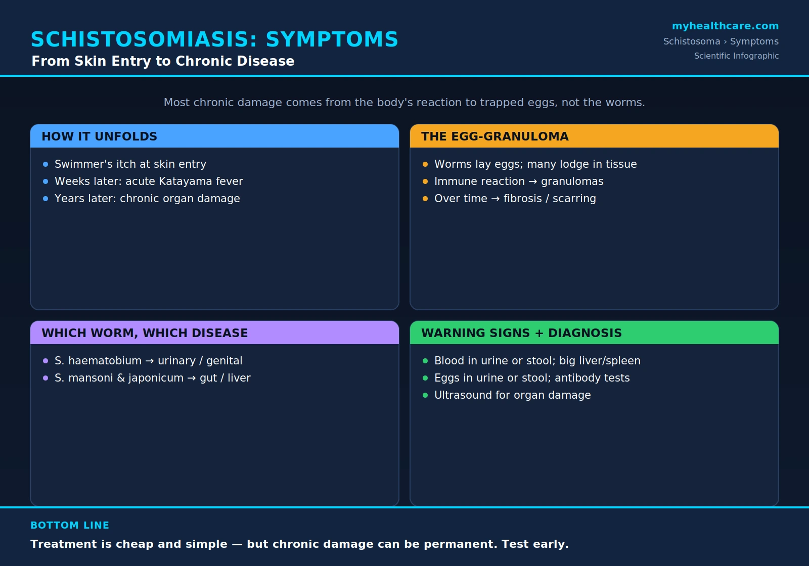
1. How Schistosomiasis Unfolds
Schistosomiasis is best understood not as a single illness but as a sequence of events that plays out over weeks, months, and years. The parasite has a complex life cycle that involves both humans and freshwater snails, and the symptoms a person experiences depend largely on which stage of that cycle is underway.
Infection begins in fresh water. The larval form of the worm, called a cercaria, is released from infected snails and swims freely until it finds human skin. Within minutes it burrows in. At the point of entry, some people develop an itchy, bumpy rash — cercarial dermatitis, popularly known as swimmer's itch. This first reaction is brief and easy to miss, and many people never notice it at all.
Several weeks later, as the maturing worms migrate through the body and begin to produce eggs, some people experience a flu-like illness called acute schistosomiasis, or Katayama fever — with fever, chills, cough, muscle aches, and fatigue. This early-reaction stage is seen most often in travelers and others encountering the parasite for the first time. For a fuller account of these early phases, see Acute Schistosomiasis & Swimmer's Itch.
The most important point to grasp is what comes next. Once adult worms are established, they can live in the blood vessels for years, steadily laying eggs. It is the slow, ongoing reaction to these eggs — not the worms themselves — that drives the chronic disease responsible for nearly all of the serious, long-term harm: scarred bladders, damaged kidneys, fibrotic livers, and more. A person can feel relatively well for a long time even as this damage accumulates quietly in the background.
2. The Egg-Granuloma Mechanism
To understand schistosomiasis, it helps to follow the eggs. Adult worms live as pairs inside the small veins of the human host — around the bladder for one species, around the intestines and liver for others. Each female worm produces large numbers of eggs every day. The eggs are equipped with a sharp spine and secrete enzymes, which help roughly half of them work their way through the vessel wall and into the bladder or bowel, so they can leave the body in urine or stool and continue the life cycle.
The trouble comes from the eggs that do not make it out. Many become trapped in the surrounding tissues — or are swept by the bloodstream to the liver and other organs, where they lodge. The body recognizes a trapped egg as foreign and mounts an immune attack against it. Immune cells surround each egg and wall it off, forming a tiny nodule of inflammation called a granuloma.
A single granuloma is harmless. But in a chronic infection, eggs are deposited continuously over years, and so countless granulomas form again and again in the same tissues. As each one heals, it leaves behind a small amount of scar tissue. Over time this repeated cycle of inflammation and healing produces widespread fibrosis — the thickening and stiffening of organs with scar tissue. This is the root of the chronic disease: a fibrotic, scarred bladder that cannot empty or store urine normally; a stiffened, scarred liver that obstructs blood flow. The worms are, in a sense, almost incidental bystanders. The lasting injury is collateral damage from the body's own prolonged immune response to the eggs.
3. Which Worm, Which Disease
Several species of Schistosoma infect humans, and a useful way to make sense of the disease is to remember that different species settle in different parts of the body. Where the adult worms live determines where the eggs are deposited — and therefore which organs are damaged and which symptoms appear.
Schistosoma haematobium lives in the veins around the bladder and the urinary and genital tract. Its eggs lodge in the bladder wall and nearby structures, producing urogenital schistosomiasis. The classic sign is blood in the urine. Over years it can scar the bladder and the tubes that carry urine, damage the kidneys, and, importantly, it is linked to an increased risk of bladder cancer. In women it can cause female genital schistosomiasis, affecting the cervix and vagina. The dedicated page on Urogenital Schistosomiasis covers this in depth.
Schistosoma mansoni and Schistosoma japonicum live in the veins draining the intestine, with eggs carried onward to the liver. They cause intestinal and hepatic schistosomiasis, with abdominal pain and bloody diarrhea early on, and, over the long term, liver fibrosis and dangerously high blood pressure in the portal vein (portal hypertension). S. japonicum, found in parts of East Asia, tends to produce the heaviest egg burden and the most severe disease. (Two further species, S. intercalatum and S. mekongi, cause similar intestinal disease in more limited regions.) The page on Intestinal & Hepatic Schistosomiasis explores these in detail.
4. Warning Signs to Know
Because schistosomiasis develops slowly and its early stages can be silent, the warning signs are easy to dismiss — yet recognizing them, especially in someone who has had freshwater contact in an affected region, can lead to early treatment. The signs depend on which organs are involved, but the most important ones to know are:
- Blood in the urine (hematuria) — the hallmark of urinary schistosomiasis from S. haematobium. In endemic areas it has historically been so common in boys that it was once mistaken for a normal sign of growing up. It should never be ignored.
- Blood in the stool, along with abdominal cramps and bouts of diarrhea — pointing toward intestinal disease.
- Abdominal pain and a sense of fullness or discomfort.
- An enlarged liver or spleen, sometimes felt as swelling or fullness on the right or left side of the upper abdomen — a sign of advancing liver involvement and portal hypertension.
- Fatigue and anemia — chronic blood loss and ongoing inflammation can leave a person tired, pale, and weak.
- Stunted growth and poor school performance in children — long-standing infection in childhood can impair physical growth, nutrition, and cognitive development, one of the most damaging public-health effects of the disease.
Many of these symptoms overlap with other conditions, so none of them proves schistosomiasis on its own. But in the right context — freshwater exposure in an endemic area, plus any of these signs — they should prompt testing.
5. How It Is Diagnosed
Diagnosing schistosomiasis combines the patient's history of freshwater exposure with laboratory tests that look for the parasite, its eggs, or the body's response to it. Several approaches are used together.
Finding the eggs under the microscope. The traditional and most direct method is to examine a sample for Schistosoma eggs. Crucially, where you look depends on the species: eggs of S. haematobium are sought in the urine, while eggs of S. mansoni and S. japonicum are sought in the stool. Each species' egg has a distinctive shape and spine, allowing the laboratory to identify it. Because egg output varies from day to day, more than one sample may be needed, and special concentration techniques improve the chances of detection. Counting eggs also gives a rough measure of how heavy the infection is.
Antibody and antigen tests. Blood tests can detect antibodies the immune system makes against the parasite — useful for travelers and for early or light infections in which eggs may be too few to find. A limitation is that antibodies can persist long after a successful cure, so a positive antibody test does not always mean active infection. Newer antigen tests instead detect molecules shed by living worms (for example, circulating cathodic antigen in urine), which more directly indicates a current, active infection.
Imaging to assess damage. Once infection is established, ultrasound is the workhorse for gauging organ injury — thickening and abnormalities of the bladder wall, blockage of the urinary tract, and the characteristic pattern of liver fibrosis and enlargement of the spleen that signals portal hypertension. Ultrasound is inexpensive, portable, and well suited to the regions where the disease is common.
Supporting clues. A raised count of a particular white blood cell, the eosinophil (eosinophilia), is a common, though nonspecific, sign of parasitic infection and can be an early hint — particularly during acute schistosomiasis. It supports the diagnosis but does not confirm it by itself.
6. Why Early Recognition Matters
The central reason to catch schistosomiasis early is a hopeful one: the treatment is simple, safe, and inexpensive, while the damage left by a long-untreated infection can be permanent. A single course of the oral drug praziquantel kills the adult worms and, taken before fibrosis has set in, can stop the disease in its tracks. It costs only a small amount per dose, which is why it can be distributed to entire communities.
But praziquantel kills worms; it cannot reverse scar tissue that has already formed. Once a bladder has been stiffened by years of granulomas, or a liver hardened by fibrosis, those changes are largely irreversible. The window in which treatment does the most good is the long, often symptom-light stretch before that permanent damage accrues — which is precisely why testing the right people at the right time is so valuable.
In practice, this means having a low threshold to test two groups: travelers who return from endemic regions after freshwater contact — even if they feel well, and especially if they have any suggestive symptom — and residents of endemic areas, particularly school-age children, who carry the greatest burden of disease. Recognizing the infection early turns a potentially disabling, lifelong condition into one that is cured with a single inexpensive treatment. For the broader strategy of treating whole populations, see Mass Drug Administration & Control and Prevention & Avoiding Freshwater.
Key Research Papers
Peer-reviewed reviews and studies on the biology, clinical features, diagnosis, and control of human schistosomiasis. Journal names appear as plain text; the year/volume/pages link opens the full citation via DOI.
- Colley DG, Bustinduy AL, Secor WE, King CH. Human Schistosomiasis. The Lancet. 2014;383(9936):2253–2264.
- Gryseels B, Polman K, Clerinx J, Kestens L. Human Schistosomiasis. The Lancet. 2006;368(9541):1106–1118.
- Ross AG, Bartley PB, Sleigh AC, Olds GR, Li Y, Williams GM, McManus DP. Schistosomiasis. New England Journal of Medicine. 2002;346(16):1212–1220.
- Steinmann P, Keiser J, Bos R, Tanner M, Utzinger J. Schistosomiasis and Water Resources Development: Systematic Review, Meta-Analysis, and Estimates of People at Risk. The Lancet Infectious Diseases. 2006;6(7):411–425.
- Mbabazi PS, Andan O, Fitzgerald DW, Chitsulo L, Engels D, Downs JA. Examining the Relationship Between Urogenital Schistosomiasis and HIV Infection. PLoS Neglected Tropical Diseases. 2011;5(12):e1396.
- Norseth HM, Ndhlovu PD, Kleppa E, Randrianasolo BS, Jourdan PM, Roald B, et al. The Colposcopic Atlas of Schistosomiasis in the Lower Female Genital Tract. PLoS Neglected Tropical Diseases. 2014;8(11):e3229.
- Lamberton PHL, Hogan SC, Kabatereine NB, Fenwick A, Webster JP. In Vitro Praziquantel Test Capable of Detecting Reduced In Vivo Efficacy in Schistosoma mansoni Human Infections. The American Journal of Tropical Medicine and Hygiene. 2010;83(6):1340–1347.
- Hotez PJ, Molyneux DH, Fenwick A, Kumaresan J, Sachs SE, Sachs JD, Savioli L. Control of Neglected Tropical Diseases. New England Journal of Medicine. 2007;357(10):1018–1027.
- Bergquist R, Utzinger J, Keiser J. Controlling Schistosomiasis with Praziquantel: How Much Longer Without a Viable Alternative? Infectious Diseases of Poverty. 2017;6(1):74.
Live PubMed Searches
Each link opens a live PubMed query so results stay current as new papers are indexed.
- Schistosomiasis symptoms diagnosis
- Schistosoma haematobium hematuria
- Schistosomiasis egg granuloma fibrosis
- Schistosomiasis circulating cathodic antigen
- Hepatosplenic schistosomiasis portal hypertension
- Female genital schistosomiasis
- Schistosomiasis ultrasound bladder liver
- Katayama fever acute schistosomiasis travelers
Connections
- Schistosoma Overview
- Urogenital Schistosomiasis
- Intestinal & Hepatic Schistosomiasis
- Acute Schistosomiasis & Swimmer's Itch
- Treatment & Prevention
- Praziquantel Treatment
- Prevention & Avoiding Freshwater
- Mass Drug Administration & Control
- All Parasites
- Urology
- Nephrology & Hepatology
- Gastroenterology
- Oncology
- Hematology
- Infectious Disease
- All Conditions