Intestinal and Hepatic Schistosomiasis
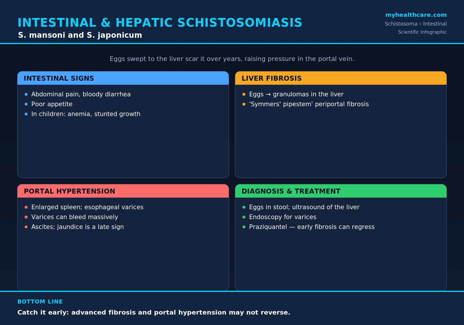
Intestinal and hepatic schistosomiasis is the form of the disease caused mainly by two blood flukes — Schistosoma mansoni and Schistosoma japonicum. The adult worms live not in the gut itself but in the small veins that drain the intestine, where the female lays hundreds of eggs each day. The eggs are the real troublemakers: many are swept by the bloodstream into the liver, where the body's reaction to them slowly scars the organ from the inside. The result, over years, is not damaged liver cells so much as a strangled blood supply — a condition that can enlarge the spleen, swell the abdomen with fluid, and cause sudden, life-threatening bleeding from veins in the esophagus. This page explains what causes the disease, how it injures the gut and the liver, why its most dangerous complication is high pressure in the portal vein rather than liver failure, how it is diagnosed, and how far treatment can turn it around.
Table of Contents
- What Causes It
- Intestinal Symptoms
- How Eggs Damage the Liver
- Portal Hypertension and Its Complications
- Hepatosplenic Disease
- Other and Severe Forms
- Diagnosis
- Treatment and Reversibility
- Key Research Papers
- Featured Videos
1. What Causes It
Intestinal and hepatic schistosomiasis is driven chiefly by two species of blood fluke. Schistosoma mansoni is found across sub-Saharan Africa, in parts of the Middle East, and in South America and the Caribbean — notably Brazil. Schistosoma japonicum is found in East Asia, including China, the Philippines, and parts of Indonesia. Two less widespread species, S. mekongi (along the Mekong River) and S. intercalatum / S. guineensis (in pockets of central Africa), cause similar but rarer intestinal disease.
People become infected the same way for every species: by contact with fresh water — wading, bathing, washing, swimming, or working in lakes, ponds, irrigation canals, and slow rivers — that harbors tiny larvae called cercariae. The cercariae are released by freshwater snails, the obligatory intermediate host, and they bore directly through intact skin. Once inside, the larvae mature into adult worms over several weeks and migrate to their final home. For the intestinal and hepatic species, that home is the veins draining the intestine — the mesenteric and portal venous system. There the worms pair up (the slender female lies in a groove along the male's body) and the female begins releasing eggs.
This is the crucial point about how the disease causes harm: the adult worms themselves provoke surprisingly little damage and can live for years — sometimes decades — cloaked from the immune system. It is the eggs that cause disease. Some eggs work their way through the intestinal wall to be passed in the stool and continue the life cycle, but a large fraction are carried backward by the blood into the tissues, above all the liver. S. japonicum is especially important here: it lays many times more eggs per worm pair than the other species, so its egg burden — and the damage that follows — tends to be greater and to appear sooner. S. japonicum is also a zoonosis: it readily infects water buffalo, cattle, pigs, dogs, and rodents, which serve as animal reservoirs. That reservoir makes S. japonicum harder to eliminate, because treating people alone leaves a large pool of infection in animals.
2. Intestinal Symptoms
Because the worms live in the veins of the bowel and force their eggs through the intestinal wall, the gut is one of the first organs affected. The eggs lodged in the wall of the intestine and colon trigger small inflammatory reactions, tiny ulcers, and patches of scarring. The typical complaints of intestinal schistosomiasis include:
- Abdominal pain and cramping, often vague and intermittent.
- Diarrhea that comes and goes, sometimes containing visible blood or mucus.
- Loss of appetite and a general sense of being unwell.
- In heavier infections, fatigue and gradual weight loss.
Many lightly infected people have few or no symptoms at all, which is part of why the disease so often goes unnoticed until it is advanced. The burden falls most heavily on children, who tend to have the most water contact and the heaviest worm loads. In children, chronic intestinal schistosomiasis — combined with the slow, steady blood loss from the inflamed bowel — commonly produces iron-deficiency anemia, persistent fatigue, and impaired growth. The anemia and tiredness, in turn, blunt concentration and energy, and infected children are documented to suffer worse school attendance and academic performance. This quiet drag on a child's development — rather than any dramatic acute illness — is one of the chief reasons schistosomiasis is treated as a major public-health problem and a target of mass treatment campaigns.
3. How Eggs Damage the Liver
To understand hepatic schistosomiasis, it helps to follow a single egg. The worm pair sits in a vein of the bowel. The blood in those veins does not return straight to the heart; instead it gathers into the portal vein and is carried first to the liver, which filters it before it rejoins the general circulation. So when a female worm releases eggs into the mesenteric veins, the bloodstream naturally sweeps many of them into the liver, where they become trapped in the small portal blood vessels.
A trapped egg is not inert. It secretes proteins that the immune system recognizes as foreign, and the body mounts a vigorous reaction around each egg, walling it off in a knot of inflammatory cells called a granuloma. A single granuloma is small. The problem is sheer numbers and time: in chronic infection, year after year of egg deposition seeds the liver with countless granulomas. As each one heals, it leaves behind a deposit of fibrous scar tissue (collagen). Because the eggs concentrate in the branching portal tracts, the scarring follows that same branching pattern, thickening the connective tissue that surrounds the portal blood vessels.
Over many years this produces the pathological signature of the disease: periportal fibrosis, classically described as "Symmers' pipestem fibrosis." The name comes from the appearance of a cut liver surface, in which the thick, pale bands of scar tissue running along the portal tracts look like the white clay stems of old smoking pipes embedded in the organ. A vital feature of this pattern distinguishes schistosomiasis from most other causes of liver scarring: the fibrosis tracks around the portal vessels, but the working liver cells (hepatocytes) and the overall liver architecture are largely preserved, at least until very late. This is fundamentally different from alcoholic or viral cirrhosis, in which the liver cells themselves are destroyed and the whole organ is remodeled into nodules.
4. Portal Hypertension and Its Complications
The preservation of the liver cells explains why hepatic schistosomiasis behaves the way it does. Because the hepatocytes keep working, the liver's chemical functions — making proteins, processing toxins, clearing bilirubin — stay relatively normal for a long time. What the dense periportal scarring does wreck is the plumbing. The fibrous cuffs squeeze and block the small branches of the portal vein inside the liver, so blood from the gut can no longer flow easily through the organ.
Blocked outflow means a backup of pressure. Pressure rises throughout the portal venous system — a condition called portal hypertension — and that elevated pressure drives the disease's most serious complications:
- Enlarged spleen (splenomegaly). The spleen sits upstream on the portal system and engorges with the backed-up blood, often growing massively. An overactive enlarged spleen can also chew up blood cells, lowering platelet and white-cell counts.
- Varices — and the danger of catastrophic bleeding. When blood cannot get through the liver, it forces open alternative detour routes back to the heart. These bypass channels run through thin-walled veins in the wall of the esophagus and stomach, which swell into fragile, dilated vessels called esophageal and gastric varices. Varices can rupture without warning and bleed massively, causing vomiting of blood or black stools; a variceal hemorrhage is a medical emergency and is the leading cause of death in advanced disease.
- Ascites. High portal pressure, sometimes together with falling blood-protein levels late in the illness, drives fluid to weep out into the abdominal cavity, producing the swollen belly of advanced disease.
The order in which these problems appear is itself a clue to the diagnosis. Because the liver cells are spared early, signs of liver-cell failure — jaundice (yellowing of the skin and eyes), confusion, easy bruising — are typically late developments, appearing only after very long-standing infection or when another insult such as viral hepatitis is also present. A patient with a huge spleen, bleeding varices, and ascites but only mild or no jaundice is, in an endemic region, a classic picture of schistosomal portal hypertension.
5. Hepatosplenic Disease
When the periportal fibrosis and portal hypertension are fully established, the illness is called hepatosplenic schistosomiasis — the advanced chronic form, and the one that does most of the disease's lasting harm in adults. The name simply combines its two defining physical findings: an abnormal liver (hepato-) and an abnormal spleen (-splenic). On examination such patients typically have:
- A firm, often enlarged liver, sometimes with a distinctively hard left lobe, reflecting the dense internal scarring rather than soft swelling.
- A large spleen, frequently palpable well below the rib margin and occasionally enormous.
- A history or active risk of variceal bleeding, the complication that most threatens life.
- In late or complicated cases, ascites and the lab findings of an overactive spleen (low platelets, low white cells).
Hepatosplenic disease develops over years and is more likely in people with heavy, repeated, untreated infection. It is also worsened by co-infection with hepatitis B or hepatitis C, which is common in endemic regions and adds true liver-cell injury on top of the schistosomal scarring — a combination that accelerates the slide toward genuine cirrhosis and liver failure. Even on its own, however, advanced hepatosplenic schistosomiasis carries real mortality, chiefly through the recurring risk of a sudden variceal hemorrhage.
6. Other and Severe Forms
Several factors can make intestinal and hepatic schistosomiasis more severe or push it in unusual directions:
- Species matters. S. japonicum tends to cause the most aggressive hepatosplenic disease, owing to its very high egg output, and it is the species classically linked with severe liver involvement and, in some studies, with liver fibrosis appearing at a younger age.
- Ectopic eggs and neuroschistosomiasis. Although eggs are meant to head for the gut and liver, some are occasionally swept to the wrong place entirely. Rarely, eggs lodge in the central nervous system and the granulomas form there. With the intestinal species, eggs most often reach the spinal cord, where the resulting inflammation can cause back pain, leg weakness, sensory loss, and bladder or bowel dysfunction — a transverse-myelitis-like picture. Eggs reaching the brain are even rarer and are more often associated with S. japonicum, where they can cause seizures or focal neurological signs. This complication, called neuroschistosomiasis, is uncommon but is a true medical emergency, because prompt treatment can prevent permanent paralysis.
- Other organs. Eggs can also seed the lungs over time, contributing to a form of pulmonary hypertension in long-standing disease.
None of these severe forms is common, but each is important to recognize because each changes how urgently and how aggressively the infection must be treated.
7. Diagnosis
Diagnosis rests on finding evidence of the parasite and gauging the damage it has done. The main tools are:
- Finding eggs in the stool. The classic, low-cost confirmatory test is microscopic examination of a stool sample (often with a concentration method such as the Kato-Katz technique, which also estimates how heavy the infection is). The two intestinal species lay eggs with characteristic shapes: S. mansoni eggs carry a prominent lateral (side) spine, while S. japonicum eggs are smaller and rounder with only a tiny lateral knob. Because egg shedding is intermittent and may be light, several specimens on different days improve the chance of detection.
- Antibody and antigen tests. Blood tests that detect antibodies against the parasite are useful, especially for travelers and others with light infections in whom eggs are hard to find — though antibodies cannot easily distinguish past from current infection. Tests that detect circulating parasite antigens (for example, the urine point-of-care CCA test) indicate active infection and are increasingly used in field settings.
- Ultrasound of the liver. Abdominal ultrasound is the workhorse for assessing chronic disease. It can directly visualize the periportal fibrosis (the thickened, bright bands along the portal tracts give a characteristic pattern), measure the enlarged spleen, and reveal signs of portal hypertension such as a widened portal vein. The World Health Organization has published standardized ultrasound grading for exactly this purpose.
- Endoscopy for varices. In patients with established portal hypertension or any history of bleeding, upper-gastrointestinal endoscopy looks directly for esophageal and gastric varices, gauges their bleeding risk, and allows them to be treated.
In practice the diagnosis often combines a relevant exposure history (freshwater contact in an endemic area), eggs or positive serology, and ultrasound or endoscopic evidence of the organ damage.
8. Treatment and Reversibility
The cornerstone of treatment is the drug praziquantel, which kills the adult worms and is safe, inexpensive, and given as a short oral course. Killing the worms stops further egg production, and that is the single most important step in halting the disease. The full details of dosing, how the drug works, and its limitations are covered on the dedicated Praziquantel page, and the broader strategy of community-wide treatment is described on the Mass Drug Administration page.
What can patients expect once the worms are gone? The encouraging news is that early liver fibrosis can regress. When infection is caught and treated before the scarring is heavily established — particularly in children and young adults — studies using ultrasound have documented that periportal fibrosis can soften and partly reverse over the months and years after cure, because the body slowly remodels scar tissue once the constant stimulus of new eggs is removed. This is a strong argument for treating early and for the mass-treatment programs that target school-age children.
The sobering counterpart is that advanced fibrosis and established portal hypertension may not reverse. Once the periportal scarring is dense and the portal system is severely obstructed, killing the worms does not undo the structural damage, and the enlarged spleen, the varices, and the bleeding risk persist. At that stage, treatment is no longer just about the parasite: it shifts to managing the complications of portal hypertension directly — for example, medications and endoscopic procedures (such as banding of varices) to prevent or stop variceal bleeding, and measures to manage ascites. The most effective approach of all, however, is to never let the disease reach that point: avoiding infested fresh water in the first place prevents the entire cascade. See the Prevention and Avoiding Freshwater page and the overarching Treatment & Prevention hub for the full picture.
Key Research Papers
Peer-reviewed reviews and studies on the biology, intestinal and hepatic disease, immunopathology, diagnosis, and treatment of schistosomiasis. Journal names appear as plain text; the year/volume/pages link opens the full citation via DOI.
- Gryseels B, Polman K, Clerinx J, Kestens L. Human Schistosomiasis. The Lancet. 2006;368(9541):1106–1118.
- Colley DG, Bustinduy AL, Secor WE, King CH. Human Schistosomiasis. The Lancet. 2014;383(9936):2253–2264.
- Ross AGP, Bartley PB, Sleigh AC, et al. Schistosomiasis. New England Journal of Medicine. 2002;346(16):1212–1220.
- Gray DJ, Ross AG, Li YS, McManus DP. Diagnosis and Management of Schistosomiasis. BMJ. 2011;342:d2651.
- Pearce EJ, MacDonald AS. The Immunobiology of Schistosomiasis. Nature Reviews Immunology. 2002;2(7):499–511.
- Steinmann P, Keiser J, Bos R, Tanner M, Utzinger J. Schistosomiasis and Water Resources Development: Systematic Review, Meta-analysis, and Estimates of People at Risk. The Lancet Infectious Diseases. 2006;6(7):411–425.
- Melman SD, Steinauer ML, Cunningham C, et al. Reduced Susceptibility to Praziquantel Among Naturally Occurring Schistosoma mansoni Isolates. The American Journal of Tropical Medicine and Hygiene. 2010;83(6):1340–1347.
- Vale N, Gouveia MJ, Rinaldi G, et al. Praziquantel for Schistosomiasis: Single-Drug Metabolism Revisited, Mode of Action, and Resistance. Infectious Diseases of Poverty. 2017;6(1):42.
- Hotez PJ, Alvarado M, Basáñez MG, et al. The Global Burden of Disease Study 2010: Interpretation and Implications for the Neglected Tropical Diseases. PLoS Neglected Tropical Diseases. 2014;8(7):e2865.
Live PubMed Searches
Each link opens a live PubMed query so results stay current as new papers are indexed.
- Intestinal schistosomiasis (S. mansoni)
- Hepatosplenic schistosomiasis
- Symmers' periportal fibrosis
- Portal hypertension and varices
- S. japonicum liver fibrosis
- Neuroschistosomiasis (spinal cord)
- Praziquantel and fibrosis regression
- Ultrasound grading of fibrosis
Connections
- Schistosoma Overview
- Symptoms & Diagnosis
- Urogenital Schistosomiasis
- Acute Schistosomiasis & Swimmer's Itch
- Treatment & Prevention
- Praziquantel Treatment
- Prevention & Avoiding Freshwater
- Mass Drug Administration & Control
- All Parasites
- Nephrology & Hepatology
- Gastroenterology
- Neurology
- Infectious Disease
- All Conditions