Urogenital Schistosomiasis
Urogenital schistosomiasis is a chronic infection of the urinary and genital organs caused by the parasitic flatworm Schistosoma haematobium. People catch it by wading, washing, swimming, or working in fresh water that carries the larval stage of the parasite, which burrows directly through unbroken skin. Once inside the body, the adult worms settle in the small veins draining the bladder and reproductive organs, and it is the eggs they lay — not the worms themselves — that cause most of the damage. The disease is widespread across sub-Saharan Africa and parts of the Middle East, and it is one of the most common parasitic infections in the world. Its hallmark sign, blood in the urine, has been recognized for thousands of years; eggs of the parasite have even been found in ancient Egyptian mummies. This page explains what urogenital schistosomiasis is, why it causes bloody urine, how it scars the bladder and raises the risk of bladder cancer, the often-overlooked toll it takes on the genital tract of women and men, and how it is diagnosed and treated.
Table of Contents
- What Is Urogenital Schistosomiasis?
- Blood in the Urine — the Classic Sign
- Chronic Bladder Damage
- Bladder Cancer Risk
- Female Genital Schistosomiasis (FGS)
- Male Genital Schistosomiasis
- Diagnosis
- Treatment and What It Can & Cannot Reverse
- Key Research Papers
- Featured Videos
1. What Is Urogenital Schistosomiasis?
Urogenital schistosomiasis is the form of schistosomiasis (also called bilharzia) caused by the blood fluke Schistosoma haematobium. It is distinct from intestinal and hepatic schistosomiasis, which is caused mainly by S. mansoni and S. japonicum and affects the gut and liver instead of the urinary tract.
The infection begins in fresh water. Eggs passed in the urine of an infected person hatch in water and release a larva that infects particular species of freshwater snails, which act as an intermediate host. Inside the snail the parasite multiplies and eventually releases swimming larvae called cercariae. When a person enters contaminated water, these cercariae penetrate the skin within minutes — no swallowing or insect bite is required. The young parasites then travel through the bloodstream, mature, and pair up as male and female worms.
In S. haematobium infection, the adult worm pairs migrate to the small veins surrounding the bladder and the pelvic organs — a network known as the vesical and pelvic venous plexus. There the female worm lays hundreds of eggs each day. Many eggs work their way through the bladder wall to be shed in the urine and continue the life cycle, but a large number become trapped in the tissues of the bladder, ureters, and genital tract. The body mounts an immune reaction against these trapped eggs, forming small inflammatory nodules called granulomas. This egg-driven inflammation, repeated over years of continuous infection, is the root of nearly all the harm the disease causes. In short, the worms themselves provoke little injury; it is the steady accumulation of eggs lodged in the urinary and genital tissues that produces the bleeding, scarring, and long-term complications described below.
2. Blood in the Urine — the Classic Sign
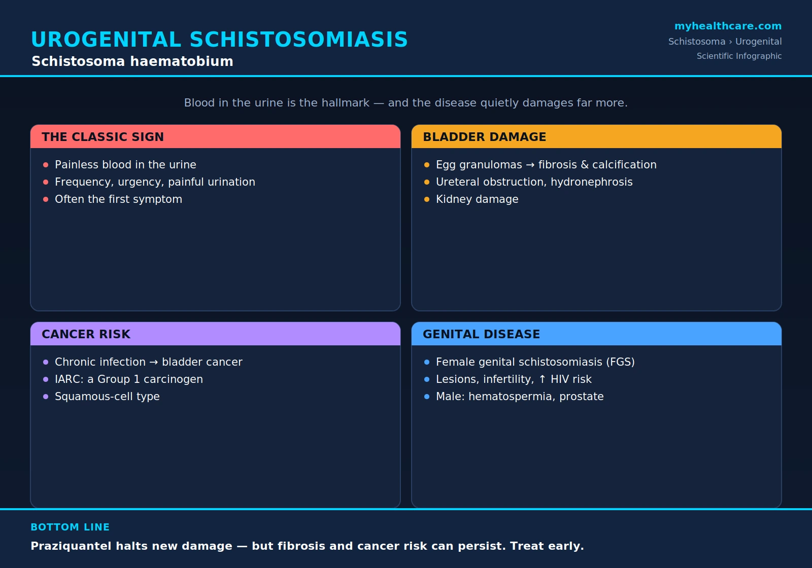
The single most characteristic symptom of urogenital schistosomiasis is blood in the urine (hematuria). As eggs push through the lining of the bladder, they cause small areas of inflammation, ulceration, and bleeding, and blood appears in the urine — often at the very end of urination. The bleeding is usually painless, and it is frequently the first sign of infection, appearing months after the original exposure to contaminated water.
In regions where the disease is common, visible blood in the urine of children has historically been so ordinary that it was sometimes mistaken for a normal event of growing up — in some communities it was wrongly regarded as a male equivalent of a girl's first menstruation, a kind of rite of passage rather than a sign of disease. In reality it is a marker of active infection that warrants treatment.
Alongside the bleeding, people often have other urinary symptoms that reflect an irritated, inflamed bladder:
- Frequency — needing to pass urine more often than usual.
- Urgency — a sudden, hard-to-ignore need to urinate.
- Dysuria — pain, burning, or discomfort during urination.
- Suprapubic discomfort — aching or pressure low in the pelvis, over the bladder.
Because these symptoms resemble an ordinary urinary tract infection, schistosomiasis is easy to overlook in travelers and migrants from areas where it is common. A history of freshwater contact in an endemic region, combined with painless bloody urine, should always raise the suspicion of S. haematobium infection.
3. Chronic Bladder Damage
When infection continues for years and eggs keep accumulating, the repeated cycles of inflammation and healing remodel the bladder and the rest of the urinary tract. The granulomas that form around trapped eggs eventually give way to scar tissue, and over time this produces a series of structural changes:
- Bladder-wall fibrosis and calcification. The wall of the bladder becomes thickened, stiff, and scarred. Dead and calcified eggs can accumulate in such numbers that the bladder wall takes on a chalky, calcified appearance — sometimes visible on imaging as a characteristic "sandy patch" or calcified rim. A stiff, scarred bladder holds less urine and empties poorly.
- Ureteral obstruction. The ureters — the tubes carrying urine from the kidneys down to the bladder — can become scarred and narrowed, especially where they enter the bladder. This blocks the normal flow of urine.
- Hydronephrosis. When urine cannot drain freely, it backs up and swells the kidneys, a condition called hydronephrosis. Persistent back-pressure stretches and damages kidney tissue.
- Kidney damage and failure. Long-standing obstruction, combined with repeated urinary infections that thrive in a damaged, poorly draining system, can gradually destroy kidney function. In severe, untreated cases this can progress to chronic kidney disease and kidney failure.
These complications develop silently over many years, which is why early detection and treatment — before scarring becomes fixed — matter so much. Once the bladder and ureters are heavily fibrosed, the structural damage may not fully reverse even after the parasite is cleared.
4. Bladder Cancer Risk
One of the most serious long-term consequences of chronic S. haematobium infection is an increased risk of bladder cancer. Decades of continuous inflammation, ulceration, and tissue repair in the bladder create conditions in which cancer is more likely to arise. The cancer associated with schistosomiasis is usually squamous-cell carcinoma of the bladder — a type that differs from the urothelial (transitional-cell) cancers more common in places where schistosomiasis is rare. In regions of Africa and the Middle East where the parasite is endemic, schistosomiasis-associated squamous-cell bladder cancer has historically been one of the leading cancers in men.
The link is well established. The International Agency for Research on Cancer (IARC), the cancer arm of the World Health Organization, classifies infection with Schistosoma haematobium as carcinogenic to humans (Group 1) — the highest level of evidence, the same category as tobacco smoke. Schistosomiasis-related bladder cancer tends to appear at a younger age than urothelial bladder cancer and often presents at an advanced stage, partly because its early symptoms — bleeding and urinary irritation — overlap with the infection itself and may be dismissed.
The practical message is that long-term, untreated urinary schistosomiasis is not a benign nuisance: it is a recognized cause of cancer. Treating the infection early and reducing the years of chronic inflammation is an important part of lowering this risk. For the broader picture of bladder and other cancers, see Oncology.
5. Female Genital Schistosomiasis (FGS)
One of the most important and most overlooked aspects of urogenital schistosomiasis is its effect on the female genital tract. Because the worms live in the pelvic veins, S. haematobium eggs are deposited not only in the bladder but also in the cervix, vagina, and vulva, and sometimes the upper genital tract. The resulting condition, female genital schistosomiasis (FGS), is thought to affect many millions of women and girls in endemic regions, yet it is frequently unrecognized and untreated.
The trapped eggs cause inflammation and characteristic lesions in the genital tissues, which can produce a wide range of symptoms:
- Genital lesions — visible sandy patches, grainy areas, or abnormal blood vessels on the cervix and vaginal walls.
- Abnormal bleeding — bleeding between periods or after intercourse, and bloody or abnormal vaginal discharge.
- Pain — pelvic pain and pain during intercourse (dyspareunia).
- Infertility and pregnancy problems — scarring of the genital tract can contribute to difficulty conceiving and to complications in pregnancy.
FGS is repeatedly mistaken for sexually transmitted infections or even genital cancer, leading to wrong diagnoses and missed treatment. Beyond its direct symptoms, FGS carries a major public-health consequence: the inflamed, broken genital surfaces it creates make it easier to acquire HIV. Studies in southern Africa have found that women with genital schistosomiasis have a substantially higher risk of HIV infection, making FGS a quietly significant driver of the HIV epidemic in some communities. For all these reasons, FGS is increasingly recognized as a serious, under-addressed women's-health problem. See also Reproductive Medicine.
6. Male Genital Schistosomiasis
Schistosomiasis of the genital tract is not limited to women. In men, S. haematobium eggs can lodge in the reproductive organs — particularly the seminal vesicles and the prostate gland, and at times the epididymis and testes. This condition is sometimes called male genital schistosomiasis (MGS), and like its female counterpart it is under-recognized.
The inflammation it causes can produce:
- Blood in the semen (hematospermia) — a frequent and telling sign of egg deposition in the seminal vesicles and prostate.
- Pelvic, perineal, or genital pain, sometimes worsened by ejaculation.
- Changes in semen and, in some men, effects on fertility, reflecting inflammation of the male reproductive glands and ducts.
As with women, these symptoms are easily attributed to other causes, and the underlying parasitic infection may be missed unless freshwater exposure in an endemic area is taken into account. Recognizing hematospermia in a traveler or resident from a schistosomiasis region as a possible sign of S. haematobium infection can lead to correct diagnosis and treatment.
7. Diagnosis
Several methods are used to diagnose urogenital schistosomiasis, often in combination. Because the eggs are the proof of infection, finding them is the foundation of diagnosis:
- Urine microscopy for eggs. The standard confirmatory test is examination of urine under a microscope for the eggs of S. haematobium, which are distinctive: oval, with a sharp terminal spine at one end (in contrast to the lateral spine of S. mansoni). Because egg shedding peaks in the middle of the day, urine is best collected around midday (roughly between 10 a.m. and 2 p.m.), sometimes after light exercise, to improve the chance of detecting eggs. The sample is often passed through a fine filter to concentrate and count the eggs.
- Urine dipstick for blood. A simple dipstick test detecting microscopic blood (hematuria) in the urine is widely used as a quick, inexpensive screening tool in endemic areas, where blood in the urine strongly suggests infection.
- Antibody and antigen tests. Blood tests that detect antibodies against the parasite, or assays that detect circulating schistosome antigens, are useful — especially in travelers and in light or early infections where eggs may be hard to find. Antibody tests cannot distinguish past from current infection, while antigen detection reflects active, living worms.
- Imaging and cystoscopy. Ultrasound of the urinary tract is the workhorse for assessing damage — bladder-wall thickening and calcification, ureteral dilation, and hydronephrosis. Cystoscopy (looking inside the bladder with a thin camera) can reveal characteristic lesions and sandy patches and allows biopsy when needed, for example to evaluate suspicious areas for cancer.
Putting these together — eggs in a midday urine sample, blood on dipstick, supportive blood tests, and imaging of the bladder and kidneys — allows both confirmation of the infection and assessment of how much damage it has already caused. For related laboratory testing of the urinary system, see Urology.
8. Treatment and What It Can & Cannot Reverse
The treatment of urogenital schistosomiasis is, fortunately, straightforward and effective at clearing the parasite. The drug of choice is praziquantel, an oral medication that kills the adult worms and rapidly halts new egg-laying. A short course is usually enough to cure the infection, and praziquantel is the backbone of both individual treatment and large-scale community control programs. Its details are covered on the dedicated Praziquantel page.
It is important, however, to understand what treatment can and cannot do. By stopping the worms from laying more eggs, praziquantel removes the source of ongoing damage, and in early disease the body can heal: bladder inflammation often subsides, recent lesions may resolve, and in children especially much of the early urinary-tract and genital change can improve or reverse with timely treatment. Killing the worms is the essential first step toward recovery.
What praziquantel cannot undo is damage that has already become permanent. Once the bladder and ureters are heavily fibrosed and calcified, that scarring tends to persist even after the parasite is gone. The drug kills worms; it does not dissolve old scar tissue or reopen badly narrowed ureters. Likewise, treatment does not erase the cancer risk built up over years of chronic inflammation, although clearing the infection and ending that inflammation is an important way to keep further risk from accumulating. People with advanced disease may still need ongoing urological care, and sometimes surgery, to manage the structural complications.
The clear lesson is that the earlier urogenital schistosomiasis is found and treated, the more of its harm can be prevented or reversed. Equally important is not getting re-infected: in endemic areas, avoiding contact with contaminated fresh water and supporting community-wide control efforts are essential. See Prevention and Avoiding Freshwater, Mass Drug Administration and Control, and the broader Treatment & Prevention hub.
Key Research Papers
Peer-reviewed reviews and reports on urogenital schistosomiasis — covering the disease overall, bladder pathology and cancer, female and male genital involvement, the burden of chronic infection, and control. Journal names appear as plain text; the year/volume/pages link opens the full citation via DOI. The IARC entry links to the World Health Organization monograph itself.
- Colley DG, Bustinduy AL, Secor WE, King CH. Human Schistosomiasis. The Lancet. 2014;383(9936):2253–2264.
- Gryseels B, Polman K, Clerinx J, Kestens L. Human Schistosomiasis. The Lancet. 2006;368(9541):1106–1118.
- Mostafa MH, Sheweita SA, O'Connor PJ. Relationship Between Schistosomiasis and Bladder Cancer. Clinical Microbiology Reviews. 1999;12(1):97–111.
- IARC Working Group. Schistosomes, Liver Flukes and Helicobacter pylori. IARC Monographs on the Evaluation of Carcinogenic Risks to Humans, Volume 61. International Agency for Research on Cancer (World Health Organization). 1994.
- Kjetland EF, Ndhlovu PD, Gomo E, et al. Association Between Genital Schistosomiasis and HIV in Rural Zimbabwean Women. AIDS. 2006;20(4):593–600.
- Kjetland EF, Leutscher PDC, Ndhlovu PD. A Review of Female Genital Schistosomiasis. Trends in Parasitology. 2012;28(2):58–65.
- Hotez PJ, Fenwick A. Schistosomiasis in Africa: An Emerging Tragedy in Our New Global Health Decade. PLoS Neglected Tropical Diseases. 2009;3(9):e412.
- King CH, Dickman K, Tisch DJ. Reassessment of the Cost of Chronic Helminthic Infection: A Meta-Analysis of Disability-Related Outcomes in Endemic Schistosomiasis. The Lancet. 2005;365(9470):1561–1569.
- Fenwick A, Webster JP, Bosque-Oliva E, et al. The Schistosomiasis Control Initiative (SCI): Present and Future Schistosomiasis Control Activities. Parasitology. 2009;136(13):1731–1737.
- Hatz CFR. The Use of Ultrasound in Schistosomiasis. Advances in Parasitology. 2001;48:225–284.
Live PubMed Searches
Each link opens a live PubMed query so results stay current as new papers are indexed.
- Urogenital schistosomiasis (S. haematobium)
- S. haematobium hematuria and bladder
- Schistosomiasis and squamous-cell bladder cancer
- Female genital schistosomiasis and HIV
- Male genital schistosomiasis and hematospermia
- S. haematobium hydronephrosis and ureter
- Praziquantel for urinary schistosomiasis
- Schistosomiasis ultrasound and urinary morbidity
Connections
- Schistosoma Symptoms & Diagnosis
- Intestinal & Hepatic Schistosomiasis
- Acute Schistosomiasis & Swimmer's Itch
- Schistosoma Treatment & Prevention
- Praziquantel Treatment
- Prevention & Avoiding Freshwater
- Mass Drug Administration & Control
- Schistosoma Overview
- All Parasites
- Urology
- Oncology
- Reproductive Medicine
- Infectious Disease
- All Conditions