Ocular Toxoplasmosis
Ocular toxoplasmosis is an infection of the back of the eye by the parasite Toxoplasma gondii. The parasite attacks the retina — the light-sensing layer that lines the inside of the eye — and the choroid beneath it, producing a localized patch of inflammation called retinochoroiditis. It is the single most common cause of posterior uveitis (inflammation of the rear segment of the eye) and the most common cause of infectious retinitis worldwide. The disease is notorious for one trait above all others: it tends to come back. A scar from a long-quiet infection can flare again years or decades later, threatening vision each time. This page explains what ocular toxoplasmosis is, how it differs depending on whether infection began before or after birth, what patients feel and what doctors see, why it recurs, when sight is genuinely at risk, and how it is diagnosed and treated.
Table of Contents
- What Is Ocular Toxoplasmosis?
- Congenital vs. Acquired
- Symptoms
- What the Doctor Sees
- Why It Comes Back
- When Vision Is Threatened
- Diagnosis
- Treatment
- Key Research Papers
- Featured Videos
1. What Is Ocular Toxoplasmosis?
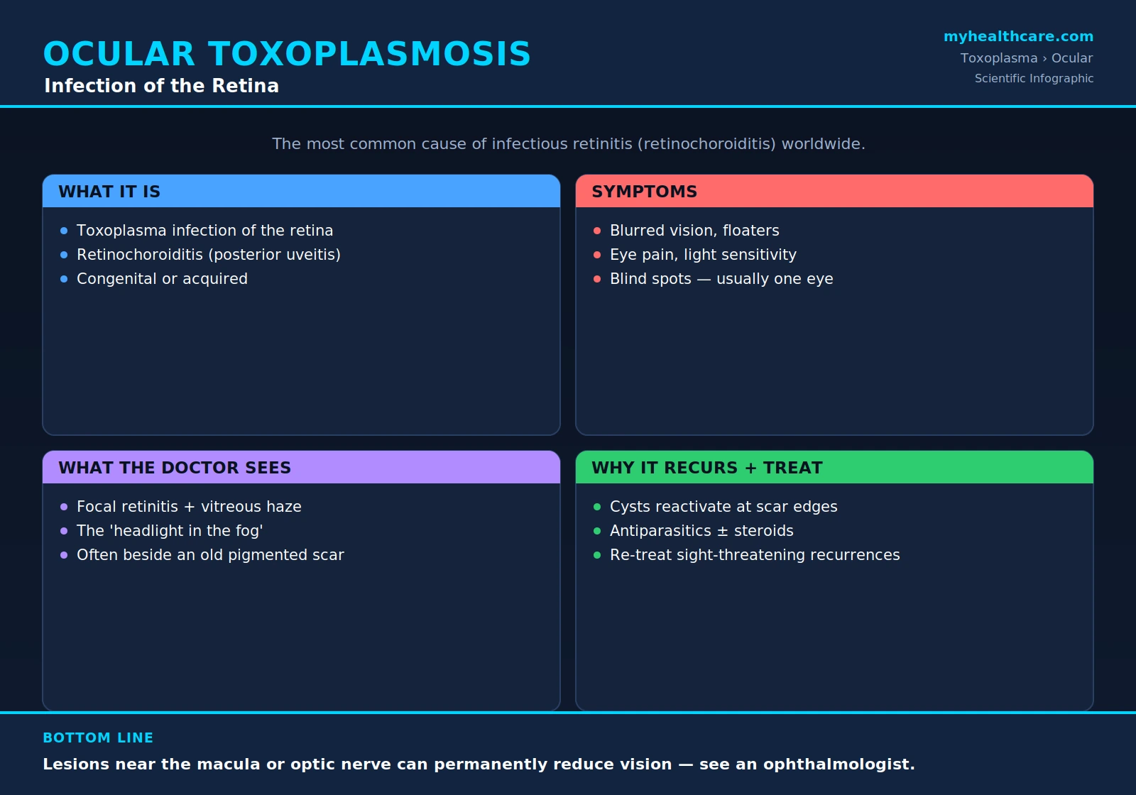
Ocular toxoplasmosis is disease of the eye caused by Toxoplasma gondii, the same single-celled parasite responsible for toxoplasmosis throughout the body. In the eye, the parasite establishes itself in the retina, the delicate layer of nerve tissue at the back of the eye that captures light and turns it into the signals the brain reads as vision. The infection inflames the retina and the underlying choroid (the vascular layer that nourishes it), which is why the classic lesion is called retinochoroiditis — inflammation of retina and choroid together.
What makes this infection so important is its sheer frequency. Among the many causes of posterior uveitis — inflammation affecting the back portion of the eye — ocular toxoplasmosis is the most common single cause worldwide. It is likewise the leading infectious cause of retinitis (inflammation of the retina). In other words, when an ophthalmologist anywhere in the world sees a patient with an inflamed lesion in the back of the eye, Toxoplasma is statistically the most likely culprit.
The infection is usually focal: it produces one (sometimes a few) discrete areas of active disease rather than inflaming the whole eye uniformly. And it typically affects one eye at a time. Because the retina does not regenerate, any tissue destroyed by an episode leaves a permanent scar — and that scar becomes the seed for future trouble.
2. Congenital vs. Acquired
Ocular toxoplasmosis can begin in two fundamentally different ways, and the distinction shapes how the disease behaves over a lifetime.
Congenital (infection before birth). If a woman acquires Toxoplasma for the first time during pregnancy, the parasite can cross the placenta and infect the fetus — a condition covered in detail on the Congenital Toxoplasmosis page. Eye involvement is one of the most common consequences. Some affected newborns already have visible retinal scars at birth; in many others, the eye looks normal at first, and the infection lies dormant only to flare up years or even decades later, often during childhood, adolescence, or early adulthood. For a long time, this congenital route was thought to account for the large majority of ocular toxoplasmosis.
Acquired (infection after birth). It is now well recognized that a substantial share of cases follow infection acquired after birth — from eating undercooked meat containing tissue cysts, or from food, water, or soil contaminated with parasite oocysts shed by cats. (See Prevention: Food and Cat Safety.) Postnatally acquired infection can also seed the retina and produce the same kind of recurring eye disease.
Strain matters. The severity of acquired ocular disease appears to depend in part on which strain of the parasite is involved. In parts of South America (notably Brazil), certain more virulent, genetically distinct strains are associated with more frequent, larger, and more aggressive eye lesions than the strains common in North America and Europe. This helps explain why ocular toxoplasmosis is both more common and often more severe in some South American populations.
3. Symptoms
The symptoms of ocular toxoplasmosis come from the inflammation in the back of the eye and from the debris (inflammatory cells) it sheds into the clear gel, the vitreous, that fills the eyeball. Because the disease is usually one-sided, symptoms are typically noticed in a single eye. They include:
- Blurred or decreased vision — the most common complaint, ranging from a slight haze to a marked drop in sharpness.
- Floaters — specks, strands, or cobweb-like shadows drifting across the field of view, caused by inflammatory cells clumping in the vitreous.
- Eye redness and pain — an ache or soreness, sometimes with a visibly red eye when inflammation spreads to the front of the eye.
- Light sensitivity (photophobia) — discomfort or glare in bright light.
- Blind spots (scotomas) — fixed gaps or dark patches in the field of vision corresponding to areas of damaged retina.
The exact mix and severity depend heavily on where the lesion sits. A small patch in the far periphery of the retina may cause only mild floaters that a person barely notices, while a lesion at the center of vision can cause a dramatic, frightening loss of sight. Symptoms usually develop over days as a recurrence becomes active, and tend to settle as the episode is treated and resolves — though any permanent scar leaves a lasting blind spot.
4. What the Doctor Sees
When an ophthalmologist examines the back of the eye during an active episode, the appearance is often strikingly characteristic. The hallmark is a focal area of necrotizing retinitis — a discrete, creamy-white or yellow-white patch where the parasite is destroying retinal tissue — with overlying inflammation in the vitreous. The cloud of inflammatory cells in the vitreous partly obscures the bright lesion beneath, producing the classic description of a "headlight in the fog": a glowing white focus seen dimly through an overlying haze.
A second, equally telling sign is the relationship of the new lesion to old damage. Active recurrences very often appear right next to an old, pigmented chorioretinal scar — the healed footprint of a previous episode. This "satellite" pattern, a fresh white lesion budding from the edge of a dark, sharply defined scar, is one of the most useful clues that an ophthalmologist has to recognize ocular toxoplasmosis on sight, even before any laboratory test.
Depending on severity, the examiner may also see inflammation of nearby blood vessels (retinal vasculitis), swelling of the optic nerve head, and cells and flare in the front chamber of the eye when the inflammation is intense. The combination of a focal retinitis, heavy vitreous reaction, and an adjacent old scar is, for many cases, diagnostic in itself.
5. Why It Comes Back
The defining feature of ocular toxoplasmosis — the thing that sets it apart from most other eye infections — is that it recurs. A single person may suffer repeated flares in the same eye across a lifetime. Understanding why requires understanding how the parasite hides.
After an episode heals, Toxoplasma does not necessarily leave. Instead, it converts into a dormant, walled-off form called a tissue cyst, packed with slowly metabolizing parasites (bradyzoites). These cysts persist silently in the retina, characteristically at the edge of the old scar. They are invisible to the immune system as long as they stay sealed, which is why a scarred eye can remain quiet for years.
A recurrence happens when one or more of these cysts at the border of an old scar reactivate — the wall breaks down and the parasites inside convert back to the fast-multiplying, tissue-destroying form (tachyzoites), spilling into the surrounding healthy retina and igniting a new patch of inflammation. This is precisely why fresh lesions so reliably appear adjacent to old scars. Because viable cysts can persist indefinitely, the risk of recurrence is lifelong. Episodes tend to cluster — a recurrence raises the short-term odds of another — and various triggers (including changes in immune status) are thought to encourage reactivation, though a flare can also occur with no identifiable trigger at all.
6. When Vision Is Threatened
Not every episode of ocular toxoplasmosis endangers sight. Many lesions sit in the peripheral retina, heal with treatment or even on their own, and leave a scar that the patient never notices because it lies outside the line of sight. The threat to vision depends on three things above all: where the lesion is, how much inflammation accompanies it, and what complications follow.
- Location near the macula. The macula is the small central zone of the retina responsible for sharp, detailed, straight-ahead vision — reading, faces, fine work. A lesion in or near the macula can leave a permanent central blind spot and a lasting drop in visual acuity, even after the infection is controlled.
- Location near the optic nerve. Lesions next to the optic nerve head (juxtapapillary disease) can damage the nerve fibers that carry vision out of the eye, threatening larger portions of the field of view.
- Heavy vitreous inflammation. A dense inflammatory reaction in the vitreous can cloud vision while it is active and, if severe, contribute to longer-term problems.
Beyond the lesion itself, complications such as retinal scarring with traction, abnormal new blood-vessel growth, swelling of the central retina (macular edema), and in some cases retinal detachment can each reduce sight permanently. It is this potential for irreversible damage — especially from centrally placed or heavily inflamed lesions — that drives the decision to treat aggressively when vision is at stake (see Treatment & Prevention).
7. Diagnosis
The diagnosis of ocular toxoplasmosis is, first and foremost, a clinical one — it rests mainly on the eye examination performed by an ophthalmologist. The characteristic combination already described — a focal necrotizing retinitis, the "headlight in the fog" of overlying vitreous inflammation, and a fresh lesion budding from the edge of an old pigmented scar — is so typical that experienced clinicians can often make the diagnosis on appearance alone.
Supporting tests are used to add confidence, especially in less typical cases:
- Serology (blood antibody tests). Detecting antibodies to Toxoplasma in the blood confirms that a person has been infected at some point. Its main value is in excluding the diagnosis: a person with no antibodies almost certainly does not have ocular toxoplasmosis. Because antibodies are common in the general population, however, a positive result alone does not prove that a given eye lesion is toxoplasmic — it must be interpreted alongside the clinical picture.
- PCR and antibody analysis of eye fluid. In atypical, severe, or diagnostically uncertain cases — for example, unusual-looking lesions, immunocompromised patients, or disease that is not responding as expected — a sample of intraocular fluid (usually aqueous humor from the front of the eye) can be tested. PCR detects parasite DNA directly, and comparison of antibody levels inside the eye versus the blood (the Goldmann–Witmer coefficient) can show that antibodies are being produced locally. These tests are reserved for difficult cases rather than used routinely.
- Imaging. Retinal photography, optical coherence tomography (OCT), and angiography help document the lesion, monitor the response to treatment, and detect complications such as macular edema or new vessel growth.
In short, the eye exam leads, serology supports, and molecular testing of eye fluid is held in reserve for the cases that do not fit the classic mold.
8. Treatment
Treatment of ocular toxoplasmosis is tailored to the threat each episode poses, and is directed by an ophthalmologist. The general principles are these:
- Antiparasitic drugs are the foundation of treatment for sight-threatening or active vision-affecting disease. They aim to suppress the multiplying parasites and limit the size of the new lesion. The conventional regimens — and how they work — are covered on the Antiparasitic Treatment page; the broader strategy is summarized under Treatment & Prevention.
- Corticosteroids are sometimes added — carefully, and generally alongside antiparasitic drugs rather than alone — to calm intense inflammation when it threatens the macula or optic nerve, since much of the damage to delicate central tissue comes from the inflammatory response itself.
- Observation is a legitimate option for small peripheral lesions in otherwise healthy people. Because such lesions often heal on their own without endangering central vision, and because the drugs carry their own risks, a clinician may reasonably choose to monitor rather than treat — a decision that always belongs to the treating ophthalmologist.
- Re-treatment for recurrences. Because the disease comes back, a patient may need a fresh course of treatment with each sight-threatening flare. Importantly, an episode-by-episode drug course treats the active infection but does not eradicate the dormant cysts, so it does not by itself prevent the next recurrence; strategies aimed at reducing recurrence frequency are a separate consideration.
The thread running through all of this is judgment: matching the intensity of treatment to the location and severity of the lesion, protecting the macula and optic nerve, and weighing the benefits of the drugs against their side effects for each individual patient.
Key Research Papers
Peer-reviewed reviews, clinical trials, and case series on ocular toxoplasmosis — covering its biology and epidemiology, recurrence patterns, diagnostic approach, and treatment. Journal names appear as plain text; the year/volume/pages link opens the full citation via DOI.
- Montoya JG, Liesenfeld O. Toxoplasmosis. The Lancet. 2004;363(9425):1965–1976.
- Park YH, Nam HW. Clinical Features and Treatment of Ocular Toxoplasmosis. Korean Journal of Parasitology. 2013;51(4):393–399.
- Holland GN. Ocular Toxoplasmosis: New Directions for Clinical Investigation. Ocular Immunology and Inflammation. 2000;8(1):1–7.
- Bosch-Driessen LEH, Berendschot TTJM, Ongkosuwito JV, Rothova A. Ocular Toxoplasmosis: Clinical Features and Prognosis of 154 Patients. Ophthalmology. 2002;109(5):869–878.
- Holland GN, Crespi CM, ten Dam-van Loon N, Charonis AC, et al. Analysis of Recurrence Patterns Associated with Toxoplasmic Retinochoroiditis. American Journal of Ophthalmology. 2008;145(6):1007–1013.
- Garweg JG, de Groot-Mijnes JDF, Montoya JG. Diagnostic Approach to Ocular Toxoplasmosis. Ocular Immunology and Inflammation. 2011;19(4):255–261.
- Garweg JG, Pleyer U. Treatment Strategy in Human Ocular Toxoplasmosis: Why Antibiotics Have Failed. Journal of Clinical Medicine. 2021;10(5):1090.
- Opremcak EM, Scales DK, Sharpe MR. Trimethoprim-Sulfamethoxazole Therapy for Ocular Toxoplasmosis. Ophthalmology. 1992;99(6):920–925.
- Pearson PA, Piracha AR, Sen HA, Jaffe GJ. Atovaquone for the Treatment of Toxoplasma Retinochoroiditis in Immunocompetent Patients. Ophthalmology. 1999;106(1):148–153.
- Soheilian M, et al.; Holland GN. Prospective, Randomized Trial of Trimethoprim/Sulfamethoxazole versus Pyrimethamine and Sulfadiazine in the Treatment of Ocular Toxoplasmosis. Ophthalmology. 2005;112(11):1882–1884.
Live PubMed Searches
Each link opens a live PubMed query so results stay current as new papers are indexed.
- Ocular toxoplasmosis
- Toxoplasmic retinochoroiditis
- Ocular toxoplasmosis recurrence
- Ocular toxoplasmosis treatment
- Ocular toxoplasmosis posterior uveitis
- Toxoplasma strain virulence retinochoroiditis
- Ocular toxoplasmosis PCR aqueous humor diagnosis
- Congenital toxoplasmosis retinochoroiditis
Connections
- Toxoplasma Overview
- Symptoms & Diagnosis
- Congenital Toxoplasmosis
- Toxoplasmosis in the Immunocompromised
- Treatment & Prevention
- Antiparasitic Treatment
- Prevention: Food & Cat Safety
- Pregnancy Screening & Prevention
- All Parasites
- Ophthalmology
- Dry Eye Disease
- Macular Degeneration
- Infectious Disease
- All Conditions