Severo Ochoa & Arthur Kornberg: The Enzymes Behind PCR, DNA Sequencing and Antiviral Drugs
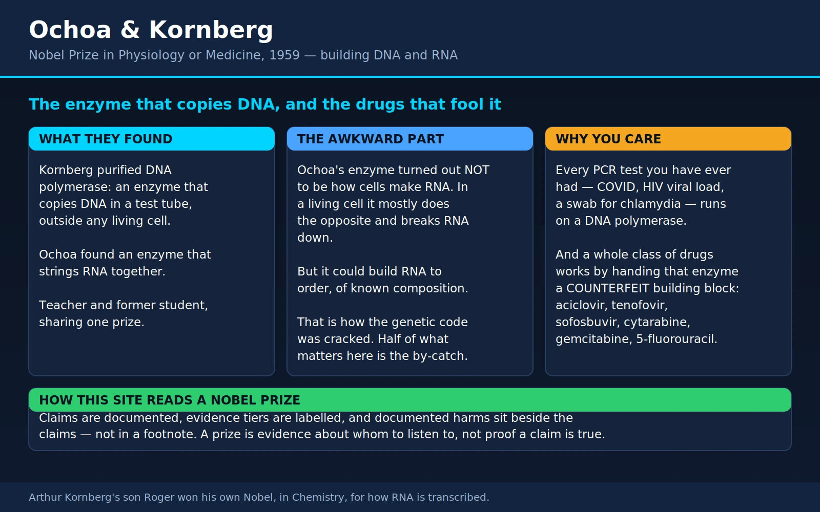
Table of Contents
- Overview
- A Teacher and His Student, Sharing a Prize
- What Ochoa Found: Polynucleotide Phosphorylase
- What Kornberg Found: DNA Polymerase
- The Awkward Part: Ochoa's Enzyme Is Not How Cells Make RNA
- Why the "Wrong" Enzyme Cracked the Genetic Code
- Kornberg's Enzyme Had Its Own Reckoning
- 1967: The "Life in a Test Tube" Headlines
- Chain Termination, Explained Plainly
- PCR: The Polymerase Inside Every Diagnostic Test
- DNA Sequencing: Reading, Not Just Copying
- What Sequencing Tells a Patient — and What It Does Not
- Antivirals That Are Counterfeit Nucleotides
- Chemotherapy That Is Counterfeit Nucleotides
- The Kornbergs: A Second Nobel in the Family
- Key Research Papers
- Connections
- Featured Videos
1. Overview
The 1959 Nobel Prize in Physiology or Medicine went jointly to Severo Ochoa and Arthur Kornberg "for their discovery of the mechanisms in the biological synthesis of ribonucleic acid and deoxyribonucleic acid." The official summary is at nobelprize.org — 1959 Prize in Physiology or Medicine. It was awarded six years after the double helix was published, and it answered the obvious next question: if DNA is a template, what actually does the copying?
Kornberg's answer has held up completely. He purified DNA polymerase — a protein that, given a strand of DNA, the four building blocks and magnesium, sits in a test tube and copies that strand. It was the first time anyone had made a nucleic acid to instructions outside a living cell. Ochoa's answer has not held up in the same way, and this page says so directly, because the honest version of the story is more interesting than the tidy one. The enzyme Ochoa was cited for, polynucleotide phosphorylase, turned out not to be how cells make RNA at all. In a living bacterium it mostly runs the other way: it takes RNA apart.
That should have been an embarrassment. Instead it was the single most useful accident in twentieth-century biology. Because Ochoa's enzyme needed no template, it would string together whatever building blocks you fed it, in whatever ratio you chose — and that is exactly what Marshall Nirenberg and Ochoa's own group needed to crack the genetic code. A synthetic RNA of known composition is a question you can ask a cell. Ochoa's "wrong" enzyme is what made the question askable.
The reason a health site covers this at all is what came out of the DNA half. Kornberg described a machine that copies DNA and can be tricked, and both halves of that sentence became medicine:
- Every diagnostic PCR test you have ever had — the COVID swab, an HIV viral load, a chlamydia or gonorrhoea NAAT, the tuberculosis GeneXpert cartridge — is a DNA polymerase copying a target sequence until there is enough of it to see.
- DNA sequencing, and therefore newborn genomic screening, tumour gene panels, pharmacogenomics and prenatal cell-free DNA screening, runs on a DNA polymerase deliberately jammed at known positions.
- A whole class of drugs — aciclovir, tenofovir, sofosbuvir, cytarabine, gemcitabine, 5-fluorouracil — works by handing the polymerase a counterfeit building block. The mechanism Kornberg described in 1956 is, quite literally, the drug target.
Two men found two enzymes. One of the enzymes was misassigned, the other was misassigned about its day job, and between them they produced the modern diagnostic laboratory and a shelf of antiviral and cancer drugs. That is a better story about how science works than the version where everyone is right the first time.
2. A Teacher and His Student, Sharing a Prize
Severo Ochoa was born in Luarca, on the Asturian coast of northern Spain, on 24 September 1905, and died in Madrid on 1 November 1993. The Spanish Civil War and the war that followed it pushed him through Heidelberg, Oxford and finally the United States; by the late 1940s he was at the New York University School of Medicine, where he spent the rest of his working life and eventually chaired the biochemistry department. He became a naturalised American citizen and, in Spain, something closer to a national figure — the country's first Nobel laureate in a laboratory science.
Arthur Kornberg was born in Brooklyn on 3 March 1918 and died on 26 October 2007. He came to enzymology by an unusual route: he trained as a physician, spent the war years at the National Institutes of Health studying rat nutrition, and concluded that if he wanted to understand vitamins he had to understand the enzymes they served. So he went and learned how to purify one. In 1946 he spent a year in Severo Ochoa's laboratory at New York University, working on enzymes of the citric acid cycle, and in 1947 a further stint with Carl and Gerty Cori at Washington University in St. Louis — two of the best enzyme-purification benches in the world.
He returned to Washington University in 1953 as chair of microbiology, and it was there, between 1953 and 1959, that the DNA polymerase work was done. In 1959 he moved to Stanford to build its biochemistry department, and in that same year the prize arrived. It is worth pausing on the shape of it: Arthur Kornberg shared the 1959 Nobel with the man who had taught him how to purify an enzyme thirteen years earlier. Both Nobel lectures are online — Kornberg's and Ochoa's.
3. What Ochoa Found: Polynucleotide Phosphorylase
The discovery was made in 1955 by Marianne Grunberg-Manago, a French postdoctoral researcher in Ochoa's laboratory, working on extracts of the soil bacterium Azotobacter vinelandii. She was not looking for an RNA-making enzyme. She was studying phosphate chemistry — how bacteria shuffle phosphate groups during energy metabolism — and she noticed that when she supplied the extract with adenosine diphosphate, phosphate came out and something large and viscous formed. The something turned out to be a long chain of adenine nucleotides: an RNA-like polymer, built in a tube.
The result was published with Ochoa in Science in 1955 and in full in Biochimica et Biophysica Acta in 1956, with Grunberg-Manago as first author on both, and the enzyme was named polynucleotide phosphorylase — PNPase. It was the first enzyme anyone had found that could assemble a nucleic acid, and in 1955 that was a genuinely startling thing to be able to do.
Two features of the reaction mattered enormously later, and both of them were, in hindsight, warnings:
- It ran on nucleoside diphosphates, not triphosphates. ADP, CDP, GDP, UDP went in; RNA and free inorganic phosphate came out. Every other biosynthetic polymerisation known then and since runs on triphosphates, because that is where the cell keeps its usable energy.
- It needed no template. Give it ADP alone and it made poly(A). Give it a mixture and it made a random copolymer whose composition simply reflected the mixture. Nothing instructed it. That is a very strange property for an enzyme that is supposed to be copying genes.
There was a third feature, and it is the one that eventually decided the question. The reaction was reversible. Add inorganic phosphate and the enzyme runs backwards, chopping RNA up into nucleoside diphosphates. Which direction it runs depends on the concentrations. In a test tube stocked with diphosphates and swept clear of phosphate, it builds. Inside a living bacterium, where inorganic phosphate is abundant, it does the opposite.
4. What Kornberg Found: DNA Polymerase
Kornberg's group at Washington University — principally Maurice Bessman, Robert Lehman and the technician Ernest Simms — went after DNA with a deliberately brutal method. They fed E. coli extracts radioactively labelled thymidine and asked one question: how much of it ends up in a form that behaves like DNA? Then they fractionated the extract, kept whatever fraction still did that, and repeated. It is unglamorous, and it is how you find an enzyme.
The first report was a two-page note in Biochimica et Biophysica Acta in 1956. The full account came in 1958 as a series in the Journal of Biological Chemistry, beginning with the preparation of the substrates and the partial purification of the enzyme from Escherichia coli, followed by the general properties of the reaction. Continuing papers in the series ran into the 1970s; by the 1967 phi-X174 paper the numbering had reached XXIV, and by 1969 it was past XXXIV.
The enzyme — now called DNA polymerase I, or informally the Kornberg enzyme — needed four things: all four deoxynucleoside triphosphates, magnesium, a free 3′ end to build from, and, decisively, a pre-existing strand of DNA to copy. Remove the template and nothing happens. That single requirement is the whole difference between what Kornberg had and what Ochoa had.
The series then took on the question that actually mattered, in the fifth paper: what is the chemical composition of the DNA the enzyme makes? If the enzyme were merely stitching nucleotides together, the product's composition would track whatever was most abundant in the tube, or whatever the enzyme happened to prefer. If it were copying, the product would match the template — adenine equalling thymine, guanine equalling cytosine, in the template's own proportions, whatever those were. It matched the template. Five years after Watson and Crick proposed that the double helix explains how genes are copied, an enzyme in a tube was observed doing it.
This is why Kornberg's half of the prize has never been seriously questioned. He did not merely find a protein that makes DNA. He established that DNA synthesis is instructed — that the sequence comes from the template and not from the machinery — which is the biochemical foundation of heredity.
5. The Awkward Part: Ochoa's Enzyme Is Not How Cells Make RNA
Between 1959 and 1961, several laboratories independently found a different enzyme. It used ribonucleoside triphosphates, not diphosphates. It required a DNA template, and the RNA it produced was a faithful copy of that template. Samuel Weiss's 1960 PNAS paper is one of the founding reports; Jerard Hurwitz's and Audrey Stevens's groups reported closely related work, and Ochoa's own laboratory at NYU joined the effort within a year. This enzyme — DNA-dependent RNA polymerase — is how cells actually make RNA. It is the enzyme the 1959 citation was reaching for.
What happened to PNPase is now settled and is stated plainly in the modern literature. A 2018 review in PLoS Genetics opens by noting that Ochoa received the Nobel Prize for the enzymatic synthesis of RNA by polynucleotide phosphorylase, and then says that subsequent work revealed the enzyme's predominant function in bacteria and eukaryotes is the reverse reaction — releasing ribonucleotides from RNA. PNPase is a real, important, thoroughly studied enzyme with a genuine role in RNA metabolism, including in RNA processing, in messenger-RNA turnover and in a growing list of what that review calls noncanonical functions. Its principal job is disassembly.
So the position is this, and there is no way to soften it that is also true: the 1959 prize cites "the biological synthesis of ribonucleic acid," and the enzyme Ochoa was cited for does not, in a living cell, principally synthesise ribonucleic acid. The reaction he and Grunberg-Manago described is real — it happens exactly as reported, in a tube, under the conditions they used. The error was not in the observation. It was in the inference from an in-vitro reaction to a cellular job, and it is one of the most instructive mistakes in the history of biochemistry because the reaction itself was never wrong.
6. Why the "Wrong" Enzyme Cracked the Genetic Code
Now the twist, and it is a large one. The very property that disqualified PNPase as the cell's RNA-maker — that it copies nothing and simply polymerises whatever you give it — made it the one tool that could break the genetic code.
By 1961 the shape of the problem was clear: a sequence of four RNA bases somehow specifies a sequence of twenty amino acids. Nobody knew how many bases per amino acid, let alone which. To find out, you needed to hand a protein-making system a piece of RNA whose composition you knew in advance and see what protein came out. There was no way to write an RNA sequence to order. But there was a way to make an RNA of known composition: feed Ochoa's enzyme exactly one kind of building block, or a mixture in an exactly known ratio.
Feed it only UDP and you get poly(U) — an RNA that is nothing but uracil. In 1961, Marshall Nirenberg and Heinrich Matthaei put poly(U) into a cell-free E. coli extract and got back a protein made of nothing but phenylalanine. UUU codes for phenylalanine. That was the first word of the genetic code, and it was read using a polymer made by Severo Ochoa's enzyme.
Ochoa's own group went straight at the rest of it. Feeding PNPase mixtures in known ratios — five parts U to one part C, say — produces a random copolymer in which the statistical frequency of every possible triplet is calculable in advance. Compare the predicted triplet frequencies against the amino acids that actually appear in the product and you can deduce each codon's base composition, even though you cannot see its order. Peter Lengyel, Joseph Speyer and Ochoa published the first of these in PNAS in December 1961, and the numbered series that followed through 1962 assigned compositions to most of the twenty amino acids in about a year. It was one of the fastest problems ever solved in biology, and it was solved statistically, with an enzyme that could not read.
The order within each codon came later, from Nirenberg and Philip Leder's ribosome-binding assay and from Har Gobind Khorana's chemically defined repeating polymers. The 1968 Nobel Prize went to Nirenberg, Khorana and Holley, not to Ochoa — he had already been recognised, in 1959, for the enzyme that made all of it possible. The irony is almost too neat: Ochoa's prize rests on an enzyme whose real cellular role is demolition, and that enzyme is why we can read the genetic code. Both of those statements are true and neither cancels the other.
7. Kornberg's Enzyme Had Its Own Reckoning
If you think Ochoa was uniquely unlucky, consider what happened to Kornberg's enzyme ten years after the prize.
In 1969, Paula De Lucia and John Cairns reported in Nature the isolation of an E. coli strain carrying a mutation that knocked out the Kornberg polymerase's activity — the strain later known as polA1. The problem was that the bacterium was alive. It grew, it divided, and it replicated its chromosome, with a tiny fraction of the enzyme activity that was supposed to be doing the replicating. (It was abnormally sensitive to ultraviolet light, which turned out to be the clue to what the enzyme really does.) Whatever DNA polymerase I was for, it was not the cell's main replication machine.
The mutant strain was also the perfect bait. Purify from a cell that has no polymerase I and whatever DNA-synthesising activity remains must be something else. In 1971 Thomas Kornberg — Arthur's son — working with Malcolm Gefter, purified that remaining activity to homogeneity and named it DNA polymerase II; it needed all four triphosphates, magnesium and native DNA, and, tellingly, it was completely unaffected by antiserum raised against polymerase I. In the same work they flagged a second, much scarcer DNA-synthesising activity in those extracts. That second activity was purified the following year and became DNA polymerase III — which is the replicase, the enzyme that actually copies the bacterial chromosome.
Kornberg's enzyme was not demoted to irrelevance. Polymerase I turned out to hold a job that is essential and rather elegant: as the replication fork runs, one strand is built in short pieces, each started by a little RNA primer. Polymerase I removes those RNA primers with a nuclease activity built into the same protein and fills the resulting gaps with DNA. It also does repair synthesis — which is why the polA1 mutant could replicate perfectly well and still be killed by ultraviolet light. The enzyme was correctly described in 1956. What was too large was the inference about which of the cell's jobs it held.
8. 1967: The "Life in a Test Tube" Headlines
In December 1967, Mehran Goulian, Arthur Kornberg and Robert Sinsheimer published a paper in PNAS titled "Enzymatic synthesis of DNA, XXIV. Synthesis of infectious phage phi-X174 DNA."
Here is what was actually done. Phi-X174 is a bacteriophage — a virus that infects bacteria — with an unusually simple genome: a single circular strand of DNA about 5,400 bases long. Kornberg's group used that natural strand as a template, ran DNA polymerase on it to build the complementary copy, and used a joining enzyme to seal the copy into a closed circle. The copy was then tested the only way that counts: they put it into E. coli. It infected the bacteria and produced phage. A DNA molecule assembled by enzymes in a tube was biologically active.
That is a genuinely major result. It demonstrated that the copy was accurate along its whole length — not approximately right, but right enough that every one of the phage's genes worked. No chemical assay of the era could have shown that; only biology could.
Here is what was not done. Nothing was designed. No sequence was chosen, written or altered — indeed nobody knew what the sequence was, because phi-X174 would not be sequenced for another decade. A natural template was copied, and a copier is not an author. Nothing was created from simple chemicals. And a virus, which cannot reproduce without a host cell to hijack, is a long way from a cell. The word for what was made is a copy of a piece of a virus.
The public account went somewhere else entirely. According to the US National Library of Medicine's archival account of the episode, Kornberg specifically cautioned journalists at the announcement that the experiment had nothing to do with "synthesizing life in a test tube" — and on the same day President Lyndon Johnson, speaking at the Smithsonian Institution, told an audience that some geniuses at Stanford University had created life in the test tube. The headlines followed the president. The archival record is at NLM Profiles in Science — "Creating Life in the Test Tube," 1959–1970.
9. Chain Termination, Explained Plainly
In 1969, a paper from Kornberg's laboratory — Atkinson, Deutscher, Kornberg, Russell and Moffatt, number XXXIV in the same long series — carried the title "Termination of chain growth by a 2′,3′-dideoxyribonucleotide." Everything that follows in the rest of this page comes out of that sentence, so it is worth understanding properly. It takes about a paragraph.
A DNA polymerase builds a strand in one direction only. Each new building block is attached to a specific chemical handle on the end of the growing chain: the 3′-hydroxyl group, an oxygen-hydrogen pair sticking off the sugar of the last nucleotide added. Every normal building block arrives carrying a fresh 3′-hydroxyl of its own, so as soon as it is attached it supplies the handle for the next one. That is what lets the chain grow indefinitely.
A dideoxynucleotide is identical to a normal one in every respect the polymerase checks — same base, same shape, same charge — except that it has no 3′-hydroxyl. The polymerase accepts it, attaches it, and then reaches for the handle that should be there. There is nothing to grab. The strand does not slow down; it stops, permanently.
The everyday version: imagine a zip in which one tooth has been manufactured without the notch that the next tooth clips into. The zip closes normally right up to that tooth, and then it can never go further, no matter how hard you pull. The defect is invisible until the moment it matters, and by then the tooth is already sewn in.
That is chain termination, and it does two completely different jobs. Aimed at a machine you want to read, it is how DNA sequencing works. Aimed at a machine you want to stop — a virus's polymerase, or a cancer cell's — it is a drug.
10. PCR: The Polymerase Inside Every Diagnostic Test
The polymerase chain reaction is one idea repeated. Take a DNA polymerase, add two short synthetic primers that flank the stretch of DNA you care about, and cycle the temperature. Heat the tube and the two strands of the target come apart. Cool it and the primers stick to their matching spots. Hold it at the polymerase's working temperature and it copies from each primer. You now have twice as much target as you started with. Repeat. Thirty cycles turns one molecule into something on the order of a billion — enough to detect.
The first published diagnostic use came in 1985, when Saiki and colleagues amplified the beta-globin gene from genomic DNA to diagnose sickle cell anaemia, reporting a roughly 220,000-fold increase in target copies and a genotype obtained in less than a day from far less than a microgram of DNA. Notice what enzyme they used: the Klenow fragment of E. coli DNA polymerase I — a trimmed-down piece of Kornberg's own enzyme.
And there was a serious problem with that. The heating step that separates the DNA strands also destroys the polymerase. Every single cycle, a technician had to open the tube and pipette in fresh enzyme by hand. Thirty cycles meant thirty interventions, thirty chances of contamination, and hours at the bench. PCR worked, but it was a technique rather than a test.
The unlock was a thermostable polymerase. In 1988 Saiki and colleagues reported PCR run with an enzyme from Thermus aquaticus, a bacterium isolated from hot springs, which had evolved a polymerase that survives near-boiling temperatures. Taq polymerase does not need replacing after the heat step. From that paper's own account, the change greatly simplified the procedure and, by allowing the whole reaction to run at higher temperatures, significantly improved specificity, yield, sensitivity and the length of DNA that could be amplified: single-copy genomic sequences were amplified more than ten million-fold, fragments up to 2,000 base pairs were routine, and a target molecule present just once in a sample of 100,000 cells could be detected.
Both halves of that mattered. Not replacing the enzyme turned PCR into something a sealed machine could do unattended, which is what made it a clinical test rather than a research procedure. And running hot mattered independently: at low temperatures primers stick to near-matches as well as exact ones, so the reaction amplifies the wrong things. Higher temperatures melt the sloppy pairings off, so what comes out is what you asked for. Thermostability bought both automation and specificity in one step, and a diagnostic test needs both.
What that turned into is on the shelf of every clinical microbiology laboratory. SARS-CoV-2 RT-PCR. HIV viral load, which is a count of virus particles per millilitre of plasma. Chlamydia and gonorrhoea nucleic acid amplification tests, which replaced culture. And the Xpert MTB/RIF cartridge for tuberculosis, which is worth quoting properly: in the 2010 multi-country study of 1,730 patients with suspected TB, a single direct test on sputum identified 551 of 561 smear-positive cases (98.2%) and 124 of 171 smear-negative cases (72.5%), and was specific in 604 of 609 patients who did not have tuberculosis (99.2%). Running three tests raised detection of smear-negative, culture-positive disease to 90.2%. The same cartridge correctly identified rifampicin resistance in 200 of 205 patients (97.6%), compared against conventional drug-susceptibility testing. A result that had required weeks of culture arrived in under two hours.
The honest caveat, because it comes up constantly. PCR detects nucleic acid. It does not detect a living organism, and it does not by itself diagnose active disease. Genetic material can persist after an infection has been treated and cleared, which is why a positive result weeks after recovery can be real and yet clinically meaningless. Sensitivity that good also means contamination shows up as a result. A PCR is a superb piece of evidence that has to be read against the clinical picture, not instead of it.
11. DNA Sequencing: Reading, Not Just Copying
In 1977, Frederick Sanger, Steve Nicklen and Alan Coulson published a method for determining nucleotide sequences that made use of, in their own description, the 2′,3′-dideoxy and arabinonucleoside analogues of the normal deoxynucleoside triphosphates, which act as specific chain-terminating inhibitors of DNA polymerase. They applied it to the DNA of bacteriophage phi-X174 and reported it faster and more accurate than the plus-and-minus method they had published two years earlier.
Read that back against the previous three sections. The chain-terminating trick is the one Kornberg's laboratory described in 1969. The enzyme doing the copying is Kornberg's polymerase. And the phage Sanger sequenced is the same phi-X174 that Kornberg had copied ten years earlier — the one the headlines called life in a test tube. Three separate strands of the same story close in a single 1977 paper.
The method itself is beautifully simple. Set up four copying reactions from the same template and the same primer. Into each, put all four normal building blocks plus a small amount of one dideoxy version. In the tube spiked with dideoxy-A, copying proceeds normally until, by chance, a dideoxy-A is used instead of a normal A — and that strand stops there. Because it is a matter of chance, you end up with a whole population of strands that stop at every single position where an A occurs. Separate them by length, shortest to longest, and each band tells you "there is an A here." Do the same in the other three tubes, line up all four ladders, and read the sequence straight off from bottom to top.
Modern instruments have changed the packaging, not the principle. The four terminators carry four different fluorescent dyes so the whole thing runs in one tube past a laser; the dominant short-read platforms use terminators whose block can be chemically removed, so the machine can stop the strand, photograph the colour, unblock it and go on to the next base. Underneath all of it is a Kornberg-type polymerase being deliberately jammed at a known letter.
12. What Sequencing Tells a Patient — and What It Does Not
Sequencing is now cheap enough to point at healthy people, which raises a question the technology cannot answer for itself: what does a sequence actually tell you about a life? The honest answer is less than the marketing suggests and more than nothing, and the amount varies enormously by context.
Newborn screening. The standard heel-prick performed on newborns in most countries is mostly biochemical, not genomic — it measures metabolites and hormones, and it targets a short list of conditions where finding them in the first days of life changes the outcome. Adding genome sequencing to that is an active research question, not established practice. The BabySeq trial gives the clearest picture so far: among 127 apparently healthy infants and 32 infants in intensive care, sequencing found 17 infants (10.7%) carrying an unanticipated monogenic disease risk. All 17 findings scored as moderately or highly actionable on a standardised scale (mean 9 on a 0–12 scale, range 7–11). In three infants the finding explained a condition the child already had; in the other fourteen it identified a future risk to watch. Thirteen findings prompted testing of relatives, and three of those relatives went on to have cancer-risk-reducing surgery. The authors are explicit that judgements about clinical utility and cost-effectiveness will need much larger datasets — which is the correct thing to say, and worth noticing that they said it.
Prenatal cell-free DNA screening. This is the example everyone should see, because it shows exactly how a better test can still mislead. In a 2014 study of 1,914 women in general obstetric practice — not high-risk pregnancies — sequencing of cell-free DNA from maternal blood beat standard serum screening decisively on false positives: 0.3% versus 3.6% for trisomy 21, and 0.2% versus 0.6% for trisomy 18. It also found every aneuploidy that was present, giving a negative predictive value of 100%. A negative result was genuinely reassuring.
And yet: the positive predictive value for trisomy 21 was 45.5%. Better than standard screening's 4.2%, and still under half. In a low-risk population, a "positive" cell-free DNA result was wrong more often than it was right. That is not a flaw in the test; it is arithmetic. When a condition is rare, even a very low false-positive rate produces more false alarms than true ones, because there are so many more unaffected pregnancies to draw them from. This is why cell-free DNA screening is a screen and a positive requires a diagnostic test — chorionic villus sampling or amniocentesis — before any decision is made.
A 2024 review sets out where the false positives come from, and the list is a good antidote to thinking of DNA as an oracle: confined placental mosaicism, where the abnormal chromosomes are in the placenta and not the baby; a vanishing twin, whose DNA is still circulating; technical failure; maternal fibroids or an unrecognised mosaicism in the mother; and, occasionally and seriously, an undiagnosed maternal cancer shedding its own abnormal DNA into her bloodstream.
Tumour and hereditary cancer panels. These tell you which variants are present. They do not tell you what will happen. A pathogenic BRCA1 variant raises lifetime risk substantially and changes surveillance and prevention decisions; it does not mean cancer is coming. And a large share of what panels return is a variant of uncertain significance — a spelling difference nobody can yet classify as harmful or harmless. A VUS is not a diagnosis, it is not a reason to act, and it is reclassified surprisingly often as databases grow.
Pharmacogenomics. Real, useful, and much narrower than direct-to-consumer testing implies. There are specific, well-evidenced gene-drug pairs where testing changes practice — HLA-B alleles that predict severe skin reactions such as Stevens-Johnson syndrome with certain drugs, thiopurine-metabolism variants before azathioprine, DPYD variants before fluoropyrimidine chemotherapy. What no panel currently delivers is a general readout of how you will respond to medicines in general; even a well-studied enzyme like CYP1A2 explains only part of the variation in one substance.
13. Antivirals That Are Counterfeit Nucleotides
This is the clearest answer to why any of this matters to a patient. A polymerase is a factory that grabs building blocks out of the surrounding soup and installs them. Hand it a building block that is subtly wrong and it will install that too — and the strand it was building fails. Kornberg's enzymology is not the background to these drugs. It is the drug target.
Aciclovir, for herpes simplex and shingles, is the founding example and still the most elegant. It is a guanine carrying a stubby acyclic side chain where the sugar ring should be — a shape close enough to fool the machinery, with no 3′-hydroxyl to continue from. Gertrude Elion's 1977 paper reported potent activity against herpes simplex virus type 1 at a 50% inhibitory dose of 0.1 µM, and laid out two independent layers of selectivity. First, the drug is inert until phosphorylated, and the enzyme that performs the first phosphorylation is a virus-specified thymidine kinase — in uninfected host cells the paper found this happens only to a very limited extent. So the drug is switched on chiefly inside cells the virus has already entered. Second, once fully phosphorylated, aciclovir triphosphate inhibits the herpes DNA polymerase 10 to 30 times more effectively than cellular DNA polymerase. The combined result is that inhibiting the host cell's growth required a 3,000-fold higher concentration than inhibiting the virus. That is why aciclovir is one of the best-tolerated antivirals ever made. Elion's story is on our page for Black, Elion and Hitchings.
The 1977 paper raised chain termination as a strong possibility. A 1984 paper settled the mechanism, and it is better than plain termination: aciclovir triphosphate is a suicide inactivator of the herpes polymerase. It does not attack the resting enzyme at all; it becomes an inactivator only while the enzyme is processing it as a substrate. The polymerase binds it tightly (two independent estimates of the dissociation constant gave 3.6 and 5.9 nanomolar), installs it, and is then locked shut. No reactivation was detectable in that work, implying an overall inhibition constant below 3 picomolar. By contrast, host DNA polymerase alpha exposed to 14 µM of the same compound was only 60% inhibited and was not inactivated at all. The virus's own enzyme destroys itself on the drug; the human enzyme walks away.
Tenofovir, a mainstay of HIV treatment and prevention and of chronic hepatitis B, is the same idea in a different chemical dress: an acyclic nucleotide analogue of adenine with no 3′-hydroxyl. HIV's reverse transcriptase is a DNA polymerase — it builds DNA from an RNA template — and when it installs tenofovir, that proviral DNA strand ends there. Because tenofovir arrives already carrying one phosphate, it skips the first activation step that limits many analogues.
Sofosbuvir, for hepatitis C, belongs to the RNA half of the 1959 prize. Hepatitis C copies itself with an RNA-dependent RNA polymerase, and sofosbuvir's active metabolite is a modified uridine triphosphate. A 2014 kinetic study measured exactly why it works, and the finding is instructive: the analogue is efficiently taken up by the viral polymerase (apparent dissociation constant 113 ± 28 µM, incorporation rate 0.67 ± 0.05 per second), but efficient incorporation was not what separated the winners from the losers. Of the 2′-modified analogues tested, only three — including sofosbuvir's — produced complete and immediate chain termination once installed. Several compounds that were incorporated more readily were worse drugs, because the polymerase could carry on past them. Getting in is not enough; the stop has to be absolute.
The clinical result was a change of era. In the 2013 phase 3 programme, twelve weeks of sofosbuvir with peginterferon and ribavirin produced a sustained virologic response in 90% of 327 previously untreated patients (95% CI 87 to 93), 98% of whom had genotype 1 or 4. In the companion trial of 499 patients with genotype 2 or 3, twelve weeks of sofosbuvir plus ribavirin matched twenty-four weeks of peginterferon plus ribavirin at 67% each — but that average hides a real split, with genotype 2 at 97% and genotype 3 at 56%. Adverse events, including fatigue, headache, nausea and neutropenia, were less common with sofosbuvir than with peginterferon. Interferon-free combinations followed within a couple of years, and modern hepatitis C regimens are more effective again; our Hepatitis C page covers current treatment.
What these drugs do not do. Aciclovir suppresses herpes outbreaks and shortens them; it does not reach the latent virus sitting in nerve ganglia, so it does not cure. Tenofovir controls HIV to undetectable levels and, at that level, prevents transmission — but stopping it lets the virus return from its reservoir, so it does not cure either. Sofosbuvir-based regimens genuinely do cure hepatitis C, which is a different category of achievement and deserves to be named as such. The difference is not drug quality; it is whether the virus keeps an archived copy of itself inside the host's own genome or its long-lived cells.
14. Chemotherapy That Is Counterfeit Nucleotides
The same trick, aimed at a far blunter target. Cancer cells divide; dividing cells must copy their DNA; so a counterfeit building block hurts them disproportionately. But the qualifier in that sentence is doing enormous work, because bone marrow, the lining of the gut and hair follicles are also full of rapidly dividing cells. The entire pattern of classical chemotherapy side effects — low blood counts, mouth sores, nausea and diarrhoea, hair loss — falls directly out of the mechanism. They are not incidental toxicity. They are the drug working exactly as designed, in the wrong tissue.
Cytarabine (ara-C) is the backbone of induction therapy for acute myeloid leukaemia. It is cytosine attached to arabinose instead of deoxyribose — a single hydroxyl group pointing the wrong way. Work comparing it with gemcitabine found that once installed, cytarabine triphosphate causes DNA polymerases alpha and epsilon to pause at the site of incorporation, and that the proofreading exonuclease built into polymerase epsilon can remove the installed cytarabine from the strand's end at about 37% of the rate at which it removes a normal nucleotide. In other words, the cell has a partial undo.
Gemcitabine, used in pancreatic, bladder, breast and lung cancer, is deoxycytidine with two fluorine atoms on the sugar, and it closes that loophole in a genuinely clever way. Its triphosphate competes with the natural building block for cytosine positions (apparent inhibition constants of 11.2 µM against DNA polymerase alpha and 14.4 µM against epsilon). But after gemcitabine is installed, the polymerase adds one more normal nucleotide and only then stalls. That extra base buries the counterfeit one position back from the end — and the proofreading exonuclease was found to be essentially unable to excise it, whether from the 3′-end or from an internal position. The block is hidden where the repair machinery cannot reach it. This is called masked chain termination, and it is why gemcitabine is harder for a cell to undo than cytarabine.
5-fluorouracil works mainly by a different lever, though it ends in the same place. Its metabolites inhibit thymidylate synthase, the enzyme that manufactures the T building block, so the cell is starved of one of the four letters it needs; other metabolites are misincorporated into RNA and DNA directly. It remains one of the most widely used cancer drugs in the world, and the standard review of its mechanisms notes that resistance is still a significant limitation on its use.
What this means at the bedside. If you or someone you care for is on one of these drugs, three things follow from the mechanism and are worth knowing. Blood counts are monitored because bone marrow is the fastest-dividing normal tissue in the body and therefore the first to be hit. The dosing schedules — days on, then a defined gap — exist to give normal tissue a window to recover, which is why shortening a gap on your own is not a small deviation. And mouth sores and gut symptoms are the drug reaching the lining of the digestive tract, which means they are predictable, manageable and worth reporting early rather than endured.
15. The Kornbergs: A Second Nobel in the Family
Arthur Kornberg married Sylvy Ruth Levy, a biochemist who worked alongside him on DNA replication and on polyphosphate synthesis and who is credited in the laboratory's work of that period. They had three sons: Roger, Thomas and Kenneth. Two of them turn up in this page already.
Thomas Kornberg is the son who purified DNA polymerase II out of the mutant strain that had embarrassed his father's enzyme, and who identified and then purified the scarce activity that became DNA polymerase III — the enzyme that actually replicates the bacterial chromosome. It is hard to think of a cleaner illustration of how a healthy field corrects itself.
Roger D. Kornberg received the 2006 Nobel Prize in Chemistry, "for his studies of the molecular basis of eukaryotic transcription" (nobelprize.org — 2006 Prize in Chemistry). His work produced atomic-resolution structures of RNA polymerase II caught in the act — the enzyme that copies DNA into messenger RNA in cells that have a nucleus, which is to say in us. He was at Stanford, in the department his father had built.
Follow the symmetry, because it is the best ending this story has. The 1959 prize was awarded for "the biological synthesis of ribonucleic acid and deoxyribonucleic acid." The DNA half was right and stayed right. The RNA half was awarded for an enzyme that, in a living cell, mostly takes RNA apart. Forty-seven years later the RNA half of that sentence finally got the answer it had been reaching for — a genuine, template-reading RNA polymerase, described down to the position of individual atoms — and the prize for it went to the son of the man who had won the DNA half. Father-and-son Nobel laureates are a very short list; a father and son whose prizes complete each other's citation is shorter still.
16. Key Research Papers
Every citation below was verified against the PubMed record. Papers marked (no abstract in PubMed) are cited for what they are about; no figure or quotation on this page is drawn from them.
- Bessman MJ, Kornberg A, Lehman IR, Simms ES. Enzymic synthesis of deoxyribonucleic acid. Biochim Biophys Acta 1956;21(1):197-8 — the first report of DNA polymerase. (no abstract in PubMed)
- Lehman IR, Bessman MJ, Simms ES, Kornberg A. Enzymatic synthesis of deoxyribonucleic acid. I. Preparation of substrates and partial purification of an enzyme from Escherichia coli. J Biol Chem 1958;233(1):163-70 (no abstract in PubMed)
- Bessman MJ, Lehman IR, Simms ES, Kornberg A. Enzymatic synthesis of deoxyribonucleic acid. II. General properties of the reaction. J Biol Chem 1958;233(1):171-7 (no abstract in PubMed)
- Lehman IR, Zimmerman SB, Adler J, Bessman MJ, Simms ES, Kornberg A. Enzymatic synthesis of deoxyribonucleic acid. V. Chemical composition of enzymatically synthesized deoxyribonucleic acid. Proc Natl Acad Sci U S A 1958;44(12):1191-6 (no abstract in PubMed)
- Grunberg-Manago M, Ortiz PJ, Ochoa S. Enzymatic synthesis of nucleic acidlike polynucleotides. Science 1955;122(3176):907-10 — the discovery of polynucleotide phosphorylase. (no abstract in PubMed)
- Grunberg-Manago M, Ortiz PJ, Ochoa S. Enzymic synthesis of polynucleotides. I. Polynucleotide phosphorylase of Azotobacter vinelandii. Biochim Biophys Acta 1956;20(1):269-85 (no abstract in PubMed)
- Grunberg-Manago M. Recollections on studies of polynucleotide phosphorylase. Biochim Biophys Acta 1989;1000:59-64 — the discoverer's own retrospective. (no abstract in PubMed)
- Weiss SB. Enzymatic incorporation of ribonucleoside triphosphates into the interpolynucleotide linkages of ribonucleic acid. Proc Natl Acad Sci U S A 1960;46(8):1020-30 — a founding report of the real RNA polymerase. (no abstract in PubMed)
- Cameron TA, Matz LM, De Lay NR. Polynucleotide phosphorylase: not merely an RNase but a pivotal post-transcriptional regulator. PLoS Genet 2018;14(10):e1007654 — the modern verdict on Ochoa's enzyme.
- Nirenberg MW, Matthaei JH. The dependence of cell-free protein synthesis in E. coli upon naturally occurring or synthetic polyribonucleotides. Proc Natl Acad Sci U S A 1961;47(10):1588-602 — poly(U) and phenylalanine. (no abstract in PubMed)
- Lengyel P, Speyer JF, Ochoa S. Synthetic polynucleotides and the amino acid code. Proc Natl Acad Sci U S A 1961;47(12):1936-42 (no abstract in PubMed)
- Goulian M, Kornberg A, Sinsheimer RL. Enzymatic synthesis of DNA, XXIV. Synthesis of infectious phage phi-X174 DNA. Proc Natl Acad Sci U S A 1967;58(6):2321-8 (no abstract in PubMed)
- De Lucia P, Cairns J. Isolation of an E. coli strain with a mutation affecting DNA polymerase. Nature 1969;224(5225):1164-6 — the polA1 mutant. (no abstract in PubMed)
- Kornberg T, Gefter ML. Purification and DNA synthesis in cell-free extracts: properties of DNA polymerase II. Proc Natl Acad Sci U S A 1971;68(4):761-4
- Kornberg T, Gefter ML. Deoxyribonucleic acid synthesis in cell-free extracts. IV. Purification and catalytic properties of deoxyribonucleic acid polymerase III. J Biol Chem 1972;247(17):5369-75
- Atkinson MR, Deutscher MP, Kornberg A, Russell AF, Moffatt JG. Enzymatic synthesis of deoxyribonucleic acid. XXXIV. Termination of chain growth by a 2',3'-dideoxyribonucleotide. Biochemistry 1969;8(12):4897-904 (no abstract in PubMed)
- Sanger F, Nicklen S, Coulson AR. DNA sequencing with chain-terminating inhibitors. Proc Natl Acad Sci U S A 1977;74(12):5463-7
- Saiki RK, Scharf S, Faloona F, Mullis KB, Horn GT, Erlich HA, Arnheim N. Enzymatic amplification of beta-globin genomic sequences and restriction site analysis for diagnosis of sickle cell anemia. Science 1985;230(4732):1350-4
- Saiki RK, Gelfand DH, Stoffel S, Scharf SJ, Higuchi R, Horn GT, Mullis KB, Erlich HA. Primer-directed enzymatic amplification of DNA with a thermostable DNA polymerase. Science 1988;239(4839):487-91 — Taq polymerase.
- Elion GB, Furman PA, Fyfe JA, de Miranda P, Beauchamp L, Schaeffer HJ. Selectivity of action of an antiherpetic agent, 9-(2-hydroxyethoxymethyl) guanine. Proc Natl Acad Sci U S A 1977;74(12):5716-20 — aciclovir.
- Furman PA, St Clair MH, Spector T. Acyclovir triphosphate is a suicide inactivator of the herpes simplex virus DNA polymerase. J Biol Chem 1984;259(15):9575-9
- Fung A, Jin Z, Dyatkina N, Wang G, Beigelman L, Deval J. Efficiency of incorporation and chain termination determines the inhibition potency of 2'-modified nucleotide analogs against hepatitis C virus polymerase. Antimicrob Agents Chemother 2014;58(7):3636-45
- Lawitz E, Mangia A, Wyles D, et al. Sofosbuvir for previously untreated chronic hepatitis C infection. N Engl J Med 2013;368(20):1878-87
- Huang P, Chubb S, Hertel LW, Grindey GB, Plunkett W. Action of 2',2'-difluorodeoxycytidine on DNA synthesis. Cancer Res 1991;51(22):6110-7 — gemcitabine's masked chain termination.
- Longley DB, Harkin DP, Johnston PG. 5-fluorouracil: mechanisms of action and clinical strategies. Nat Rev Cancer 2003;3(5):330-8
- Boehme CC, Nabeta P, Hillemann D, et al. Rapid molecular detection of tuberculosis and rifampin resistance. N Engl J Med 2010;363(11):1005-15 — Xpert MTB/RIF.
- Bianchi DW, Parker RL, Wentworth J, et al. DNA sequencing versus standard prenatal aneuploidy screening. N Engl J Med 2014;370(9):799-808
- Raymond Y, Fernando S, Menezes M, et al. Placental, maternal, fetal, and technical origins of false-positive cell-free DNA screening results. Am J Obstet Gynecol 2024;230(4):381-389
- Green RC, Shah N, Genetti CA, et al. Actionability of unanticipated monogenic disease risks in newborn genomic screening: findings from the BabySeq Project. Am J Hum Genet 2023;110(7):1034-1045
- Kornberg RD. The molecular basis of eukaryotic transcription. Proc Natl Acad Sci U S A 2007;104(32):12955-61 — the 2006 Chemistry Nobel work. (no abstract in PubMed)
Live PubMed Searches
- DNA polymerase I — discovery and history
- Polynucleotide phosphorylase and RNA degradation
- Nucleoside analogue chain termination in antivirals
- Thermostable DNA polymerase in diagnostic PCR
- Cell-free DNA screening and positive predictive value
Connections
- All Notable Doctors
- Nobel Prize in Physiology or Medicine — the full run of awards, year by year
- Watson, Crick and Wilkins — the double helix, which posed the question this prize answered
- Nirenberg, Khorana and Holley — the genetic code, cracked with RNA that Ochoa's enzyme made
- Arber, Nathans and Smith — restriction enzymes, the scissors to Kornberg's copier
- Roberts and Sharp — split genes and RNA splicing
- Carl and Gerty Cori — where Kornberg learned enzyme purification
- Black, Elion and Hitchings — rational drug design, and aciclovir
- Baltimore, Temin and Dulbecco — reverse transcriptase, the polymerase tenofovir targets
- Alter, Houghton and Rice — discovering hepatitis C, which sofosbuvir cures
- Barré-Sinoussi and Montagnier — discovering HIV
- Svante Pääbo — ancient DNA, which is PCR and sequencing pushed to their limit
- Nobel Prizes That Aged Badly — why the 1959 award does not belong on that list
- Herpes Simplex — the infection aciclovir was designed against
- HIV and AIDS — viral load testing by PCR, and tenofovir
- Hepatitis C — the first chronic viral infection routinely cured by a chain terminator
- Tuberculosis and Mycobacterium tuberculosis — where PCR replaced weeks of culture
- Acute Myeloid Leukaemia — cytarabine, a counterfeit nucleotide in daily clinical use
- Stevens-Johnson Syndrome — one of the few places pharmacogenomic testing genuinely changes practice
- Lab Tests — how to read a result, including the screening-versus-diagnostic distinction
- DNA to Protein — interactive visualization — watch transcription and translation run