Roberts and Sharp: Genes Come in Pieces, and What That Meant for Children with SMA
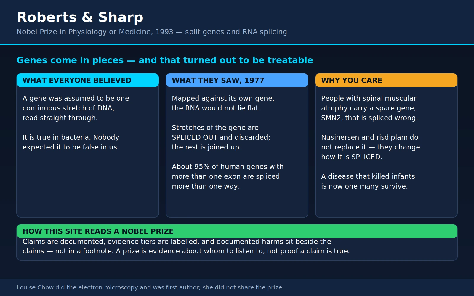
Table of Contents
- The Assumption That Turned Out to Be Wrong
- The 1977 Experiment: A Gene That Would Not Lie Flat
- Exons, Introns, and the Spliceosome
- Why Cells Bother: One Gene, Many Proteins
- When Splicing Goes Wrong
- Spinal Muscular Atrophy: The Spare Copy That Almost Works
- Newborn Screening, and Why the Clock Matters
- What It Costs, and Who Actually Gets It
- Other Splice-Targeting Drugs — and Thinner Evidence
- Introns and the Rest of the Genome
- Roberts' Second Act
- Where Mainstream Medicine Agrees — and What Remains Debated
- Key Research Papers
- Connections
- Featured Videos
1. The Assumption That Turned Out to Be Wrong
To understand why 1977 was a shock, you have to know what everyone believed the day before.
By the mid-1970s molecular biology had a clean, confident picture of how a gene works. DNA is a long text. A gene is a continuous stretch of that text. An enzyme reads it from one end to the other and copies it into a messenger molecule — RNA — and the cell then reads the RNA and builds the protein. Gene, message, protein: three colinear things, the same length, in the same order. You could line the gene up against its message and they would match end to end, like a photocopy against its original.
This was not a guess. It had been demonstrated, repeatedly and rigorously, in bacteria. In Escherichia coli the gene really is continuous, the message really is a faithful copy, and the ribosome can even start translating the front of a message while the back of it is still being transcribed. Bacteria were the workhorse of the field, and there was no obvious reason to expect anything else in a human cell. The reigning assumption of the era, often stated out loud, was that what was true for E. coli would be true for the elephant.
So when Richard J. Roberts, working at Cold Spring Harbor Laboratory on Long Island, and Phillip A. Sharp, working at the Massachusetts Institute of Technology, independently set out in 1977 to map where a particular messenger RNA came from on a viral chromosome, nobody expected the map to be interesting. They expected a line. What they got was a knot.
The 1993 Nobel Prize in Physiology or Medicine went to the two of them "for their discoveries of split genes." The phrase is worth pausing on. Not a split gene — split genes, as a category. What they had found was not an oddity in one virus. It was the ordinary architecture of nearly every gene in every animal, plant and fungus on the planet, and it had been sitting in plain sight the whole time.
2. The 1977 Experiment: A Gene That Would Not Lie Flat
Both groups were working on adenovirus 2, a common respiratory virus. That choice matters. Adenovirus makes a lot of messenger RNA very quickly inside an infected human cell, and it does so using the host cell's own machinery — so whatever the virus was doing, the human cell was doing it for the virus. And the viral chromosome is small enough that you could actually map things on it in 1977, which the three-billion-letter human genome was not.
The technique was RNA–DNA hybridisation viewed under an electron microscope. The idea is simple enough to picture. Take the mature messenger RNA that the cell finished making. Take the DNA it supposedly came from, and separate the two DNA strands. Mix them and let the RNA find its matching DNA sequence and stick to it. Then coat the whole thing in a heavy-metal stain, put it under an electron microscope, and look at the shape.
If the textbook picture were right, the shape would be boring: a straight double-stranded segment where the RNA had paired with the DNA, with plain single-stranded DNA running off both ends. That is what a colinear gene looks like.
That is not what either group saw. The pictures were the result. There was no follow-up assay that revealed the answer; the answer was the shape of the molecules on the film.
Sharp's group — the paper's first author was Susan Berget, with Claire Moore and Sharp — purified the messenger RNA for hexon, the major structural protein of the virus particle, and hybridised it to single-stranded fragments of the viral DNA. What they saw were branched forms. The bulk of the RNA lay neatly along the DNA, but about 160 nucleotides at the front end of the RNA hung loose, unpaired, with nothing on that DNA fragment to match it. They then went hunting for where that loose front end did match, and found it in three separate places elsewhere on the same strand of the viral genome — at roughly 17, 20 and 27 map units. Their conclusion, stated plainly in the 1977 paper: four separate segments of RNA are joined together to make one finished hexon message, and a model was proposed in which a longer precursor RNA is cut and spliced during maturation.
Roberts' group — Louise T. Chow as first author, with Richard Gelinas, Thomas Broker and Roberts — came at it from the other direction, using whole viral DNA rather than small fragments, and the R-loop method. Here the RNA invades the double-stranded DNA, pairs with one strand, and pushes the other strand out into a visible bubble. Because they had the entire genome in the frame, they could see the whole architecture at once: the front ends of many late viral messages were built from short sequences encoded at three widely separated positions on the genome — about 16.6, 19.6 and 26.6 map units — joined together into a leader of 150 to 200 nucleotides. The DNA lying between those joined segments had no partner in the RNA at all. It had nowhere to go but out, and it looped out as a strand of unmatched DNA. Chow and colleagues titled the paper, with no false modesty, An amazing sequence arrangement at the 5' ends of adenovirus 2 messenger RNA, and concluded that the findings implied a new mechanism for making messenger RNA in mammalian cells.
Those unmatched loops of DNA are the whole discovery in one image. The gene is longer than its message. Something in the middle gets thrown away.
The two papers appeared within weeks of each other — Berget, Moore and Sharp in the Proceedings of the National Academy of Sciences in August 1977, Chow, Gelinas, Broker and Roberts in Cell that September. Neither group scooped the other; they had converged on the same impossible picture from two directions, which is a large part of why the result was believed so fast.
3. Exons, Introns, and the Spliceosome
The vocabulary arrived a few months later. In a short 1978 piece in Nature titled Why genes in pieces?, Walter Gilbert gave the two kinds of sequence the names we still use:
- Exons — the segments that are kept and expressed. They end up in the finished message and, for protein-coding genes, they carry the instructions the ribosome reads.
- Introns — the intervening segments. They are transcribed along with everything else, and then cut out and discarded before the message leaves the nucleus.
The order of operations matters and is easy to get backwards. The cell does not skip over the introns while copying the DNA. It copies the entire gene, introns and all, into a long precursor RNA. Only afterwards is that precursor edited — the introns snipped out, the exons stitched together end to end — to produce the mature messenger RNA. That editing step is called splicing, and it happens in the nucleus, before the message is exported to the cytoplasm to be translated.
The machine that does the cutting and joining is the spliceosome. It is not a single enzyme. It is a large, dynamic assembly built from five small nuclear ribonucleoproteins — U1, U2, U4, U5 and U6, each a short RNA bundled with proteins — plus well over a hundred additional proteins that come and go as the reaction proceeds. Joan Steitz's laboratory later showed that U1 works by base-pairing directly with the start of the intron, which is how the spliceosome knows where to cut: it reads the RNA sequence with another RNA.
The precision required is difficult to overstate. A human intron can be thousands or tens of thousands of nucleotides long. The spliceosome has to find both of its ends in that haystack, cut at exactly the right nucleotide on each side, and rejoin the two exons without gaining or losing a single letter. One letter out and the reading frame shifts and the protein is garbage from that point onward. The cell does this correctly, tens of thousands of times, in every cell, every day.
And — this is the detail that turns out to matter enormously in section 6 — the spliceosome can be persuaded to make a different choice. That is not a bug. It is the point.
4. Why Cells Bother: One Gene, Many Proteins
If introns were merely rubbish to be removed, splicing would be an expensive way to achieve nothing. The reason the arrangement survived hundreds of millions of years of evolution is alternative splicing: the same precursor RNA can be cut and rejoined in more than one way, producing several different messages — and therefore several different proteins — from a single gene.
The mechanics are ordinary once you accept that exon choice is a decision rather than a fixed fact. An exon can be included in one version of the message and skipped in another. A splice site can be used at one position in one tissue and a few nucleotides away in a different tissue. Two mutually exclusive exons can be offered and exactly one selected. The protein that results differs accordingly — sometimes trivially, sometimes to the extent that the two versions do opposite things.
How common is this? When high-throughput RNA sequencing finally made it possible to count, the answer was: nearly universal. Analysing human tissues, Pan and colleagues estimated in 2008 that transcripts from approximately 95% of human genes with more than one exon undergo alternative splicing, with on the order of 100,000 reasonably abundant alternative splicing events across major tissues. Alternative splicing is not a special case. It is the normal operating mode of the human genome.
The gene-count point, stated accurately
This is where a genuine finding regularly gets flattened into a folk story, so it is worth stating carefully.
The accurate part. Before the human genome was sequenced, published and widely circulated estimates of the number of human genes ran to many tens of thousands, and figures approaching 100,000 were often quoted. When the International Human Genome Sequencing Consortium published the finished euchromatic sequence in 2004, the abstract noted the result almost apologetically: the human genome "seems to encode only 20,000–25,000 protein-coding genes." Current annotations put the figure at the lower end of that range, and this is genuinely fewer genes than a great many people expected. It is also not dramatically more than far simpler animals carry.
The part that gets overstated. The folk version says that alternative splicing explains human complexity — that we get away with 20,000 genes because splicing multiplies them into hundreds of thousands of proteins, and that this is what makes us more complicated than a worm. That claim goes further than the evidence supports, for three reasons. First, alternative splicing is not a human speciality; it is widespread across animals and plants, including organisms with far fewer cell types than we have. Second, many detected splice variants are present at very low abundance, and it is still debated how many of them produce a stable, functional protein rather than being noise or a route to degradation. Third, "complexity" is not a measured quantity that a gene count or a splice-variant count can be divided into.
What the low gene count really demolished was something simpler and more important: the assumption of one gene, one protein. That assumption had already been broken in 1977. The genome sequence just made it impossible to ignore. A gene is not a blueprint for a protein. It is a set of parts and a set of rules for which parts get used, and the rules are as informative as the parts.
5. When Splicing Goes Wrong
Here is the practical consequence, and it is the hinge of this entire page.
If you are looking for the genetic cause of a disease and you only inspect the coding sequence — the exons, the letters that specify amino acids — you will miss a whole class of mutations. A change can leave every amino acid instruction intact and still ruin the gene, by altering where the spliceosome cuts.
There are several ways this happens:
- A canonical splice-site mutation. The first two and last two nucleotides of nearly every intron are almost invariant. Change one and the spliceosome no longer recognises that boundary. The exon may be skipped entirely, or the intron may be retained in the finished message.
- A new splice site created where none belonged. A single-letter change deep inside an intron can create a sequence that looks like a splice site to the machinery, which then dutifully uses it and splices in a chunk of intron as if it were an exon.
- Loss of a splicing enhancer or gain of a splicing silencer. Exons carry short sequences that recruit regulatory proteins telling the spliceosome "include me." These can sit inside the coding sequence. A mutation that changes one of them may not change the amino acid at all — a so-called silent change — and yet cause the exon to be dropped. This mechanism is the whole story of spinal muscular atrophy, below.
How large a share of inherited disease works this way? The honest answer is that it depends entirely on how you count, and the estimates vary widely. Counting only mutations that fall exactly on the canonical intron boundaries gives a modest figure. Counting every mutation that measurably perturbs splicing — including the silent exonic changes that disrupt enhancers — gives a much larger one. In 2005, López-Bigas and colleagues asked the question directly in a paper titled Are splicing mutations the most frequent cause of hereditary disease? and concluded that, contrary to the prevailing assumption that disease-causing point mutations act mainly by changing amino acids, a great many of them may actually be acting on splicing. A decade later, Scotti and Swanson's review RNA mis-splicing in disease (Nat Rev Genet 2016;17(1):19-32) surveyed a still-growing list of conditions in which mis-splicing is the mechanism. What is not in dispute is that the share is substantial and that it is considerably larger than the textbooks of the 1980s assumed.
Beta-thalassaemia: the first clear human example
Beta-thalassaemia is an inherited anaemia in which the body makes too little of the beta-globin chain of haemoglobin. Depending on severity it ranges from a mild, symptom-free finding on a blood count to a transfusion-dependent illness from infancy. It has been intensively studied for a century, partly because it is common around the Mediterranean, the Middle East, South and Southeast Asia, and partly because globin was one of the first human genes anyone could clone.
In 1983, Treisman, Orkin and Maniatis cloned five beta-thalassaemia genes from patients and worked out what each mutation actually did. Some of them were transcription defects — the gene was not being copied properly in the first place. But others were RNA splicing defects: the gene was transcribed perfectly well and then edited wrongly. In some patients the mutation sat in an intron and created a competing splice site, so that a fraction of the messages came out with extra intron sequence embedded in them. Those messages produce no usable beta-globin.
This was the proof of principle that mattered clinically. A person could carry a gene whose protein-coding letters were entirely normal and still have a serious inherited disease, because the editing step had been sabotaged. Once that was established, the obvious question followed: if a mutation can push splicing in the wrong direction, can a drug push it back?
6. Spinal Muscular Atrophy: The Spare Copy That Almost Works
Spinal muscular atrophy is the clearest answer to that question that medicine has produced. It is also, for a small number of families, the difference between burying a child and watching that child learn to walk.
What the disease is
SMA is a genetic disease of the motor neurons — the nerve cells in the spinal cord that carry the command to move from the brain out to the muscles. The muscles themselves are healthy. The nerves that drive them die. Without that input the muscle wastes, which is what "muscular atrophy" refers to, but the primary loss is neurological.
It is recessive: a child develops SMA only by inheriting a non-working copy from both parents, each of whom is an unaffected carrier. Carrier frequency is commonly estimated at around one person in fifty, though it varies by ancestry, and the disease occurs in something on the order of one birth in ten thousand, again with estimates varying by population. Most carriers never know.
Severity has traditionally been described in types. The most severe, type 1, begins in the first six months of life: the baby is floppy, never gains the ability to sit unsupported, and develops progressive difficulty swallowing and breathing, because the muscles between the ribs fail before the diaphragm does. Type 2 begins later, and these children sit but never walk independently. Type 3 begins later still and these children walk, though many lose that ability over years. The types are a spectrum, not four separate diseases, and — as you will see — they are the reason you must read every SMA trial carefully before believing a headline.
Two genes, one silent letter
In 1995, Lefebvre and colleagues identified the gene responsible and named it SMN, for survival motor neuron. The SMN protein turned out to have a job that is almost too neat for a page about splicing: it is required for the assembly of the small nuclear ribonucleoprotein particles that build the spliceosome. The protein whose loss kills motor neurons is itself part of the machinery Roberts and Sharp discovered.
Then comes the twist that makes SMA treatable. Human chromosome 5 carries the SMN region in a duplicated arrangement, and humans have two nearly identical copies of the gene:
- SMN1 — the working gene. In about 95% of people with SMA, both copies of SMN1 are deleted or disabled. This is the cause of the disease.
- SMN2 — a near-identical duplicate, present in humans and not in the mice and other animals used to model the disease. Everyone with SMA still has at least one SMN2, and many have several. Copy number varies from person to person, and more copies of SMN2 generally means milder disease — which was itself the first strong hint that SMN2 is doing something useful.
SMN1 and SMN2 differ at only a handful of nucleotides, and — critically — the crucial difference does not change the protein at all. In 1999 Lorson, Hahnen, Androphy and Wirth engineered a series of hybrid SMN genes to test each difference in turn, and found that a single C-to-T change in exon 7, which is translationally silent and alters no amino acid, was by itself necessary and sufficient to change how the gene is spliced. In their words, the failure of SMN2 to compensate for the loss of SMN1 comes down to a nucleotide exchange that weakens an exonic splicing enhancer.
The practical result: when the spliceosome processes an SMN2 transcript, it mostly skips exon 7. The resulting protein, usually written SMNΔ7, is short, unstable, poor at binding to itself, and rapidly degraded. Only a minority of SMN2 transcripts are made full-length, so SMN2 produces a small amount of good protein — enough to soften the disease when you have several copies, never enough to prevent it.
Cartegni and Krainer showed in 2002 that the affected sequence is an exonic splicing enhancer that recruits the splicing factor SF2/ASF; other groups have argued the same nucleotide change instead creates a binding site for a splicing repressor. Both mechanisms may contribute, and the debate is not settled. What is settled is the consequence: exon 7 goes missing.
So people with SMA are in a very unusual position. They already carry a spare copy of the gene they need, and it very nearly works. It is not missing. It is not broken in its instructions. It is being edited wrongly. And an editing decision is something a drug can reach.
The two splicing drugs: nusinersen and risdiplam
Nusinersen (brand name Spinraza; approved by the FDA in December 2016) is an antisense oligonucleotide — a short, chemically stabilised piece of synthetic nucleic acid, 18 units long, designed to stick to one specific spot on the SMN2 precursor RNA. That spot is a splicing silencer in intron 7. When the drug occupies it, the repressor proteins that would normally bind there cannot, and the spliceosome includes exon 7 instead of skipping it. The drug does not edit DNA, does not add a gene, and does not stay in the body permanently. It changes one splicing decision, for as long as it is present.
Its major practical drawback follows from its chemistry. Oligonucleotides of this kind do not cross from the bloodstream into the central nervous system, so nusinersen must be delivered intrathecally — injected into the spinal fluid by lumbar puncture. After a loading schedule, that means a procedure roughly every four months, indefinitely. For a child with significant scoliosis or spinal fusion, which many older SMA patients have, the injection itself can require imaging guidance and is not trivial.
Risdiplam (brand name Evrysdi; approved by the FDA in August 2020) attacks the same problem from a completely different chemical direction. It is a small molecule, taken by mouth once a day as a liquid. It works by stabilising the interaction between the U1 particle and the splice site at the end of exon 7 — nudging the spliceosome, at the moment it is deciding, toward inclusion. Because it is a small molecule that distributes through the body, it reaches the central nervous system without a lumbar puncture and also reaches tissues outside the nervous system, which may matter since SMN is needed everywhere.
Both are splicing drugs. Both take a gene the patient already has and change how it is read.
Onasemnogene abeparvovec is a different thing entirely
The third approved treatment is frequently discussed in the same breath as the other two, and it should not be. Onasemnogene abeparvovec (brand name Zolgensma; FDA approved May 2019) is gene replacement, not splicing modification. It uses a modified adeno-associated virus, serotype 9, as a delivery vehicle to carry a working copy of the SMN1 coding sequence into cells, given as a single intravenous infusion. It does not touch SMN2 and does not alter any splicing decision. It supplies the missing gene product from a new source.
The delivered gene does not integrate into the child's chromosomes; it persists as a separate episome inside the nucleus. That raises a real and still-open question about durability in a growing child, since cells that divide will dilute it. It also carries a distinct risk profile: liver injury is the main one, and corticosteroid cover is part of the standard protocol. In the first published trial of fifteen infants (Mendell and colleagues, N Engl J Med 2017;377(18):1713-1722), four had raised liver enzymes that responded to prednisolone; in the later phase 3 trial, two of twenty-two had raised liver enzymes reported as serious adverse events, and one had hydrocephalus.
Keeping these three straight matters clinically, because they are not interchangeable and the choice between them turns on age, weight, prior treatment and route of administration — not on which one is "newest."
The trial evidence, with the populations attached
This is the part where reporting most often goes wrong. SMA trials studied very different populations, and their results do not transfer between them. A result obtained in symptomatic infants with type 1 disease says nothing reliable about a fifteen-year-old with type 3, and vice versa. Read each of the following with its population attached:
- ENDEAR (Finkel and colleagues, 2017) — nusinersen in symptomatic infants with infantile-onset (type 1) SMA, randomised against a sham procedure. In the final analysis, 37 of 73 infants on nusinersen (51%) achieved a motor-milestone response, against 0 of 37 controls. The hazard ratio for death or permanent assisted ventilation was 0.53 (P=0.005) and for death alone 0.37 (P=0.004). The trial was stopped early at the interim analysis because the difference was already unambiguous. The authors also reported that infants with a shorter disease duration at screening were more likely to benefit — a finding that becomes the entire argument of section 7.
- CHERISH (Mercuri and colleagues, 2018) — nusinersen in 126 children whose symptoms began after 6 months of age, a much less severe population than ENDEAR. The measure here was the Hammersmith Functional Motor Scale-Expanded, scored 0 to 66. At 15 months the nusinersen group had gained 4.0 points and the control group had lost 1.9, a difference of 5.9 points (95% CI 3.7 to 8.1). In the final analysis, 57% of treated children versus 26% of controls gained at least 3 points. Note what this is and is not: a meaningful functional gain in children who were already sitting, not a survival result.
- FIREFISH part 2 (Darras and colleagues, 2021) — risdiplam in 41 infants with type 1 SMA aged 1 to 7 months, open-label, compared against natural-history controls rather than a randomised placebo group. After 12 months, 12 infants (29%) could sit unsupported for at least 5 seconds — a milestone type 1 SMA does not otherwise produce. 85% survived without permanent ventilation, against 42% for the upper boundary of the historical comparison.
- SUNFISH part 2 (Mercuri and colleagues, 2022) — risdiplam in 180 non-ambulant patients aged 2 to 25 with type 2 or type 3 SMA, properly randomised 2:1 against placebo. At 12 months the treatment difference on the 32-item Motor Function Measure was 1.55 points (95% CI 0.30 to 2.81, P=0.016). That is a real, statistically significant result and it is also a modest average one; the authors' own exploratory analysis suggested function generally improved in younger participants and stabilised in older ones, and they explicitly flagged that this needs confirmation. Anyone quoting SMA drug results should be able to hold both the FIREFISH number and this one in mind at the same time.
- STR1VE (Day and colleagues, 2021) — onasemnogene abeparvovec in 22 symptomatic infants under 6 months with type 1 SMA and one or two SMN2 copies, single-arm, against an untreated natural-history cohort. 13 of 22 (59%) sat independently for at least 30 seconds at the 18-month visit, against 0 of 23 untreated. 20 of 22 (91%) survived free of permanent ventilation at 14 months, against 6 of 23 (26%) untreated.
- NURTURE (De Vivo and colleagues, 2019) — nusinersen in 25 infants who were diagnosed genetically and treated before any symptoms appeared. This is the different-in-kind study. At a median age of 34.8 months, past the age at which type 1 or type 2 would have declared itself, all 25 were alive, none had needed a tracheostomy or permanent ventilation, all 25 could sit without support, 23 of 25 could walk with assistance, and 22 of 25 could walk independently.
What actually changed
To see the size of the shift you need the baseline, and the baseline is well documented. In a prospective natural-history study of type 1 SMA published in 2014 — before any of these drugs existed — Finkel and colleagues followed infants with the disease and found the median age at reaching the combined endpoint of death or requiring at least 16 hours a day of ventilation support was 13.5 months, with an interquartile range of 8.1 to 22.0 months. Note that this endpoint is death or near-continuous ventilation, not death alone; it is the honest way to describe an illness where survival became possible only with intensive respiratory support. The same study found that baseline electrophysiological measurements already showed substantial motor neuron loss, and that having two copies of SMN2 predicted worse outcomes than having three.
The untreated comparison groups in the later trials tell the same story from a different angle: 26% ventilation-free survival at 14 months in the natural-history cohort used by STR1VE, and 8% event-free survival at 20 months in the historical cohort used by the first gene-replacement study.
Against that baseline, the trials above describe a genuinely different disease. Infants with type 1 SMA who would not have sat up are sitting up. Children who would have died or been ventilated before their second birthday are surviving. Children treated before symptoms began are, in a substantial majority, walking.
Two honest qualifications belong immediately alongside that. First, these are not cures. They increase the amount of SMN protein; they do not restore motor neurons that have already died, and treated children generally still have a neuromuscular disease requiring physiotherapy, respiratory monitoring and orthopaedic care. Second, the longest follow-up available is still short relative to a human lifetime. We do not yet know what a 40-year-old treated as a newborn looks like, because there is not one yet.
7. Newborn Screening, and Why the Clock Matters
Every result in the previous section points in one direction, and it is not a subtle direction. Treating earlier works better, and treating before symptoms appear works dramatically better.
The reason is biological, not administrative. Motor neurons that die do not come back. There is no treatment, existing or foreseeable, that regrows them. The SMA drugs work by giving the surviving motor neurons enough SMN protein to stay alive and functional. That means the ceiling on how well a child can do is set by how many motor neurons are still there on the day treatment starts. In type 1 SMA the loss is fast and it begins before the parents notice anything is wrong — recall that Finkel's natural-history cohort already showed substantial motor neuron loss on baseline testing. By the time a floppy baby prompts a referral, a neurology appointment, a genetic test and a funding decision, months of irreplaceable neurons are gone.
Genetic newborn screening breaks that sequence. The homozygous deletion of SMN1 exon 7 that causes about 95% of SMA can be detected from the same dried blood spot already taken from every newborn's heel, using a straightforward assay, at very low marginal cost. A positive result puts the child in a neuromuscular clinic in the first days of life, before any symptom.
The best real-world evidence for what that is worth comes from Germany, Austria and Switzerland. Schwartz and colleagues compared 234 children with genetically confirmed SMA born between 2018 and 2021 — 44 identified by a newborn screening pilot in two German federal states, and 190 diagnosed the old way, after symptoms appeared, all within the same health system and receiving the same standard of care (JAMA Pediatr 2024;178(6):540-547). The results:
- Mean age at starting treatment: 1.3 months in the screened group versus 10.7 months in the clinically diagnosed group.
- Gained the ability to sit independently: 40 of 44 (90.9%) screened, versus 141 of 190 (74.2%) clinically diagnosed.
- Achieved independent walking: 28 of 40 (63.6%) screened, versus 28 of 190 (14.7%) clinically diagnosed.
Those are the same drugs in both arms. The only variable is when they started.
SMA was added to the United States' recommended newborn screening panel in 2018 and adopted state by state thereafter, and many other countries have followed or are piloting. Where it exists, it works as designed.
Why this is not an argument for screening in general
It is tempting to draw a general lesson — find disease earlier, always — and that lesson would be wrong. Screening is only justified when finding the condition early changes what happens to the person. SMA meets that bar about as cleanly as any condition in medicine ever has: the disease is severe, progressive and certain to declare itself; the test is cheap and accurate; effective treatment exists; and the benefit of early treatment is measured and large.
Compare that with thyroid cancer, which is the subject of our page on Theodor Kocher and the surgical history of the thyroid gland. When ultrasound screening for thyroid cancer was rolled out at population scale, enormous numbers of small papillary cancers were found — and the death rate from thyroid cancer did not fall. What screening had detected, in large part, were tumours that would never have caused symptoms or shortened a life. The people found by that screening were not saved; they were converted into cancer patients, with surgery, lifelong hormone replacement and the anxiety that comes with a diagnosis. Several countries have since pulled back from it.
The two cases point in opposite directions, and both conclusions are correct. The variable is not enthusiasm for early detection. It is whether early detection changes the outcome. For SMA it changes the outcome enormously. For asymptomatic thyroid nodules it mostly does not. Anyone who reads the SMA story as a general argument for more screening has taken exactly the wrong lesson from it.
8. What It Costs, and Who Actually Gets It
A page that described the trial results and stopped there would be misleading, because for most of the world's children with SMA the binding constraint is not biology. It is price.
These are among the most expensive medicines ever marketed. Onasemnogene abeparvovec was launched in the United States in 2019 at a list price above two million dollars for the single infusion, which made it, at the time, the most expensive drug in the world. Nusinersen and risdiplam are priced as ongoing therapy at figures in the hundreds of thousands of dollars per patient per year. Real prices paid by health systems are lower than list, are usually confidential, and vary by country.
Why so high? The stated logic is that the fixed cost of developing a drug has to be recovered from a very small number of patients — an ultra-rare disease has no volume to spread it over — and that a one-time treatment which prevents decades of intensive care is worth more than its price. Both halves of that argument are contestable and both are contested. What is not in dispute is that the arithmetic produces numbers that no individual family can pay, which makes access entirely a question of what the health system will fund.
The published economic analyses give a sense of the scale. A systematic review by Dangouloff and colleagues (Orphanet J Rare Dis 2021;16(1):47) found that even the standard of care for SMA, before any of these drugs, cost between $75,047 and $196,429 per year for type 1, and between $27,157 and $82,474 per year for the later-onset forms. For the drugs themselves, the incremental cost per quality-adjusted life year gained ranged from $210,095 to $1,150,455 for nusinersen in type 1, and from $32,464 to $251,403 for onasemnogene abeparvovec. For the later-onset forms the range ran from $275,943 to over $8 million per quality-adjusted life year. Health systems that use a cost-effectiveness threshold to decide what to fund do not usually approve things at the top of that range.
What that looks like in practice is worth one concrete example. In Brazil, families sought court orders compelling the Ministry of Health to supply onasemnogene abeparvovec. Kretzschmar and colleagues examined every such lawsuit filed between January 2019 and September 2022 (Rev Saude Publica 2024;58:36): 136 lawsuits, 113 of them (83%) decided in the patient's favour, at a cost of R$944.8 million — 2.45% of the health system's entire medicines budget. Only 6 of those 113 grants would have met the criteria the national health-technology committee had set, and R$146 million went to supplying the drug to children over the age of two, outside the manufacturer's own labelled indication.
That is not a simple story with a villain. It is what happens when an effective treatment exists, a public system cannot afford it for everyone, and desperate parents have a legal route open to them. The families who sued were not wrong to sue. The committee that set restrictive criteria was not being cruel. Access to these drugs today depends on where a child is born to a degree that has no medical justification at all, and pretending otherwise does patients no favours.
9. Other Splice-Targeting Drugs — and Thinner Evidence
Antisense oligonucleotides are now an established drug class. They are short synthetic nucleic acids, chemically modified so the body does not destroy them immediately, designed to base-pair with a chosen RNA. Depending on the chemistry they can trigger the destruction of that RNA, block a protein from binding to it, or — as with nusinersen — redirect a splicing decision. The appeal is that the target is a sequence, so in principle a drug can be designed for any gene whose sequence is known, including targets that no conventional small molecule can reach.
The most prominent splice-targeting application after SMA is Duchenne muscular dystrophy, and the evidence there is genuinely weaker. It is important to say so.
Duchenne is caused by mutations in the dystrophin gene, which is enormous — 79 exons — and produces a protein that anchors the muscle-cell membrane during contraction. Many Duchenne mutations are deletions that shift the reading frame, so translation terminates early and no functional dystrophin is made at all. The exon-skipping strategy is elegant: use an antisense oligonucleotide to make the spliceosome skip an additional exon, restoring the reading frame. The resulting dystrophin is shorter than normal and missing a chunk in the middle, but if it is partly functional it might convert a severe disease into a milder one. There is a natural precedent — Becker muscular dystrophy, a milder condition, is caused by in-frame deletions producing exactly this kind of shortened dystrophin.
The strategy is mutation-specific, and this is a serious practical limit. An oligonucleotide that skips exon 51 helps only those Duchenne patients whose particular deletion happens to be corrected by skipping exon 51 — a substantial minority, but a minority. A different deletion needs a different drug.
Eteplirsen, which skips exon 51, was granted accelerated approval by the FDA in 2016, and this is where the story diverges sharply from SMA. The pivotal published trial (Mendell and colleagues, Ann Neurol 2013;74(5):637-647) enrolled twelve boys, four per group. It reported that dystrophin-positive muscle fibres rose to 23% of normal at 24 weeks in one dose cohort, with larger increases at 48 weeks, and that "ambulation-evaluable" treated patients walked 67.3 metres further on a six-minute walk test than the placebo/delayed group. That phrase — ambulation-evaluable — is doing a great deal of work: it means the walking-distance comparison was made after excluding boys who had already lost the ability to walk, in a trial with four patients per arm. The paper drew several published critiques in the same journal.
The review that led to approval was contentious and included a high-profile meeting of an external advisory committee. Kesselheim and Avorn set out the case against it in a JAMA viewpoint the same year (JAMA 2016;316(22):2357-2358). A fuller account of the aftermath appears in a 2023 review by Bendicksen and colleagues (Ann Intern Med 2023;176(9):1251-1256), which is worth reading in full and which records the following: the approval rested on a study of twelve boys; FDA leadership concluded that very small increases in dystrophin were "reasonably likely to predict clinical benefit"; further drugs in the same class were then approved by the same route; the confirmatory trial the FDA required by November 2020 had still not been completed as of 2023; and the relationship between the level of truncated dystrophin produced and any clinical outcome remains uncertain.
The contrast with SMA is the point of this section. Nusinersen was approved on the basis of randomised, sham-controlled trials with unambiguous endpoints — motor milestones, survival, ventilation — in which the difference was large enough that the trials were stopped early for benefit. Eteplirsen was approved on a biochemical surrogate measured in twelve boys. Both are antisense oligonucleotides that redirect splicing; the mechanism is not what separates them. What separates them is the quality of the evidence, and the two should never be cited together as though they carried equal weight.
None of that means exon skipping cannot work in Duchenne. It means we do not yet know whether these particular drugs do, a decade after the first of them was approved, and the families paying for them deserve to know that.
10. Introns and the Rest of the Genome
A brief section, deliberately, because this is an area where confident overstatement is common.
Introns are not empty. Some carry enhancers and other regulatory sequences — stretches of DNA that influence when and how strongly the surrounding gene is switched on. Some host entire functional genes for small RNAs: many small nucleolar RNAs are encoded within introns and released when the intron is excised, and a substantial number of microRNAs are found in introns as well. From the cell's point of view, transcribing an intron and then cutting it out is a way of producing two different products from one transcription event.
Splicing also feeds into quality control. Signals deposited on a message at the junctions where exons were joined help the cell recognise transcripts carrying a premature stop codon and destroy them, which prevents a truncated and potentially harmful protein from being made.
What should not be claimed is that every intron does something, or that intron content explains organismal complexity, or that the discovery of function in some non-coding DNA means the concept of non-functional DNA has been overturned. Intron numbers and lengths vary enormously between species with no tidy relationship to anything, some introns look very much like the remnants of mobile genetic elements that inserted themselves and were tolerated, and the question of how much of the human genome is functional in any meaningful sense remains genuinely open and genuinely argued over. Bacteria, which mostly lack spliceosomal introns, manage without them entirely — which is precisely why the 1977 result was so surprising.
The defensible summary is narrower and still remarkable: the sequence that gets thrown away is not inert, the act of throwing it away is itself informative to the cell, and a gene is a more complicated object than a stretch of coding text.
11. Roberts' Second Act
Richard Roberts did not retire into the prize. He had joined New England Biolabs in 1992, the year before the Nobel, and became its chief scientific officer; he has continued publishing on restriction and modification enzymes ever since, and maintains REBASE, the reference database of restriction enzymes and their recognition sequences, which is a piece of infrastructure the whole of molecular biology uses without thinking about it. A scientist's most durable contribution is not always the one that won the prize.
He has also been an unusually visible public advocate on scientific policy, and the most prominent of those campaigns concerns agricultural biotechnology. Roberts organised open letters signed by large numbers of Nobel laureates supporting the use of genetically modified crops, most publicly a 2016 letter addressed to Greenpeace, to the United Nations and to governments, urging an end to opposition to genetically modified crops in general and to vitamin-A-enriched "Golden Rice" in particular. More than a hundred laureates signed. He has continued to argue the case since; he is a co-author of a 2024 comment piece in Nature Plants arguing for public–private partnership to deliver agricultural biotechnology in developing countries (Nat Plants 2024;10(1):2-5).
These campaigns have been welcomed by some and criticised by others — on the merits of the underlying science, on the question of whether a Nobel Prize in an unrelated field confers authority in agronomy or development economics, and on whether the debate over GM crops is really a debate about safety at all rather than about seed patents, corporate control and who decides what farmers grow. This page takes no position on any of that. It is outside our subject matter and outside our competence, and the reader is better served by being told plainly what Roberts did and left to investigate the arguments than by being handed our opinion of them.
Roberts has separately campaigned for open access to publicly funded research literature, a position with rather broader support among working scientists.
12. Where Mainstream Medicine Agrees — and What Remains Debated
Settled
- Genes in animals, plants and fungi are split into exons and introns, and introns are removed from the precursor RNA after transcription. This is not controversial anywhere and has not been for four decades.
- Alternative splicing is the norm for human genes with more than one exon, and a single gene routinely gives rise to more than one protein.
- Mutations that affect splicing are a substantial cause of inherited disease, including mutations that change no amino acid at all.
- SMA is caused by loss of SMN1, and the severity is modified by how many copies of SMN2 the person carries.
- Nusinersen and risdiplam work by forcing inclusion of SMN2 exon 7; onasemnogene abeparvovec works by delivering a new copy of the gene. These are different mechanisms.
- All three treatments change the course of SMA, and the effect in infantile-onset disease is large compared with an extremely well-documented natural history.
- Earlier treatment produces better outcomes, and treatment before symptoms produces the best outcomes recorded so far.
Genuinely open
- How durable the benefit is. The oldest treated children are still children. Whether a single dose of gene replacement holds up over decades in tissue that grew after the infusion is not known, and cannot be known yet.
- Whether combining treatments helps. Adding a splicing drug to gene replacement, or switching between them, is done in practice and is not well supported by randomised evidence.
- How much benefit there is in older and less severely affected patients. SUNFISH found a real but modest average difference in patients aged 2 to 25, and its own authors called for confirmation of the age-related pattern.
- Whether SMN needs to be restored outside the nervous system. SMN is required by every cell. Whether the heart, liver and other tissues matter to long-term outcomes — and therefore whether a systemically distributed drug has an advantage over one confined to the spinal fluid — is unresolved.
- The precise molecular explanation for the SMN2 defect — loss of a splicing enhancer, gain of a splicing silencer, or both.
- Whether exon skipping works in Duchenne muscular dystrophy at all. See section 9. This is a live and unresolved question, not a settled success.
- What these drugs should cost and how societies should pay for them. There is no scientific answer to this. It is a distributional question, and it is being decided court case by court case in some countries.
- How much of the genome outside the exons is functional, and what most introns are for, if anything.
13. Key Research Papers
- Chow LT, Gelinas RE, Broker TR, Roberts RJ. An amazing sequence arrangement at the 5' ends of adenovirus 2 messenger RNA. Cell 1977;12(1):1-8
- Berget SM, Moore C, Sharp PA. Spliced segments at the 5' terminus of adenovirus 2 late mRNA. Proc Natl Acad Sci U S A 1977;74(8):3171-3175
- Gilbert W. Why genes in pieces? Nature 1978;271(5645):501
- International Human Genome Sequencing Consortium. Finishing the euchromatic sequence of the human genome. Nature 2004;431(7011):931-945
- Pan Q, Shai O, Lee LJ, Frey BJ, Blencowe BJ. Deep surveying of alternative splicing complexity in the human transcriptome by high-throughput sequencing. Nat Genet 2008;40(12):1413-1415
- Wang ET, Sandberg R, Luo S, et al. Alternative isoform regulation in human tissue transcriptomes. Nature 2008;456(7221):470-476
- López-Bigas N, Audit B, Ouzounis C, Parra G, Guigó R. Are splicing mutations the most frequent cause of hereditary disease? FEBS Lett 2005;579(9):1900-1903
- Treisman R, Orkin SH, Maniatis T. Specific transcription and RNA splicing defects in five cloned beta-thalassaemia genes. Nature 1983;302(5909):591-596
- Lefebvre S, Bürglen L, Reboullet S, et al. Identification and characterization of a spinal muscular atrophy-determining gene. Cell 1995;80(1):155-165
- Lorson CL, Hahnen E, Androphy EJ, Wirth B. A single nucleotide in the SMN gene regulates splicing and is responsible for spinal muscular atrophy. Proc Natl Acad Sci U S A 1999;96(11):6307-6311
- Cartegni L, Krainer AR. Disruption of an SF2/ASF-dependent exonic splicing enhancer in SMN2 causes spinal muscular atrophy in the absence of SMN1. Nat Genet 2002;30(4):377-384
- Finkel RS, McDermott MP, Kaufmann P, et al. Observational study of spinal muscular atrophy type I and implications for clinical trials. Neurology 2014;83(9):810-817
- Finkel RS, Mercuri E, Darras BT, et al. Nusinersen versus sham control in infantile-onset spinal muscular atrophy (ENDEAR). N Engl J Med 2017;377(18):1723-1732
- Mercuri E, Darras BT, Chiriboga CA, et al. Nusinersen versus sham control in later-onset spinal muscular atrophy (CHERISH). N Engl J Med 2018;378(7):625-635
- Darras BT, Masson R, Mazurkiewicz-Bełdzińska M, et al. Risdiplam-treated infants with type 1 spinal muscular atrophy versus historical controls (FIREFISH part 2). N Engl J Med 2021;385(5):427-435
- Mercuri E, Deconinck N, Mazzone ES, et al. Safety and efficacy of once-daily risdiplam in type 2 and non-ambulant type 3 spinal muscular atrophy (SUNFISH part 2). Lancet Neurol 2022;21(1):42-52
- Day JW, Finkel RS, Chiriboga CA, et al. Onasemnogene abeparvovec gene therapy for symptomatic infantile-onset spinal muscular atrophy in patients with two copies of SMN2 (STR1VE). Lancet Neurol 2021;20(4):284-293
- De Vivo DC, Bertini E, Swoboda KJ, et al. Nusinersen initiated in infants during the presymptomatic stage of spinal muscular atrophy: interim results from the Phase 2 NURTURE study. Neuromuscul Disord 2019;29(11):842-856
Additional sources cited inline above: Scotti and Swanson on RNA mis-splicing in disease (section 5); Mendell and colleagues on the first gene-replacement study (section 6); Schwartz and colleagues on newborn screening outcomes (section 7); Dangouloff and colleagues on the economics of SMA, and Kretzschmar and colleagues on court-ordered access in Brazil (section 8); Mendell and colleagues on eteplirsen, and Kesselheim and Avorn and Bendicksen and colleagues on the accelerated approval that followed (section 9); and Itam and colleagues, with Roberts among the authors, on agricultural biotechnology (section 11).
Live PubMed Searches
- RNA splicing and the discovery of split genes
- SMN2 exon 7 splicing
- Nusinersen and risdiplam outcomes in SMA
- SMA newborn screening and presymptomatic treatment
- Antisense exon skipping in Duchenne muscular dystrophy
14. Connections
- All Notable Doctors
- The Nobel Prize in Physiology or Medicine — every prize, in order, and what each one actually established
- Watson, Crick & Wilkins — the structure of DNA, and the confident model of the gene that split genes complicated
- Nirenberg, Khorana & Holley — cracking the genetic code, the work that made a gene readable in the first place
- Barbara McClintock — jumping genes: the earlier discovery that a genome is not a fixed text, ignored for decades
- Brenner, Horvitz & Sulston — programmed cell death and the nematode; Brenner also helped establish messenger RNA itself
- Fire & Mello — RNA interference, the other route by which short RNAs became medicines
- Karikó & Weissman — modified messenger RNA, and how far chemistry can push a nucleic acid into being a drug
- Shinya Yamanaka — induced pluripotent stem cells, now used to grow patient-derived motor neurons for SMA research
- Capecchi, Evans & Smithies — gene targeting in mice, the technique behind the animal models used to test SMA drugs
- Theodor Kocher — thyroid surgery, and the screening story that reaches the opposite conclusion from SMA
- Prizes That Aged Badly — Nobel awards the science later contradicted, and why 1993 was not one of them