Niels Finsen: Light as Medicine, and What Light Therapy Can Actually Do
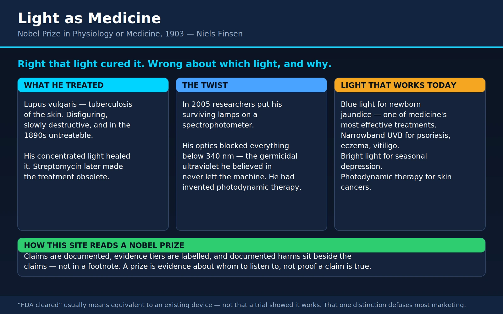
Niels Ryberg Finsen won the 1903 Nobel Prize in Physiology or Medicine for curing a disease with light. Not for a metaphor, not for a wellness claim — for a machine that pointed concentrated light at a disfiguring facial infection that had no other treatment, and made it heal. He was 43 when he died the following year, having spent most of his adult life seriously ill himself.
This page tells that story, and then does the thing the story earns: it is this site's reference page for light therapy in general. Light is now the basis of several genuinely excellent treatments — and also of an enormous consumer market of red-light panels, masks, helmets and beds whose claims run far ahead of the evidence. Both facts are true at once. Sorting one from the other is what the second half of this page is for.
Table of Contents
- The Man Who Studied Light Because He Was Ill
- What Lupus Vulgaris Was
- How the Finsen Light Worked — and What Did Not Hold Up
- Light Therapy That Is Genuinely Established Today
- Red Light and Photobiomodulation: The Claim at Its Strongest
- Red Light, Tiered by Indication
- The Dosing Problem That Makes This Literature Hard to Read
- Safety, and the One Real Precaution
- What "FDA Cleared" Means — and Does Not Mean
- Sunlight, Vitamin D, and the Honest Trade-Off
- Three Different Claims That Get Blurred Together
- How to Read a Light-Device Claim
- Where Mainstream Medicine Agrees, and What Remains Debated
- Finsen's Institute and Legacy
- Key Research Papers
- Connections
- Featured Videos
1. The Man Who Studied Light Because He Was Ill
Niels Ryberg Finsen was born in 1860 in Tórshavn, in the Faroe Islands — a treeless North Atlantic archipelago where the winter sun barely clears the horizon and summer daylight runs almost around the clock. His family was Icelandic; his father became governor of the Faroes. He was sent to school in Denmark, then to Iceland, then back to Copenhagen to study medicine, qualifying in 1890.
He was not an outstanding student. He was, by the accounts that survive, slow, stubborn and unusually literal-minded — the sort of person who notices something ordinary and then refuses to stop noticing it. What he noticed was sunlight.
The illness, stated carefully. From around 1883, in his early twenties, Finsen began to develop symptoms of a chronic illness that would define the rest of his life: progressive weakness, fluid accumulation in the abdomen, an enlarged liver and spleen, and eventually near-total immobility. He spent his final years in a wheelchair, on a severely restricted diet, having fluid drained repeatedly. He died in September 1904, aged 43, one year after receiving the Nobel Prize.
The medical-history literature most often attributes this to Niemann-Pick disease, a hereditary lipid-storage disorder, and that is the diagnosis given in the standard dermatological biography of him. It is worth being honest about what that attribution is: a retrospective diagnosis, made from nineteenth-century case notes, physical descriptions and autopsy findings, using disease categories that did not exist when he was alive. His own physicians described a constrictive disease of the pericardium — the sac around the heart — with consequent congestion of the liver. Retrospective diagnoses of historical figures are inherently uncertain, and this one has been argued both ways. What is not in doubt is that he was chronically, progressively and severely ill for roughly the entire twenty years of his working life.
Why that matters to the science. Finsen said his interest in light began with his own body. Living in a north-facing house in Copenhagen, he found that sitting in direct sunlight made him feel measurably better — more energetic, less unwell — and he moved to rooms with southern exposure to get more of it. That is a single-patient observation of a subjective effect, which is the weakest kind of evidence there is, and it would be easy to sneer at it. But it is also exactly how a great many real discoveries start: someone notices something about themselves and, crucially, does not stop at the noticing. Finsen went from "sunlight seems to help me" to a laboratory, to instruments, to controlled comparisons, to a treatment that other physicians could reproduce. The observation was the beginning of the work, not a substitute for it. That distinction is the whole subject of this page.
He began experimenting in the early 1890s — on tadpoles and salamander larvae, on the pigmentation of skin, on the healing of smallpox pustules — and in 1896 he established the Medical Light Institute in Copenhagen, funded initially by a Danish industrialist. By 1903 the Nobel Committee awarded him the prize "in recognition of his contribution to the treatment of diseases, especially lupus vulgaris, with concentrated light radiation, whereby he has opened a new avenue for medical science." He was too ill to travel to Stockholm to collect it.
2. What Lupus Vulgaris Was
Almost nobody encounters this term today, and without it the achievement is unreadable. So: lupus vulgaris is tuberculosis of the skin.
It is not the autoimmune disease people now mean when they say "lupus" (systemic lupus erythematosus). The name is much older and comes from the Latin for wolf — because the lesion was said to devour the face. It is caused by Mycobacterium tuberculosis, the same organism Robert Koch had identified in 1882, seeded into the skin either from an infection elsewhere in the body or directly through a break in the surface.
The disease is slow and relentless. It typically begins on the face — often around the nose, cheeks or ears — as a small, soft, brownish-red nodule. Pressed with a glass slide, the nodules show the characteristic "apple-jelly" colour that dermatologists still use as a clinical sign. Left alone, and it was almost always left alone, the plaque spreads outward over years and decades while its centre ulcerates and scars. It destroys cartilage. It eats away the nose, erodes the eyelids, contracts the mouth, perforates the palate. Patients were disfigured in the most socially visible way possible, slowly, over a working lifetime, while remaining otherwise well enough to be fully aware of it. A minority went on to develop squamous cell carcinoma in the long-standing scars.
In the 1890s there was no treatment. There were no antibiotics of any kind — not for another four decades. The options were surgical destruction of the tissue (scraping, cautery, caustic pastes, arsenic paste, X-rays once those arrived), which meant trading the disease's disfigurement for the surgeon's. Koch's tuberculin, launched in 1890 amid enormous excitement as a tuberculosis cure, had failed publicly and damagingly. Lupus vulgaris was, in the plainest sense, hopeless.
Into that vacuum, Finsen put a lamp. Patients sat for an hour at a time, day after day, with a pressure lens held hard against the lesion to squeeze the blood out of the skin so the light could reach the tissue underneath. Treatment ran for months, sometimes years. It was tedious, uncomfortable and staffed by trained nurses working in shifts.
And it worked. Reported cure rates from the Finsen Institute and from the institutes that copied it ran to roughly half or better of treated patients, in a disease with no alternative — and Finsen's own published series was one of the first times a dermatological treatment had been reported with systematic follow-up rather than as a handful of triumphant case reports. Within a few years around forty Finsen institutes had opened across Europe and the United States. Queen Alexandra, herself of Danish birth, sent a Finsen lamp to the London Hospital.
And then it became obsolete — which is the honest ending. In 1943, in Selman Waksman's laboratory at Rutgers, streptomycin was isolated from a soil actinomycete; it was the first antibiotic effective against M. tuberculosis. Combination chemotherapy followed. A disease that had taken two years of daily light treatment could now be cured with tablets and injections, and the Finsen institutes closed or converted to other work. See Selman Waksman for that half of the story.
This arc — a real cure, genuinely effective, completely superseded — is worth sitting with, because it is the opposite of the two stories usually told about historical medicine. It is not a fraud unmasked, and it is not a suppressed cure. It is a treatment that was the best available, that helped real people, and that a better treatment replaced. Most of medicine's honest history looks like this.
3. How the Finsen Light Worked — and What Did Not Hold Up
The apparatus. The Finsen lamp was built around a carbon arc — two carbon electrodes struck to produce a blindingly bright, ultraviolet-rich discharge, the same technology then lighting streets and film studios. That raw output was useless as it stood: it was hot enough to burn, and diffuse. Finsen's engineering solved both problems at once.
The light was collected by a set of large quartz or glass lenses arranged in a telescope-like tube, one tube per patient, with four tubes radiating from a single central arc so four patients could be treated simultaneously. Between the lens elements ran circulating cooled water, sometimes tinted with a dye, acting as a heat filter — water absorbs infrared strongly, so the beam that emerged was intense but comparatively cool. At the patient end sat a hollow, water-cooled compression lens of rock crystal or quartz, pressed firmly against the lesion. This last piece was the clinically decisive one: blood is a powerful absorber of ultraviolet and violet light, so squeezing the skin bloodless let the beam reach the infected tissue rather than being soaked up in the capillaries.
What Finsen believed was doing the work. He was convinced the active agent was short-wavelength ultraviolet — what he called the "chemical rays" — killing the mycobacteria directly. That belief was not unreasonable. UV is genuinely germicidal: it is absorbed by nucleic acids, and short-wave UV (UVC, roughly 200–280 nm) produces thymine dimers and other DNA lesions that stop bacteria replicating. That mechanism is real, it is why UV is used to disinfect water and air, and it was the obvious explanation available in 1900.
What a modern reconstruction found. In 2005, dermatologists at Bispebjerg Hospital in Copenhagen — the successor institution to Finsen's own — took the surviving original lamps and actually measured them. They put the historical lens systems and filters on a spectrophotometer and asked what wavelengths could physically have reached the patient.
The answer was a surprise. The optics blocked essentially everything below 340 nm. The lenses were glass, not quartz, and glass absorbs UVB and UVC. The dye filters absorbed below 340 nm as well. The methylene-blue solution used as a heat filter blocked below 340 nm and between 550 and 700 nm. In other words, the germicidal short-wave ultraviolet Finsen thought he was delivering never left the machine.
So what did the healing? The same team looked for endogenous photosensitisers in the bacterium and found them: M. tuberculosis fluoresced in a way indicating porphyrins, and chemical analysis of a related mycobacterium confirmed coproporphyrin III. Porphyrins absorb strongly in the violet-blue region around 400 nm — which the Finsen lamp did transmit — and when they do, they hand that energy to molecular oxygen and generate singlet oxygen, a highly reactive species that destroys nearby biological structures.
Which means the most plausible reading of Finsen's cure is that he had accidentally invented photodynamic therapy: violet light exciting a photosensitiser that the pathogen was already carrying, killing it with the reactive oxygen that followed. He was right that light cured lupus vulgaris. He was wrong about which light and wrong about the mechanism. The treatment did not care.
The part of his record that did not hold up. Finsen's other major claim concerned smallpox. He argued that the pitting and scarring of smallpox was caused not by the disease alone but by ultraviolet light acting on the pustules, and that nursing patients in a room lit only by red light — red curtains, red glass, "the red-light treatment" — would prevent the suppuration stage and leave patients unscarred. He published on it in 1893, and it was taken up widely; red-light smallpox wards operated in several countries.
It did not survive scrutiny. The apparent benefit came from uncontrolled comparisons, selection of milder cases, and the general improvement in nursing that came with any new protocol. Controlled work found no effect on scarring, and the practice was abandoned. It is not a small footnote: it was a substantial part of his reputation at the time, and it was simply wrong.
Two things follow from putting these side by side, and both matter for the rest of this page. First, the same investigator produced one durable result and one that evaporated, using the same instincts and the same enthusiasm — which is why "he was right about that, so he is probably right about this" is never a good argument. Second, and more pointedly for the modern market: the claim of Finsen's that failed was the red-light one.
4. Light Therapy That Is Genuinely Established Today
Here is the practical inventory. These are the light-based treatments that a hospital or a dermatology clinic actually uses, with a fair statement of how strong each one is. Nothing here is fringe.
4a. Neonatal jaundice phototherapy — the strongest of them all
If light therapy has a flagship, this is it. Roughly six in ten newborns become visibly jaundiced in the first week of life as they clear the surplus red blood cells they were born with. The yellow pigment is bilirubin, and the newborn liver is slow to process it. In most babies this resolves harmlessly. In a small number, bilirubin climbs high enough to cross into the brain, where it is toxic to specific nuclei — producing acute bilirubin encephalopathy and, if it progresses, kernicterus: permanent athetoid cerebral palsy, hearing loss, gaze palsy, in the worst cases death. It is devastating and it is entirely preventable.
The mechanism is beautiful and it is real chemistry. Unconjugated bilirubin is fat-soluble, which is exactly the problem — the body cannot excrete it in urine or bile without first attaching a sugar to it in the liver. But bilirubin is also a pigment, and pigments absorb light. Blue light in roughly the 460–490 nm band is absorbed by bilirubin sitting in the skin and subcutaneous tissue, and the absorbed energy rearranges the molecule. The dominant reaction is configurational isomerisation — a double bond flips from Z to E geometry, which breaks the internal hydrogen bonds that were hiding the molecule's water-loving groups. There is also structural isomerisation to lumirubin, which is irreversible and cleared faster still. Both products are water-soluble enough to leave in bile and urine without needing the liver to conjugate them. The baby simply excretes the problem.
This is not a treatment that nudges a biomarker. It reliably lowers total serum bilirubin, it prevents exchange transfusions, and it has effectively eliminated kernicterus in settings where jaundice is monitored and phototherapy is available. The American Academy of Pediatrics rebuilt its entire hyperbilirubinemia guideline around it in 2022, with treatment thresholds that vary by gestational age, hours of life and risk factors. What matters clinically is irradiance in the right waveband, and the amount of skin exposed — not "brightness" in any general sense. Modern LED units deliver narrow-band blue at high irradiance; "intensive" phototherapy means at least 30 µW/cm²/nm over the maximum body surface area. Eyes are shielded. Side effects are minor and manageable: temperature instability, loose stools, water loss.
Two things to take from this section. One, light therapy at its best is this good — specific molecule, specific waveband, measurable dose, hard clinical endpoint, guideline-defined thresholds. Two, notice how much specification that took. Keep that standard in mind for section 5.
4b. Narrowband UVB and PUVA for psoriasis, eczema and vitiligo
This is Finsen's direct descendant, and it is ordinary dermatological care.
Narrowband UVB (NB-UVB) uses lamps emitting a tight band around 311–313 nm — a range chosen because it retains the therapeutic immunological effect on skin while minimising the erythema (sunburn) response, which peaks at shorter wavelengths. Sessions run two or three times a week, starting at a low dose set by the patient's skin type or minimal erythema dose, and increasing incrementally. It works by suppressing skin-resident T cells, inducing local immune tolerance and slowing the runaway keratinocyte turnover of psoriasis. For vitiligo it does something different again — it stabilises the autoimmune attack on melanocytes and stimulates repigmentation from melanocyte reservoirs in the hair follicles, which is why repigmentation classically appears as small dots that spread and merge.
PUVA pairs UVA (320–400 nm) with psoralen, a plant-derived photosensitising drug taken orally or applied topically. Psoralen intercalates into DNA and, on UVA absorption, cross-links it — a far more aggressive intervention. PUVA is more potent than NB-UVB for stubborn disease and carries a clearly higher long-term skin-cancer risk, so it has largely been displaced by NB-UVB as first-line phototherapy.
The joint American Academy of Dermatology and National Psoriasis Foundation guidelines cover this in detail, and phototherapy sits solidly among recommended psoriasis treatments. It is also used for atopic dermatitis, cutaneous T-cell lymphoma, and several other conditions.
The supervision and the dose ledger. This is the honest caveat, and it is not optional. Ultraviolet radiation is a complete carcinogen — it both initiates and promotes skin cancer — and phototherapy exposure is cumulative over a lifetime. Clinics therefore keep a running total: number of treatments, total joules delivered, lifetime PUVA sessions. There are thresholds beyond which the risk calculus changes, and patients on long-term phototherapy get skin surveillance. Eyes are protected. Genitalia are shielded in men. This bookkeeping is the price of a treatment that is otherwise remarkably well tolerated.
How large is that risk in practice? For PUVA it is well documented, dose-dependent and real, particularly for squamous cell carcinoma. For NB-UVB it appears considerably smaller, and the data are more reassuring than many patients expect: a meta-analysis of five retrospective cohorts covering more than 228,000 patients with vitiligo — about 110,000 of whom had received UV phototherapy — found no significant increase in either non-melanoma skin cancer or melanoma, including in the NB-UVB-specific subgroup, and no relationship with the number of sessions. That is genuinely reassuring, and it is also retrospective cohort data in one disease population, which is why the dose ledger and the skin checks stay in place regardless.
Home units exist, by prescription. Home NB-UVB is an established option for patients who cannot make two or three clinic trips a week — a real barrier that causes people to abandon effective treatment. A prescribed home unit is dose-controlled, comes with a treatment protocol and a logbook, and is supervised by the prescribing dermatologist. It is a different object from a tanning bed in every respect that matters: waveband, dosimetry, and the fact that someone is counting.
4c. Bright light therapy for seasonal affective disorder and some non-seasonal depression
Seasonal affective disorder is a recurrent depression that arrives with the shortening days of autumn or winter and lifts in spring. Prevalence rises with latitude — roughly 1.5% to 9% depending where you are — and the symptoms lean toward hypersomnia, carbohydrate craving, weight gain and profound daytime lethargy rather than the insomnia and appetite loss of classic melancholic depression.
The standard protocol is specific, and the specificity is the point: a 10,000 lux white or broad-spectrum light box, filtered to exclude ultraviolet, positioned at roughly arm's length and angled down toward the eyes from above eye level, used for about 30 minutes, in the morning, shortly after waking, with eyes open but not looking directly at the source. Lower-intensity boxes need proportionally longer sessions (2,500 lux for around two hours). Timing matters as much as intensity: the effect depends on light reaching intrinsically photosensitive retinal ganglion cells that signal to the suprachiasmatic nucleus, and the direction of the resulting circadian phase shift depends on when in the cycle the light arrives. Evening light shifts the clock the other way. This is why "I put the lamp on at night while I watch television" is not the treatment.
The evidence is real. The most influential meta-analysis, published in the American Journal of Psychiatry in 2005, pooled randomised controlled trials and found an effect size of 0.84 for bright light treatment in seasonal affective disorder, 0.73 for dawn simulation in SAD, and 0.53 for bright light in non-seasonal depression — effect sizes the authors described as comparable to those seen in most antidepressant drug trials. Two caveats from that same paper, which honest reporting requires: only 13% of the studies they screened met their methodological inclusion criteria, and bright light used purely as an add-on to antidepressant medication in non-seasonal depression was not effective in the trials available then.
One further nuance that is often lost. Treating an episode of SAD and preventing one are different questions with different evidence. A 2019 Cochrane review looked specifically at light therapy started before symptoms begin, in people with a history of SAD who were currently well. From 3,745 citations screened, exactly one eligible trial existed, with 46 participants. That is not a negative finding — it is an absence of evidence, and it is worth knowing that the preventive use is far less studied than the treatment use.
Practical notes: benefit usually appears within one to two weeks; the box needs to be a purpose-built therapy unit with a stated lux rating at a stated distance, not a bright room lamp; people with bipolar disorder should not start light therapy unsupervised, because it can precipitate mania; and anyone with retinal disease, or taking a photosensitising drug, should ask first. The connection between light, the retina and the internal clock is the subject of our page on Hall, Rosbash and Young, who won the 2017 Nobel Prize for working out the molecular machinery of that clock.
4d. Photodynamic therapy — light plus a drug
PDT is the deliberate, engineered version of what Finsen stumbled into. You give a photosensitiser — a molecule that is inert in the dark but generates reactive oxygen when it absorbs light of the right wavelength — wait for it to accumulate preferentially in the target tissue, then illuminate that tissue. The drug does nothing where there is no light; the light does nothing where there is no drug. The selectivity comes from the overlap.
In dermatology, the standard agents are 5-aminolevulinic acid (ALA) and its ester methyl aminolevulinate (MAL). Neither is itself a photosensitiser: they are precursors that cells convert into protoporphyrin IX, and rapidly dividing abnormal keratinocytes accumulate it faster than normal skin does. Illumination with red or blue light then destroys them. The main dermatological indication is actinic keratosis — the rough, scaly sun-damaged patches that are precursors to squamous cell carcinoma — and superficial basal cell carcinoma and Bowen's disease.
The numbers are strong. A Cochrane review of 83 randomised trials in actinic keratosis found complete clearance significantly favoured PDT over placebo-PDT: risk ratio 6.22 for ALA with blue light, 5.94 for ALA with red light, and 4.46 for MAL with red light. ALA-PDT also outperformed cryotherapy (RR 1.31). Roughly 530–660 patients per 1,000 cleared completely with ALA or MAL PDT, against 89–147 per 1,000 with placebo-PDT. It stings considerably during illumination, and the treated area goes red and crusts for a week or two, but the cosmetic result is generally better than freezing or surgery.
Beyond the skin, PDT is used in oesophageal and non-small-cell lung cancer, in bladder cancer, in Barrett's oesophagus with high-grade dysplasia, and — with verteporfin — in certain retinal conditions. Antimicrobial PDT is an active research area in dentistry and wound care.
4e. Blue light for acne — the weakest entry on this list
The rationale is genuinely elegant. Cutibacterium acnes, the bacterium involved in acne, produces endogenous porphyrins as part of its metabolism — the same class of molecule that turned out to explain Finsen's lupus cure. Illuminate them with blue light around 415 nm and they generate singlet oxygen inside the bacterium. It is, in principle, the Finsen mechanism aimed at a different organism, and it is the reason the LED face mask exists.
The clinical evidence is thinner than the mechanism suggests. The most careful systematic review and meta-analysis, in Annals of Family Medicine in 2019, pooled 14 trials with 698 participants. Most were small, most ran under 12 weeks, and most were at high risk of bias. Investigator-assessed improvement favoured blue light in three of the five trials reporting it quantitatively. But the harder outcome — actual lesion counts — showed no statistically significant difference between blue light and comparator for non-inflammatory lesions at any timepoint or overall (mean difference 3.47, 95% CI −0.76 to 7.71), and likewise no significant difference for inflammatory lesions. The authors concluded that methodological and reporting limitations prevent firm conclusions about effectiveness at all.
So the fair statement is: plausible mechanism, mild and inconsistent measured benefit, low-quality evidence base, low risk of harm. If someone has mild acne, dislikes topical retinoids, and wants to avoid oral antibiotics — a real motivation given resistance concerns — blue light is a defensible thing to try. It is not a substitute for treatment that works in moderate or severe disease, and the marketing routinely oversells it.
5. Red Light and Photobiomodulation: The Claim at Its Strongest
This is the section this page exists for. Red-light therapy is now a large consumer market — masks, panels, wands, helmets, beds, whole-body cabins — and the discussion around it is usually either uncritical enthusiasm or blanket dismissal. Neither is accurate. Let me first state the case as strongly and as fairly as its best advocates would, because a reader cannot evaluate a claim they have only seen strawmanned.
The technical name is photobiomodulation (PBM), which replaced the older and less accurate "low-level laser therapy" (LLLT) once it became clear that lasers were not required and that the effects were not thermal. It uses red and near-infrared light, roughly 600–1000 nm, at intensities far too low to heat tissue.
Why that particular band. This is not arbitrary. Biological tissue has an "optical window" in the red and near-infrared. Below about 600 nm, haemoglobin and melanin absorb light heavily and it barely penetrates. Above about 1100 nm, water absorbs it. Between those, absorption by the major tissue chromophores is at its minimum, and light genuinely does travel further — millimetres into skin and soft tissue, with near-infrared reaching deeper than red. Whether it reaches a joint capsule, a muscle belly or the brain in useful quantity is a separate and much more contested question, but the basic physics of the window is not in dispute.
The leading proposed mechanism. The dominant hypothesis is that the primary photoacceptor is cytochrome c oxidase, complex IV of the mitochondrial electron transport chain — the enzyme that hands electrons to oxygen at the end of cellular respiration. Its copper and haem centres absorb in the red and near-infrared. The proposal is that photon absorption there displaces inhibitory nitric oxide from the enzyme, increases electron transport and membrane potential, raises ATP production, and produces a brief, modest burst of reactive oxygen species which acts as a signal — activating redox-sensitive transcription factors such as NF-κB, altering gene expression toward reduced inflammation, increased growth factor production and improved cell survival. Some versions of the hypothesis add light-gated ion channels and changes in the ordering of mitochondrial water.
The biochemistry being invoked here is genuine and well characterised in its own right — it is the machinery of oxidative phosphorylation, the field that Hans Krebs and his successors built. Cytochrome c oxidase really does absorb in this band. Mitochondria really are central to cell function. The steps that remain genuinely unsettled are the ones in between: how much light actually arrives at mitochondria in a living human at consumer device intensities, whether the enzyme's absorption translates into a sustained functional change, and whether that change is large enough to matter clinically. Some of the mechanistic work is elegant cell-culture science; extrapolating from a dish of cells under a calibrated laser to a person standing in front of a panel is a long jump.
The steelman, in one paragraph. There is a plausible and specific photoacceptor with the right absorption spectrum. There is a physical window that lets the light in. There are hundreds of cell and animal studies showing consistent-direction effects on ATP, inflammatory markers and wound healing. There are randomised, sham-controlled human trials with positive results in several indications. There is a formal clinical practice guideline from an international supportive-care body recommending it in specified settings. And the treatment is non-invasive, painless and, at the doses used, remarkably free of side effects. That is a real case. It deserves to be evaluated indication by indication rather than as a single verdict — which is what the next section does.
6. Red Light, Tiered by Indication
"Does red light therapy work?" is not answerable, in the same way "do drugs work?" is not answerable. Here is the evidence tiered by what it is being asked to do, strongest first.
Tier 1 — Oral mucositis in cancer therapy: in guidelines
This is the strongest indication by a clear margin, and most people who buy a red-light panel have never heard of it.
Oral mucositis is one of the most feared complications of chemotherapy and head-and-neck radiotherapy. The lining of the mouth and throat breaks down into confluent ulcers. Patients cannot eat, cannot drink, cannot swallow their own saliva; they need opioids, feeding tubes and parenteral nutrition; and in severe cases treatment for the cancer itself has to be interrupted or dose-reduced, which affects survival. It is a serious problem with few good answers.
The Multinational Association of Supportive Care in Cancer / International Society of Oral Oncology (MASCC/ISOO) maintains the international clinical practice guidelines for mucositis. Their methodology assigns a level of evidence to each intervention in each specific treatment setting and issues one of three verdicts: Recommendation, Suggestion, or No Guideline Possible. In the 2019 systematic review and guideline update devoted specifically to photobiomodulation, the group issued recommendations for PBM to prevent oral mucositis and its associated pain in patients undergoing haematopoietic stem cell transplantation, head-and-neck radiotherapy without chemotherapy, and head-and-neck radiotherapy with chemotherapy. The full MASCC/ISOO mucositis guidelines, published in Cancer in 2020, carry these forward alongside the other interventions.
Three details from that guideline deserve emphasis, because they are the model for how the rest of the field ought to work:
- The recommendation is for prevention, not treatment. For treating mucositis that has already developed, and for chemotherapy-related mucositis outside the transplant setting, the evidence was judged inadequate — the verdict was "No Guideline Possible".
- Specific protocols are named. For each setting the group specified one or two clinically effective protocols and stated that the clinician should adhere to all parameters of the protocol selected. Wavelength, power, irradiance, energy per point, treatment schedule, delivery geometry — all of it. "Use red light" is not the recommendation. A defined dose delivered a defined way is the recommendation.
- Even here, the reviewers complained about heterogeneity. Their own words: the reported clinical settings were extremely variable, limiting data integration. This is the field's chronic problem showing up even in its best-supported use.
Tier 2 — Androgenetic alopecia: multiple randomised trials, modest effect
Pattern hair loss — see Alopecia — is the consumer indication with the most randomised evidence behind it, and the devices (combs, caps, helmets) are cleared by the FDA.
A 2019 meta-analysis in Lasers in Medical Science pooled 11 double-blind randomised controlled trials from 8 studies and found a significantly greater increase in hair density with low-level laser therapy than with sham devices, with a standardised mean difference of 1.32 (95% CI 0.99–1.64). Benefit appeared in both sexes and with both comb-type and helmet-type devices. Intriguingly, the subgroup treated less frequently did better than the high-frequency subgroup — a hint at the biphasic dose behaviour discussed below. The authors explicitly flagged that their review was limited by heterogeneity between trials.
How impressed should you be? Some caution is warranted, and it is not about the statistics. Hair density measured in a marked scalp region is not the same as what a person sees in the mirror. Standardised mean differences around 1.0 sound large but are expressed in pooled units, not hairs; trials were mostly short; sham devices in this field are hard to blind convincingly, since the active unit often emits visible red light; and several trials were manufacturer-sponsored. The reasonable summary is: a genuine, reproducible, modest effect on measured hair density in mild-to-moderate pattern loss, well short of what finasteride or minoxidil achieve, with the advantage of essentially no systemic side effects. As an adjunct to established treatment, it is defensible. As a replacement, it is not.
Tier 3 — Skin: wrinkles, collagen and wound healing
The best-known controlled trial here randomised 136 volunteers to red or red-plus-near-infrared LED treatment or to an untreated control, twice weekly for 30 sessions. The treated groups reported significantly better satisfaction with skin appearance and showed measured improvement in fine lines, wrinkles and skin roughness, plus increased intradermal collagen density assessed by ultrasonographic measurement — an objective outcome rather than a questionnaire, which is what makes this trial worth citing at all.
Fair reading: the effects are real and measurable, and they are modest. The comparator was no treatment rather than a convincing sham, which in a cosmetic endpoint driven partly by self-assessment matters. And "improves wrinkles" in a trial like this means a detectable change in an instrument reading and a satisfaction score — not the result of a resurfacing laser or a retinoid used consistently for a year. Photobiomodulation is also used adjunctively in wound healing, where the cell-biology rationale is strongest and the clinical trial base is patchy.
Tier 4 — Musculoskeletal pain and tendinopathy: mixed, with some strong positives
The best-publicised positive result is a 2009 systematic review and meta-analysis in The Lancet covering 16 randomised trials and 820 patients with neck pain. It reported a relative risk of 1.69 for pain improvement in acute neck pain, 4.05 in chronic neck pain (from trials reporting categorical outcomes), and a pooled pain reduction of 19.9 mm on a 100 mm visual analogue scale, with relief persisting for up to 22 weeks after treatment ended and side effects no different from placebo. (A published correction to this paper appeared in The Lancet in March 2010, and it drew several published comment letters and author replies — a normal sign of a contested result in a high-profile journal.)
Against that: the field as a whole is inconsistent. Reviews of PBM in knee osteoarthritis, low back pain, tendinopathy and myofascial pain reach conflicting conclusions, and the conflicts track closely with which dose parameters the included trials happened to use. Some negative trials used doses now considered far too low or far too high to be in the effective range at all. So the honest position is not "it works" or "it doesn't" — it is that a subset of adequately-dosed trials find clinically meaningful analgesia, the pooled literature is muddied by trials that were never going to detect anything, and there is no consensus protocol a patient can simply follow.
Tier 5 — Not established: fat loss, testosterone, athletic performance, general "mitochondrial health"
These are the claims that sell devices, and they are the claims with the least behind them.
- Fat loss / body contouring. Some low-level laser devices carry FDA clearance for circumference reduction, and small trials report a few centimetres of change. There is no persuasive evidence of actual fat mass loss as opposed to transient fluid shifts, no long-term follow-up, and no plausible energy-balance mechanism by which shining light on skin removes stored triglyceride from the body.
- Testosterone. The claim circulating online traces to small, old and methodologically weak work, mostly not in humans and not with the devices being sold. There is no credible randomised evidence that red-light exposure to the body raises testosterone in men.
- Athletic performance and recovery. This one deserves precision, because the localised-PBM literature is not empty — there are positive trials of pre-exercise irradiation of specific muscle groups, largely from a small cluster of research groups. But the consumer product is a whole-body panel or bed, and that specific question has been looked at directly: a 2025 systematic review of whole-body photobiomodulation for exercise performance and recovery found five studies with 105 participants in total, and none of the five reported any benefit on fatigue biomarkers or exercise performance. Two reported better subjective sleep quality. That is the state of the evidence for the thing people are actually buying.
- General "mitochondrial health", energy, longevity, detoxification. These are not findings; they are mechanism-language repurposed as marketing. A proposed effect on cytochrome c oxidase in a cell culture is not a demonstration that a person becomes healthier. There are no endpoints, no trials and nothing to evaluate.
An emerging area worth flagging separately: transcranial photobiomodulation for brain conditions is under genuine investigation, with early-phase trials in traumatic brain injury, stroke recovery and depression. It is legitimately interesting and it is not yet established, and the question of how much near-infrared light penetrates the scalp and skull to reach cortex in useful quantity is unresolved. Treat current claims accordingly.
7. The Dosing Problem That Makes This Literature Hard to Read
If you take one technical idea away from this page, make it this one, because it explains why the red-light literature looks so contradictory and why you cannot resolve the contradiction by counting studies.
A "dose" of light is not one number. To specify what a person received, you need at minimum: the wavelength (in nm, and the spectral width around it); the power output (watts); the beam area (cm²); the irradiance or power density (mW/cm²) at the tissue, which is power divided by area at the actual treatment distance; the exposure time; the resulting fluence or energy density (J/cm²); whether the light is continuous or pulsed and at what frequency; the number and spacing of sessions; and the geometry — contact or non-contact, and at what distance, since irradiance falls off steeply with distance from an LED array.
Change any one of those and you have changed the experiment. Two trials both described as "red light therapy for knee pain" can differ by an order of magnitude in delivered energy while using the same three words.
How badly is this reported? It has been measured. A 2016 systematic review in Lasers in Medical Science, memorably titled "The dark art of light measurement", examined 74 papers from the photobiomodulation literature and audited what they actually reported. The results are worth reading slowly:
- 73% gave no information whatsoever about how the light was measured, relying instead on manufacturer-stated values.
- Radiant energy was missing from 74% of papers.
- Pulse frequency was missing from 52%.
- Irradiance was missing from 43%.
- Power was missing from 41%; beam area from 40%.
- Exposure time was missing from 16%, fluence from 16%, light source type from 8%, and even wavelength from 3%.
The reviewers also found frequent misuse of radiometric terminology — irradiance confused with fluence, "dose" miscalculated — and concluded that these errors undermine repeatability between laboratories and could degrade the effectiveness of patient treatment. Their recommendation was blunt: research teams need a physicist or a suitably skilled engineer, and journals should reject papers that do not report beam measurement properly.
What follows from that, logically. A negative trial in a field with this level of parameter chaos does not clearly mean "light does not work for this condition". It may mean "this particular unmeasured dose did not work". And a positive trial does not tell you what to buy, because you often cannot reconstruct what was delivered. This cuts in both directions, and that symmetry is the point — it is not a rescue clause for believers, and it is not a licence for dismissal. It is a statement that a large part of this literature is, strictly speaking, uninterpretable.
The biphasic dose-response. Layered on top is a phenomenon that would be merely interesting in another field and is genuinely destructive here. Photobiomodulation appears to follow a biphasic dose-response curve — sometimes called the Arndt-Schulz curve — in which too little light does nothing, an intermediate dose produces the effect, and too much light produces less effect than the intermediate dose, or none, or the opposite. This has been described for both energy density and power density, and it has been reviewed in detail in the photobiomodulation literature.
Think about what that does to a research field. In pharmacology, more drug generally means more effect until you hit toxicity, so a dose-finding study has an obvious direction to search. Here, a trial can fail by overdosing just as easily as by underdosing, and the failures look identical in the results table. It also demolishes the intuition every consumer brings to a device purchase — that a more powerful panel, used longer, closer, must be better. On this model, it may be worse.
And consumer devices mostly do not tell you. Marketing copy quotes total wattage of the unit, or lists wavelengths, or advertises "medical grade". Almost none state irradiance in mW/cm² at a specified distance, which is the number you would need to compute your delivered dose in J/cm² from a session length. Where irradiance figures are given, they are frequently measured at implausibly close range or taken from the LED manufacturer's datasheet rather than the assembled product. So even a reader who has understood everything in this section usually cannot apply it to the thing in the box.
8. Safety, and the One Real Precaution
Credit where it is due: at consumer intensities, red and near-infrared photobiomodulation has a good safety record. Across the trial literature, adverse events are uncommon and mild, and where sham-controlled trials report harms they typically do not differ from control. There is no ionising radiation, no ultraviolet in a properly built red/NIR device, no drug, no systemic exposure. Occasional transient effects are reported: mild warmth, temporary redness, headache, and short-lived worsening of pain in some musculoskeletal use. This is genuinely different from the risk profile of ultraviolet phototherapy, and it is one of the honest reasons the field is worth taking seriously.
The exception is the eyes, and it is not trivial.
- Any device marketed as a laser can cause retinal injury. Do not look into the aperture. Do not skip the goggles supplied.
- Near-infrared is invisible, which removes the blink reflex — the eye has no aversion response to light it cannot see, so there is no natural protection against overexposure.
- High-irradiance LED panels used at close range are bright enough to be uncomfortable and should not be stared into. Use the eye protection that comes with the unit, or keep eyes closed and averted.
- Facial and periocular use — masks in particular — warrants care. Follow the manufacturer's eye-protection instructions rather than deciding they are excessive.
Other sensible cautions: avoid treating over undiagnosed skin lesions or suspected skin cancers; be careful if you are taking a photosensitising drug (doxycycline, some diuretics, amiodarone, St John's wort, retinoids and others) — this matters more for UV and blue devices than for red, but the principle holds; avoid direct application over the thyroid or a pregnant abdomen in the absence of any safety data either way; and if you have a photosensitivity disorder, ask a dermatologist before using any light device.
The real risk of most consumer light devices is not injury. It is substitution and cost. A panel that displaces effective treatment for a treatable condition, or several hundred pounds spent on a claim with no evidence behind it, is the realistic harm.
9. What "FDA Cleared" Means — and Does Not Mean
If you read only one section of this page before buying anything, read this one. This single distinction defuses most of the marketing you will encounter, and it applies far beyond light devices.
The US Food and Drug Administration has two quite different routes by which a medical device can reach the market, and the words used for them are not interchangeable.
"FDA approved" (Premarket Approval, PMA). This is the demanding route, reserved for the highest-risk (Class III) devices — implanted defibrillators, heart valves, and similar. The manufacturer must submit valid scientific evidence of safety and effectiveness, generally including clinical trial data in humans, for that device and that indication. The FDA reviews the evidence and approves or does not. This is the standard people imagine when they see any FDA marking.
"FDA cleared" (510(k) premarket notification). This is the route almost every light-therapy device on the market takes, and it asks an entirely different question. The manufacturer does not have to show the device works. They have to show it is substantially equivalent to a device already legally on the market — a "predicate" — in intended use and technological characteristics. If it is sufficiently similar to something already sold, it may be sold too.
Read that again, because everything follows from it. 510(k) clearance is a comparison to a predecessor, not a verdict on whether the device helps anyone. It is a market-access mechanism. Its logic is that a device much like one already in use presumably carries similar risk, so the regulatory burden can be lighter. It is a reasonable policy for managing risk on a huge and heterogeneous device market. It is not, and was never intended to be, evidence of clinical benefit.
Some consequences that matter in practice:
- Predicate chains drift. A device cleared in 2024 may be equivalent to one from 2015, which was equivalent to one from 2004, which was equivalent to something on the market before the modern rules applied. Nobody along that chain necessarily ran a trial.
- Clearance is indication-specific, and marketing rarely respects that. A device cleared "for the temporary relief of minor muscle and joint pain" has been cleared for that. It has not been cleared for hair growth, skin rejuvenation, fat loss, inflammation, sleep or mitochondrial function, whatever the packaging suggests.
- "Registered with the FDA" or "FDA listed" means even less. Establishment registration and device listing are administrative filings. Every facility making devices files them. It is a mailing address, not an evaluation.
- Some general-wellness products are not regulated as devices at all. A product making only low-risk wellness claims may fall outside device regulation entirely.
- Clearance says nothing about dose. Two cleared panels can deliver wildly different irradiance. Clearance does not standardise the parameters that determine whether you get a therapeutic dose, no dose, or an overdose on a biphasic curve.
The practical translation: when you see "FDA cleared" on a light device, mentally substitute "the FDA agreed this is similar enough to an existing product to be sold". Then go and ask the separate question of whether there is trial evidence for the specific thing you want it to do. Sometimes — laser combs for pattern hair loss — there is some. Often there is none.
The same logic applies, with local variation, to CE marking in Europe and UKCA in Britain: conformity assessment against safety and performance requirements is not the same as demonstrated clinical efficacy for a marketed claim.
10. Sunlight, Vitamin D, and the Honest Trade-Off
Any page about therapeutic light has to deal with the sun, because the sun is the source everyone actually gets, and because the vitamin D argument is used to justify deliberate UV exposure.
The mechanism is real. UVB in roughly the 290–315 nm band strikes 7-dehydrocholesterol in the skin and converts it to previtamin D3, which isomerises to vitamin D3. That is then hydroxylated in the liver to 25-hydroxyvitamin D — the form measured in blood — and again in the kidney to the active hormone. For most of human history, and for most people today, sunlight is the dominant source. Vitamin D matters for calcium absorption and bone mineralisation; deficiency causes rickets in children and osteomalacia in adults.
The problem is that the same waveband is the carcinogen. Solar ultraviolet radiation is classified by the International Agency for Research on Cancer as carcinogenic to humans, and it is the principal cause of basal cell carcinoma, squamous cell carcinoma, and a major contributor to melanoma — as well as of essentially all the visible features of photoageing: wrinkling, leathery texture, mottled pigmentation, telangiectasia. There is no wavelength that makes vitamin D without also causing DNA damage, because it is the same photons doing both. Any "safe sun exposure for vitamin D" advice is negotiating a trade-off, not escaping one.
So what is the sensible position? Supplementation is the low-risk route to vitamin D sufficiency. An oral supplement delivers a known dose, works in winter, works at high latitudes, works for people with darker skin who need substantially longer UV exposure for the same synthesis, works for anyone who covers up for cultural or medical reasons, works for the housebound and for care-home residents — and carries none of the carcinogenic or photoageing cost. There is no benefit that deliberate UV exposure provides for vitamin D that a supplement does not provide more reliably. Getting outdoors has real and separate benefits — exercise, daylight for the body clock, mood, being in the world — and those are excellent reasons to be outside. "Topping up my vitamin D" is not the strongest of them.
What about sunscreen blocking vitamin D? This is a persistent worry and the evidence does not support it. A 13-expert international consensus panel spanning endocrinology, dermatology, photobiology, epidemiology and biological anthropology reviewed the literature and concluded that broad-spectrum sunscreens that prevent erythema are unlikely to compromise vitamin D status in healthy populations, even when applied under optimal conditions. The panel took a serum 25(OH)D of at least 50 nmol/L as a target for everyone, and recommended screening and supplementation specifically for people at genuine risk of deficiency — notably patients with photosensitivity disorders who require rigorous photoprotection. In practice, people apply far less sunscreen than the test conditions use, so real-world transmission is higher still.
Tanning beds. There is no ambiguity here at all. In 2009 an IARC Working Group reviewed the evidence and classified UV-emitting tanning devices as Group 1 — carcinogenic to humans, the same category as tobacco smoke and asbestos. That classification reflects the strength of the evidence that the exposure causes cancer, not the size of the risk, but the risk is real and it is concentrated in the young: the association with melanoma is strongest for use beginning before around age 35, and risk rises with the number of sessions. Several countries and many US states now prohibit tanning-bed use by minors. A tanning bed is not a health device, it is not phototherapy, and it is not a way to get vitamin D.
And to close the loop with section 4b: a prescribed narrowband UVB unit and a tanning bed are not the same object wearing different labels. NB-UVB emits a tight therapeutic band, delivers a calculated dose, tracks cumulative exposure, treats a diagnosed disease and is supervised by a dermatologist. A tanning bed emits a broad, mostly UVA output at high intensity for cosmetic purposes with no dose ledger and no clinical indication.
11. Three Different Claims That Get Blurred Together
The wellness market talks about "light" as one topic. It is at least three, with different wavelengths, different biology, different evidence bases and different verdicts. Conflating them is how a well-supported claim lends undeserved credibility to a weak one.
Claim 1 — Bright light shifts the body clock
What it is: Broad-spectrum or blue-enriched white light, at high intensity (thousands of lux), entering the eyes, at a specific time of day.
The biology: A subset of retinal ganglion cells containing the photopigment melanopsin sends signals not to the visual cortex but to the suprachiasmatic nucleus of the hypothalamus, the body's master clock. That input entrains the clock to the external day. The molecular gearing of the clock itself was worked out by Hall, Rosbash and Young. Morning light advances the clock; evening light delays it.
The verdict: Well established. This is the basis of bright light therapy for SAD (section 4c), of treatment for circadian rhythm sleep-wake disorders such as delayed sleep phase, and of practical countermeasures for jet lag and shift work. It is also why sleep advice emphasises getting daylight in the morning.
Claim 2 — Red and near-infrared light acts on tissue
What it is: Red/NIR light, 600–1000 nm, aimed at skin, scalp, mucosa or joints — not at the eyes, and with no particular time of day.
The biology: The proposed cytochrome c oxidase mechanism in mitochondria (section 5). Entirely different pathway, different organ, different photoreceptor.
The verdict: Tiered, as set out in section 6. Guideline-level for preventing oral mucositis in specified cancer treatment settings; modest and real for pattern hair loss and skin; mixed for musculoskeletal pain; unsupported for fat loss, testosterone, whole-body performance and general wellness.
The key point: Claim 2 borrows credibility from Claim 1 constantly, and it is not entitled to any. A red-light panel does not shift your circadian rhythm, and a light box for SAD is not photobiomodulation. Evidence for one is not evidence for the other.
Claim 3 — Blocking blue light improves sleep
What it is: Amber or orange glasses, screen filters and "night mode" settings intended to remove short-wavelength light from evening exposure.
The biology: Sound in principle — melanopsin is most sensitive around 480 nm, so evening blue light is the most effective wavelength at suppressing melatonin and delaying the clock. Removing it should reduce that effect.
The verdict: The weakest of the three, and much weaker than its ubiquity implies. Trials are small, short and heterogeneous, use different lens tints with different actual transmission spectra, and report inconsistent effects on sleep outcomes. Reviews of blue-blocking lenses for sleep and for eye strain have generally found the evidence insufficient to support the marketing. The more robust observation is that total evening light exposure and screen-driven behaviour (stimulating content, delayed bedtime) matter more than the spectral composition of what a phone emits.
A summary you can carry: bright white light in the morning, into the eyes, is the well-evidenced one. Red light on tissue is a separate claim with a genuine but narrow evidence base. Blue-blocking in the evening is a third claim with the least support of the three.
12. How to Read a Light-Device Claim
A short practical checklist, applicable to any device you are considering.
- Which of the three claims is this? Circadian, photobiomodulation, or blue-blocking. If the marketing mixes them, that is itself informative.
- What wavelength, and is it stated as a number? "Red light" is not a specification. 660 nm and 850 nm are. A device that will not state its wavelengths in nanometres is telling you something.
- What irradiance, at what distance? This is the question that separates the serious manufacturers from the rest. You want mW/cm² at a stated treatment distance, ideally measured on the assembled product. Total unit wattage is not the same thing and is often quoted precisely because it sounds bigger.
- Can you compute a dose? Irradiance (mW/cm²) × time (seconds) ÷ 1000 = fluence in J/cm². If you cannot get to a number, you cannot know whether you are under, over, or on the biphasic curve.
- What exactly is it cleared for? Not "FDA cleared" — cleared for what indication. Then check whether that matches what you want it to do.
- Is there trial evidence for this indication, or only mechanism talk? Cytochrome c oxidase appearing in the sales copy is not a clinical result.
- Who ran the trials? Manufacturer-sponsored trials are not automatically wrong, but independent replication is what makes a result trustworthy.
- What is the sham? In light research, blinding is genuinely hard, because visible red light is visible. A trial with no credible sham is measuring expectation as well as light.
- What is it displacing? The most important question. A red-light panel alongside proven treatment is a low-risk experiment. A red-light panel instead of proven treatment is the actual danger.
13. Where Mainstream Medicine Agrees, and What Remains Debated
Settled, and not controversial anywhere
- Blue light phototherapy for neonatal jaundice works, prevents kernicterus, and is standard care worldwide with guideline-defined thresholds.
- Narrowband UVB and PUVA are effective, guideline-supported treatments for psoriasis, atopic dermatitis, vitiligo and cutaneous T-cell lymphoma, delivered with cumulative-dose tracking.
- Bright light therapy is an effective treatment for seasonal affective disorder, with effect sizes comparable to antidepressant trials, and has evidence in non-seasonal depression too.
- Photodynamic therapy clears actinic keratoses and superficial skin cancers with strong randomised evidence, and is used in several internal cancers.
- Photobiomodulation prevents oral mucositis in specified cancer-treatment settings, under named protocols, per MASCC/ISOO guidelines.
- Solar UV and tanning devices cause skin cancer. IARC Group 1. Not disputed.
- Finsen's light genuinely cured lupus vulgaris, and streptomycin and combination chemotherapy superseded it.
Genuinely unsettled
- The precise mechanism of photobiomodulation. Cytochrome c oxidase is the leading candidate; the quantitative chain from absorption to clinical effect in a living human is not nailed down.
- Optimal dosing parameters for almost every PBM indication. The biphasic response makes this hard, and the reporting standards make it harder.
- PBM for musculoskeletal pain. Positive high-profile meta-analyses coexist with negative trials, and the disagreement is entangled with dosing.
- Whether consumer devices deliver therapeutic doses at all. Without published irradiance at treatment distance, unanswerable for most products.
- Transcranial photobiomodulation for brain injury, stroke and depression — interesting early work, penetration to cortex unresolved.
- Optimal vitamin D targets and how much, if any, deliberate sun exposure is worth its cost.
- Blue-blocking eyewear for sleep — plausible mechanism, unconvincing trials.
Claimed but not supported
- Red light for fat loss as opposed to transient circumference change.
- Red light raising testosterone in humans.
- Whole-body PBM improving exercise performance or recovery.
- Light devices for "detoxification", "cellular energy" or general longevity as marketed endpoints.
- Red light for smallpox scarring — Finsen's own failed claim, and a useful reminder that this exact idea has been tried and did not work.
14. Finsen's Institute and Legacy
The Medical Light Institute, founded in Copenhagen in 1896 and later renamed the Finsen Institute, was funded initially by the industrialist Vilhelm Jensen and subsequently supported by the city and by donations that followed the Nobel Prize. Finsen directed it until his death. It grew into a research and treatment centre and eventually merged into the Copenhagen University Hospital system; the Bispebjerg Hospital dermatology department in Copenhagen is its institutional descendant, and it was that department's researchers who, a century later, put the original lamps on a spectrophotometer and worked out what Finsen's light had really been doing.
He also, in a detail that says something about him, donated more than half his Nobel Prize money back to the institute and to a Danish sanatorium.
What he actually left behind. Not the lamp — the lamp is a museum object. Three things outlasted it:
- The demonstration that light is a therapeutic agent with specific, physical, dose-dependent effects on tissue. Before Finsen, "heliotherapy" was a vague hygienic notion about fresh air and sunshine. After him, light was something you could focus, filter, measure and prescribe. Every treatment in section 4 rests on that premise.
- Photodermatology and photobiology as disciplines. The Finsen Institute trained a generation, and the journals, societies and clinics that grew from it are why narrowband UVB exists at all. The Finsen Medal is still awarded in photobiology.
- A model of how to introduce a new therapy. He built the apparatus, defined a protocol, treated a defined disease, followed patients systematically, and published the outcomes — including failures — in an era when the alternative was anecdote. That is a large part of why the Nobel Committee honoured him.
And the cautionary half. The same man published the red-light smallpox treatment, which was wrong; hospitals adopted it anyway on the strength of his reputation; and it took controlled work to undo. Both halves are his legacy, and a reader in 2026 standing in front of a wall of red LED panels is looking at a market that has, in a sense, revived the losing half of Finsen's record and attached it to the winning half's prestige.
The fair conclusion is neither cynical nor credulous. Light really is medicine — specific wavelengths, at measured doses, for defined conditions, with evidence attached. That is exactly what Finsen proved, and it is exactly the standard by which the current market should be judged. Some of it passes. Most of it has not been tested.
For the device-focused companion to this section, see our page on Red-Light Therapy.
15. Key Research Papers
A note on Finsen's own publications. Finsen wrote in Danish and German in the 1890s and early 1900s — his key work on concentrated chemical light rays appeared in 1896, and an English-language collection of his papers, Phototherapy, was published in London in 1901. None of it is indexed in PubMed, which begins in the mid-twentieth century, and it is not reliably available through DOI resolution either. It is described in prose above rather than cited below. The historical papers listed here are modern scholarship about him; everything else is contemporary clinical literature. Every citation below was verified against its PubMed record for author list, journal, year, volume and pages.
- Grzybowski A, Pietrzak K. From patient to discoverer — Niels Ryberg Finsen (1860–1904) — the founder of phototherapy in dermatology. Clin Dermatol 2012;30(4):451-5
- Møller KI, Kongshoj B, Philipsen PA, Thomsen VO, Wulf HC. How Finsen's light cured lupus vulgaris. Photodermatol Photoimmunol Photomed 2005;21(3):118-24 — the spectrophotometric reconstruction of the original lamps; concluded the germicidal short-wave UV Finsen believed in was blocked by his own optics, and that porphyrin-mediated singlet oxygen at around 400 nm is the plausible mechanism.
- Kemper AR, Newman TB, Slaughter JL, et al. Clinical Practice Guideline Revision: Management of Hyperbilirubinemia in the Newborn Infant 35 or More Weeks of Gestation. Pediatrics 2022;150(3):e2022058859 — the current American Academy of Pediatrics guideline; phototherapy thresholds by gestational age, hours of life and risk factors.
- Maisels MJ, McDonagh AF. Phototherapy for neonatal jaundice. N Engl J Med 2008;358(9):920-8 — the clinical-practice review of the photochemistry: configurational and structural isomerisation of bilirubin into water-soluble forms.
- Elmets CA, Lim HW, Stoff B, et al. Joint American Academy of Dermatology–National Psoriasis Foundation guidelines of care for the management and treatment of psoriasis with phototherapy. J Am Acad Dermatol 2019;81(3):775-804 (a published correction appeared in J Am Acad Dermatol 2020;82(3):780).
- Wu YH, Chou CL, Chang HC. Risk of skin cancer after ultraviolet phototherapy in patients with vitiligo: a systematic review and meta-analysis. Clin Exp Dermatol 2022;47(4):692-9 — note that despite the title, the finding was no significant increase in non-melanoma skin cancer or melanoma across five retrospective cohorts totalling 228,607 patients.
- Golden RN, Gaynes BN, Ekstrom RD, et al. The efficacy of light therapy in the treatment of mood disorders: a review and meta-analysis of the evidence. Am J Psychiatry 2005;162(4):656-62 — effect size 0.84 for bright light in SAD, 0.73 for dawn simulation, 0.53 for bright light in non-seasonal depression; bright light as an add-on to antidepressants was not effective.
- Nussbaumer-Streit B, Forneris CA, Morgan LC, et al. Light therapy for preventing seasonal affective disorder. Cochrane Database Syst Rev 2019;3(3):CD011269 — a near-empty review: one eligible trial with 46 participants out of 3,745 citations screened. The emptiness is the finding.
- Gupta AK, Paquet M, Villanueva E, Brintnell W. Interventions for actinic keratoses. Cochrane Database Syst Rev 2012;12(12):CD004415 — 83 randomised trials, 10,036 participants; complete clearance favoured ALA-PDT (blue light RR 6.22; red light RR 5.94) and MAL-PDT (red light RR 4.46) over placebo-PDT, and ALA-PDT over cryotherapy (RR 1.31).
- Scott AM, Stehlik P, Clark J, et al. Blue-light therapy for acne vulgaris: a systematic review and meta-analysis. Ann Fam Med 2019;17(6):545-53 — 14 trials, 698 participants, mostly small, short and at high risk of bias; no significant difference in inflammatory or non-inflammatory lesion counts.
- Zadik Y, Arany PR, Fregnani ER, et al. Systematic review of photobiomodulation for the management of oral mucositis in cancer patients and clinical practice guidelines. Support Care Cancer 2019;27(10):3969-83 — MASCC/ISOO recommendations for prevention in stem cell transplantation and head-and-neck radiotherapy with or without chemotherapy; "No Guideline Possible" for treating established mucositis; named protocols, all parameters to be adhered to.
- Elad S, Cheng KKF, Lalla RV, et al. MASCC/ISOO clinical practice guidelines for the management of mucositis secondary to cancer therapy. Cancer 2020;126(19):4423-31 — the umbrella guideline, drawing on 1,197 publications.
- Liu KH, Liu D, Chen YT, Chin SY. Comparative effectiveness of low-level laser therapy for adult androgenic alopecia: a systematic review and meta-analysis of randomized controlled trials. Lasers Med Sci 2019;34(6):1063-9 — 11 double-blind RCTs; standardised mean difference 1.32 (95% CI 0.99–1.64) for hair density versus sham; limited by heterogeneity between trials.
- Wunsch A, Matuschka K. A controlled trial to determine the efficacy of red and near-infrared light treatment in patient satisfaction, reduction of fine lines, wrinkles, skin roughness, and intradermal collagen density increase. Photomed Laser Surg 2014;32(2):93-100 — 136 volunteers; measured improvement in skin roughness and ultrasonographically assessed collagen density, against an untreated rather than sham control.
- Chow RT, Johnson MI, Lopes-Martins RA, Bjordal JM. Efficacy of low-level laser therapy in the management of neck pain: a systematic review and meta-analysis of randomised placebo or active-treatment controlled trials. Lancet 2009;374(9705):1897-908 (a published correction appeared in Lancet 2010;375(9718):894, and the paper drew several published comment letters with author replies).
- Álvarez-Martínez M, Borden G. A systematic review on whole-body photobiomodulation for exercise performance and recovery. Lasers Med Sci 2025;40(1):55 — five studies, 105 participants; none reported benefit for exercise performance or recovery; two reported improved subjective sleep quality.
- Hadis MA, Zainal SA, Holder MJ, et al. The dark art of light measurement: accurate radiometry for low-level light therapy. Lasers Med Sci 2016;31(4):789-809 — audit of 74 photobiomodulation papers: 73% reported no light-measurement method; radiant energy missing in 74%, pulse frequency in 52%, irradiance in 43%, power in 41%, beam area in 40%.
- Huang YY, Chen AC, Carroll JD, Hamblin MR. Biphasic dose response in low level light therapy. Dose Response 2009;7(4):358-83 — the standard reference for the idea that too much light produces less effect than the right amount.
- El Ghissassi F, Baan R, Straif K, et al. A review of human carcinogens — part D: radiation. Lancet Oncol 2009;10(8):751-2 — the IARC Monograph Working Group summary that classified UV-emitting tanning devices as Group 1, carcinogenic to humans.
- Passeron T, Bouillon R, Callender V, et al. Sunscreen photoprotection and vitamin D status. Br J Dermatol 2019;181(5):916-31 — 13-expert consensus: broad-spectrum sunscreens preventing erythema are unlikely to compromise vitamin D status in healthy populations; 25(OH)D of at least 50 nmol/L as a target for all.
Live PubMed Searches
- Phototherapy for neonatal jaundice
- Narrowband UVB for psoriasis
- Bright light therapy for depression
- Photobiomodulation for oral mucositis
- Low-level laser therapy for androgenetic alopecia
16. Connections
- All Notable Doctors — the full index of physicians and scientists profiled on this site.
- Nobel Prize in Physiology or Medicine — the complete roll of laureates, 1901 to the present; Finsen was the third.
- Robert Koch — identified Mycobacterium tuberculosis in 1882; lupus vulgaris is cutaneous tuberculosis, so Koch named the organism Finsen's lamp was killing.
- Selman Waksman — streptomycin, the first antibiotic effective against tuberculosis, which made Finsen's treatment obsolete.
- Hall, Rosbash and Young — the molecular clock that morning bright light entrains; the biology behind light therapy for seasonal affective disorder.
- Wald, Hartline and Granit — how the eye converts light into a biological signal in the first place; the visual-pigment chemistry that underlies every claim on this page involving light entering the eye.
- Hans Krebs — the citric acid cycle and cellular respiration; the actual biochemistry behind the mitochondrial claim that photobiomodulation rests on.
- Dermatology — the specialty Finsen effectively handed a new instrument to.
- Red-Light Therapy — the device-focused companion page on photobiomodulation.
- Psoriasis — the flagship indication for narrowband UVB phototherapy.
- Eczema — atopic dermatitis, also treated with phototherapy in moderate to severe disease.
- Vitiligo — where narrowband UVB drives repigmentation from follicular melanocyte reservoirs.
- Acne — the blue-light indication, and the weakest of the established light treatments.
- Actinic Keratosis — the main dermatological target of photodynamic therapy.
- Alopecia — pattern hair loss, the consumer photobiomodulation indication with the most randomised evidence.
- Melanoma — the cancer at the sharp end of the ultraviolet trade-off.
- Seasonal Affective Disorder — the condition bright light therapy treats.
- Depression — where bright light has a smaller but real evidence base outside the seasonal pattern.
- Circadian Rhythm Sleep-Wake Disorders — treated by timed light exposure.
- Sleep Hygiene — where morning daylight and evening light exposure fit into practical sleep advice.
- Vitamin D3 — what UVB makes in the skin, and why supplementation is the lower-risk route to it.