Hermann Muller: X-Rays, Mutation, and How Much Radiation Medical Imaging Delivers
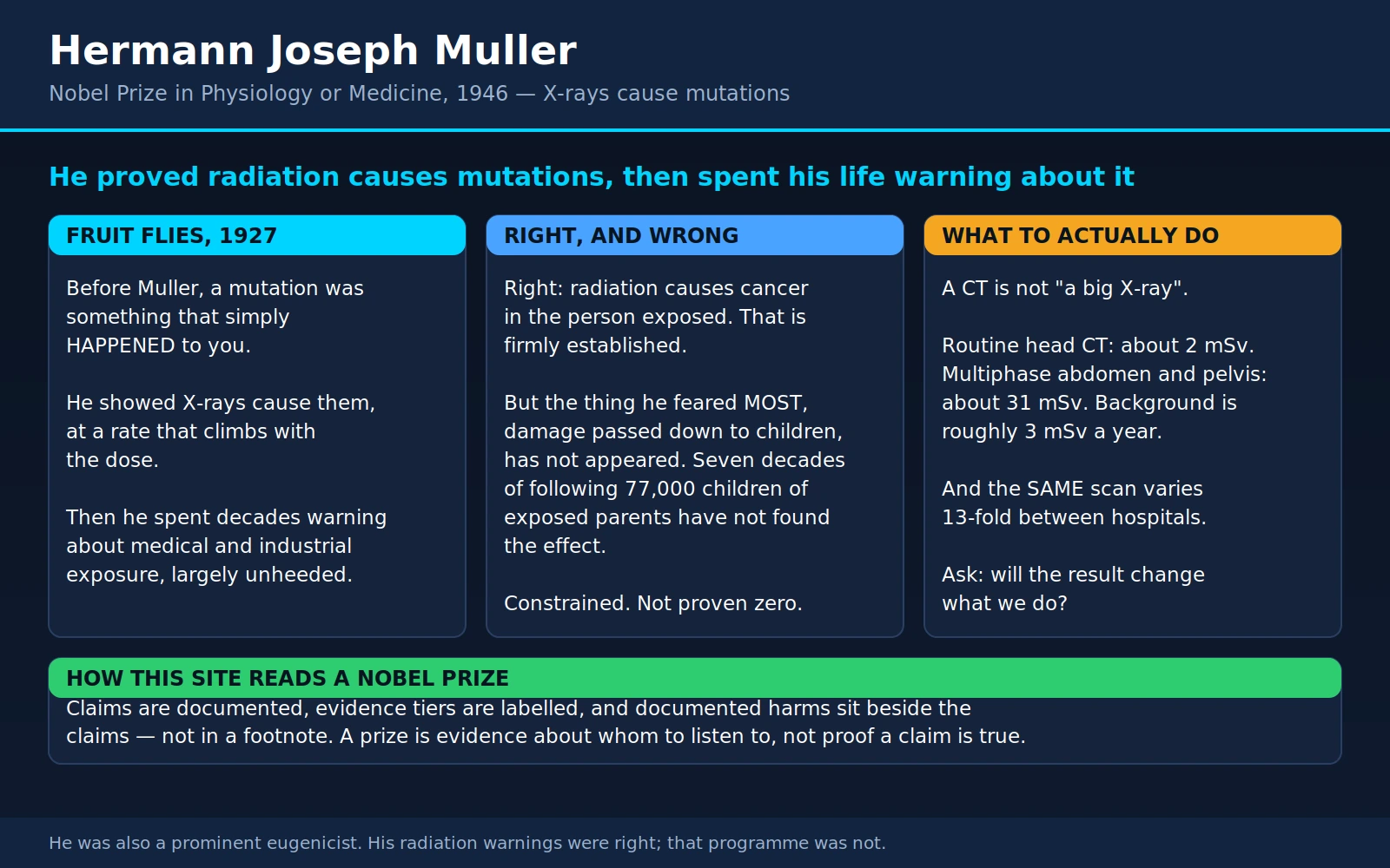
Table of Contents
- Overview
- Before 1927: Mutation Was Something That Just Happened
- The Experiment: Flies, X-Rays, and a Booby-Trapped Chromosome
- Proportional to Dose, With No Floor in Sight
- The Warnings Nobody Heeded
- The Irradiated Children: Ringworm and the Thymus
- The Radium Girls
- Hiroshima and Nagasaki: The Backbone of Every Risk Estimate
- The Linear No-Threshold Model, in Plain Language
- Is LNT Actually Right? The Honest Answer
- Medical Imaging: The Largest Exposure You Control
- A CT Is Not "a Big X-Ray"
- Children Are Not Small Adults
- The Pearce Cohort and Its Critics
- What to Actually Do
- Radon: The Other Everyday Exposure
- Muller the Man: Right About Radiation, Wrong About People
- Key Research Papers
- Connections
- Featured Videos
1. Overview
Hermann Joseph Muller (1890–1967) was an American geneticist who received the Nobel Prize in Physiology or Medicine in 1946 — the citation reads "for the discovery of the production of mutations by means of X-ray irradiation." He trained in Thomas Hunt Morgan's fruit-fly laboratory at Columbia, did the prize-winning work at the University of Texas in 1926 and 1927, and spent his final two decades at Indiana University. The official record is at nobelprize.org — 1946 Prize in Physiology or Medicine.
What he did sounds modest and is not. Before Muller, a mutation was something that happened to an organism — rare, spontaneous, unexplained, essentially an act of nature. After Muller, a mutation was something you could cause, deliberately, in a laboratory, in numbers, using a machine that thousands of hospitals and shoe shops already owned. He turned heredity into an experimental science, and in the same stroke he turned the X-ray tube from a marvel into a hazard.
Muller understood the second implication immediately, and he spent the rest of his life saying so. He was largely ignored for decades, while medicine used radiation to treat acne, ringworm of the scalp, enlarged tonsils and enlarged thymus glands in infants, and while shoe shops let children put their feet under fluoroscopes for fun. The cohorts of people this created are still being followed today, and they are a large part of how we know what radiation does to a human body.
This page is therefore mostly about radiation safety, not about a man. That is deliberate. Muller's biography is interesting; the practical question of how much radiation you are getting from medical imaging, and what to do about it, is useful. The single most important sentence on this page is in section 15, and it is not "avoid scans." It is ask whether the scan will change what happens next.
2. Before 1927: Mutation Was Something That Just Happened
By the early 1920s the fruit fly Drosophila melanogaster had made the gene a real object rather than a bookkeeping device. Morgan's group had shown that genes sit on chromosomes in a linear order, and that they occasionally change — a red eye becomes white, a normal wing becomes vestigial — and that the change is then inherited. Those spontaneous changes were the raw material of the whole science.
The problem was that they were maddeningly rare. A geneticist could keep bottles of flies for years and harvest a handful of new mutations. Nobody could make one on purpose. Investigators had tried heat, chemicals, centrifugation and radiation, and had produced a literature of unrepeatable claims. Without a way to generate mutations at will, the gene could be mapped but not manipulated, and its physical nature stayed out of reach.
Muller's contribution had two halves, and the second is the one that made the first believable. The first half was the idea that X-rays might do it. The second was a method for counting mutations reliably enough that a rate could be measured rather than asserted.
3. The Experiment: Flies, X-Rays, and a Booby-Trapped Chromosome
Muller built a genetic trap. He assembled a specially engineered fruit-fly X chromosome carrying three features at once: a crossover suppressor (a chromosomal inversion that prevents the chromosome from recombining with its partner, so it passes down intact), a recessive lethal gene, and the dominant Bar eye marker that made carriers visually identifiable. Geneticists call the stock ClB, after those three elements.
The trap works like this. Female flies carrying this chromosome are mated to X-irradiated males. Because a male fly has only one X chromosome — inherited from his mother — any new recessive lethal mutation induced anywhere on the father's X will be unmasked in his grandsons. Set the cross up correctly and one entire class of expected male grandchildren simply fails to appear. You do not have to find the mutation, describe it, or even know what it is. You count empty bottles. That converted mutation from an anecdote into a number.
With the counting method in hand, the X-ray result was unambiguous: irradiated males produced lethal mutations at a rate that dwarfed the spontaneous background, and the induced changes were heritable, transmitted to descendants who had never been anywhere near the machine. Muller announced the finding in Science in 1927 under the title "Artificial transmutation of the gene."
4. Proportional to Dose, With No Floor in Sight
The finding that mattered for the next hundred years of public health was not simply that X-rays cause mutations. It was the shape of the relationship: within the range studied, the number of induced mutations rose in proportion to the dose delivered. Double the dose, double the mutations. There was no dose at which the effect visibly switched on, and no sign of a floor beneath which nothing happened.
If mutation is proportional to dose all the way down, then there is no such thing as a completely safe exposure — only a small one. Muller drew exactly that conclusion, and he drew it in public, repeatedly, for forty years. He served on the United States National Academy of Sciences committee whose 1956 report on the genetic effects of atomic radiation carried the idea into policy, and that report is the direct ancestor of modern radiation protection.
It is worth pausing on how large a jump this was. Muller's flies were irradiated at doses enormously higher than anything a person encounters from nature or from a hospital. In a later re-examination of Muller's own data, the toxicologist Edward Calabrese calculated that the total dose used in the 1927 experiments was on the order of 95 million times the average natural background exposure — and argued that extrapolating from there down to background was an over-reach that shaped radiation policy for decades. That criticism is contested, and section 10 gives the other side of it. But the underlying observation is fair and worth carrying with you: the dose-response line that governs radiation protection was anchored at one end by experiments conducted very far indeed from the doses it is now used to regulate.
5. The Warnings Nobody Heeded
Muller's warnings were aimed at two things: weapons fallout, which the public eventually took seriously, and medical and industrial radiation, which for a long time it did not.
The medical uses of the period read now like a catalogue of avoidable harm. X-rays were used to treat acne. They were used to remove hair. They were used to treat tinea capitis — ringworm of the scalp — a nuisance fungal infection of children, because the radiation made the hair fall out and the fungus went with it. They were used to shrink the thymus in infants, a gland that was widely and wrongly believed to cause sudden infant death when enlarged, and which in fact is simply large in healthy babies. They were used on tonsils. Fluoroscopy — a live X-ray movie, delivering dose continuously rather than in a single snap — was used casually and repeatedly, including in the long-term monitoring of tuberculosis patients.
And outside medicine entirely, from the 1920s into the 1950s, shoe shops in Britain and North America installed shoe-fitting fluoroscopes: a cabinet a customer stood at while an X-ray beam passed through their feet, so that parent, child and salesperson could all look at the bones inside the shoe. Children queued to use them. They were a novelty. They were withdrawn over the following decades as the profession's understanding caught up, but the point is that they existed at all, for thirty years, in the middle of a shopping street, while a Nobel laureate was explaining what ionising radiation does to chromosomes.
None of this was malice. Radiation genuinely worked on some of these conditions, its harms appeared decades later rather than immediately, and the specific idea that a modest dose today can produce a cancer in 1985 was exactly the idea Muller was trying to establish. But the result is that the twentieth century ran a series of very large, very unethical experiments on children, and then had the decency to follow them up. The next two sections are what those follow-ups found.
6. The Irradiated Children: Ringworm and the Thymus
The ringworm cohort. Israeli researchers followed 10,834 people who had been irradiated as children for tinea capitis in the 1950s, against two matched unirradiated comparison groups — the general population, and their own siblings. Thyroid doses were low to moderate, in the range of 4.5 to 49.5 centigray (roughly 45 to 495 mGy). Over follow-up extending as long as 54 years, 159 thyroid cancers were diagnosed. The excess relative risk worked out at 20.2 per gray (95% CI 11.8–32.3), and the excess absolute risk at 9.9 cases per gray per 10,000 person-years (95% CI 5.7–14.7). Risk rose with dose and fell with age at exposure — the younger the child, the worse the outcome. The excess became significant 10 to 19 years after treatment, peaked at 20 to 30 years, and although it fell substantially after 40 years it was still significantly elevated then.
The thymus cohort. Children irradiated in infancy for a supposedly enlarged thymus between 1926 and 1957 were re-surveyed between 2004 and 2008, again alongside their unexposed siblings. Thyroid cancer occurred in 50 irradiated subjects (mean thyroid dose 1.29 Gy) against 13 unirradiated siblings across 334,347 person-years. After adjustment, the rate ratio was 5.6 (95% CI 3.1–10.8), the excess relative risk 3.2 per gray (1.5–6.6), and the excess absolute risk 2.2 cases per gray per 10,000 person-years (1.4–3.2). Median follow-up was 57.5 years, and the authors' conclusion was that increased risk "remains a lifelong concern in those exposed to lower doses of medical radiation during early childhood."
Two features of these studies do most of the work on this page. The first is latency: the harm showed up twenty to thirty years later, which is precisely why it was not noticed at the time and precisely why the practice continued. The second is age: in both cohorts, the younger the child at exposure, the higher the eventual risk. That is not a statistical artefact, and it is the reason section 13 exists. Our Thyroid Cancer and Thyroid Nodules pages cover the disease end of this.
7. The Radium Girls
The other great natural experiment of the era was occupational and involved no X-ray tube at all. In the 1910s and 1920s, young women were employed in the United States painting luminous radium dials onto watches and instruments. To keep a fine point on the brush, they shaped it with their lips. Radium is chemically similar to calcium, so what they swallowed was deposited in bone, where it stayed, irradiating the surrounding tissue continuously for the rest of their lives.
The consequences — necrosis of the jaw, anaemia, and bone sarcomas — emerged over the following years, and the recognition of the pattern and the establishment of its cause is one of the founding episodes of occupational epidemiology. As a review of the U.S. dial-worker studies puts it, health-effects investigations of these women began in the early 1920s and continued for the rest of the century, and it was the demonstration of a causal association in this group that led to the development of radiation protection standards. Our Osteosarcoma page covers the bone tumours themselves.
The dial painters matter to a page about medical imaging for a reason that is easy to miss. Their exposure was internal and chronic — an emitter lodged in the skeleton delivering dose continuously for decades, which is a completely different exposure pattern from a scan lasting seconds. Anyone reasoning from the Radium Girls straight to a CT scan is comparing two things that differ by orders of magnitude in dose and entirely in kind. They belong on this page as history and as the origin of protection standards, not as a dose comparison.
8. Hiroshima and Nagasaki: The Backbone of Every Risk Estimate
Almost every number anyone quotes about radiation and cancer risk — including the ones your radiology department uses — traces back to one cohort. The Life Span Study (LSS) has followed the survivors of the 1945 atomic bombings since 1950, with individual dose estimates reconstructed from where each person was standing and what was between them and the blast. It comprises roughly 120,000 subjects, with two further cohorts alongside it: about 3,600 people exposed in utero, and about 77,000 children conceived after their parents' exposure, followed since 1945.
The mortality analysis covering 1950–2003 found the risk of death positively associated with dose, with the additive radiation risk for solid cancers continuing to increase throughout life. The sex-averaged excess relative risk was 0.42 per gray (95% CI 0.32–0.53) for all solid cancer at age 70 following exposure at age 30. Risk rose by about 29% for each decade younger at exposure (95% CI 17%–41%) — the same age pattern as the irradiated-children cohorts. The lowest dose range showing a statistically significant excess was 0 to 0.20 Gy, and a formal threshold analysis put the best estimate of the threshold at zero.
There is a second, quieter finding that belongs on a page about Muller specifically. The effect he feared most, and campaigned hardest about, was heritable damage — mutations passed to children who were never exposed. In the survivors themselves, radiation-related cancer risk is firmly established. In their children, as the Radiation Effects Research Foundation summarised it, no increased risk from parental exposure has been observed for malignancies or other diseases — with the honest caveat that investigations continue, because those cohorts are still relatively young.
9. The Linear No-Threshold Model, in Plain Language
The linear no-threshold model (LNT) is the rule that radiation protection runs on worldwide. It makes two claims:
- Linear — the extra risk of cancer is proportional to the dose. Twice the dose, twice the extra risk. Half the dose, half the extra risk.
- No threshold — the line runs all the way down to zero dose without hitting a floor. There is no amount of radiation small enough to be certainly harmless; there is only an amount small enough that the risk is negligible.
Everything downstream follows from those two sentences. It is why radiographers stand behind a screen, why dose limits for workers are set the way they are, why lead shielding exists, and why the governing principle of the field is ALARA — As Low As Reasonably Achievable. If there were a safe threshold, the job would be to stay under it and stop worrying. Because LNT assumes there is not, the job is instead to justify every exposure and then minimise it.
LNT is the direct descendant of Muller's dose-proportional mutation curve. It is also, and this is the part usually left out, a policy choice as much as a scientific finding — a deliberately cautious default adopted because it is prudent, simple, and additive, not because the shape of the curve at very low doses has been measured. It cannot be measured directly. The excess risk from a single scan-sized dose is far too small to detect against a lifetime cancer risk of roughly one in two or three, in any study of any feasible size.
10. Is LNT Actually Right? The Honest Answer
At moderate and high doses, essentially nobody disputes the relationship. The argument is about the bottom of the curve, and it is a real argument between serious people.
The case for LNT. The U.S. National Council on Radiation Protection and Measurements ran a full review of the low-dose and low-dose-rate epidemiology, assessing 29 studies or groups of studies for methods, dosimetry and statistical modelling. The committee's published conclusion was that the risks at these doses are "small and uncertain," that the available epidemiological data were "broadly supportive of the LNT model," and that "no alternative dose-response relationship appears more pragmatic or prudent for radiation protection purposes." The atomic-bomb mortality data agree: the formal threshold analysis returned zero.
The case that it is unsettled. The most recent solid-cancer incidence analysis of the same atomic-bomb cohort, covering 1958–2009 with 22,538 first primary solid cancers, found something more complicated. In women the dose response was consistent with a straight line (ERR 0.64 per gray, 95% CI 0.52–0.77). In men there was significant upward curvature, so a linear-quadratic model was used instead, giving an ERR of 0.20 at 1 Gy (0.12–0.28) but only 0.010 at 0.1 Gy (−0.0003 to 0.021). The shapes differed significantly between the sexes (P = 0.02), and the authors reported the results were "not fully consistent with those previously reported, raising unresolved questions." Their own summary is the fairest sentence anyone has written on this: "uncertainties in the shape of the dose response preclude definitive conclusions to confidently guide radiation protection policies."
The historical argument. There is also a long-running and sharp dispute about how LNT came to be adopted in the first place. Calabrese, a proponent of radiation hormesis — the hypothesis that very low doses may be harmless or even protective — has argued across many papers that Muller and the 1956 National Academy genetics panel mischaracterised the research record. Beyea reviewed the same historical material and reached the opposite verdict: that the contemporaneous evidence overwhelmingly favoured a genetics LNT, that no calculations were suppressed, that Calabrese's claims fail chiefly on statistical grounds, and — the detail most people miss — that the outlier studies of the period were more likely to favour supra-linearity, meaning more risk at low dose, not less. Both papers appeared in the same journal and both are cited below. We are not adjudicating between them.
11. Medical Imaging: The Largest Exposure You Control
Natural background radiation is unavoidable and roughly fixed. Occupational exposure applies to a small minority. For most people in a developed country, medical imaging is the single largest radiation exposure they have any say over — and by the late 2000s medical uses had become the largest source of exposure to the U.S. population, having grown very rapidly over the preceding decade.
A published catalogue of effective doses gives the honest shape of the landscape. Ordinary radiographic examinations — the plain films — span a range of more than a thousandfold, from about 0.01 to 10 mSv depending on what is being imaged. CT examinations sit in a narrower but much higher band, roughly 2 to 20 mSv. Diagnostic nuclear-medicine procedures run about 0.3 to 20 mSv. Interventional procedures — the long fluoroscopically-guided ones — are the highest, around 5 to 70 mSv. For comparison, the same source gives average annual effective dose from natural background radiation as about 3 mSv.
Those ranges, rather than single numbers, are the truthful presentation. Notice what they imply: a plain film at the bottom of the radiography range is a rounding error against a year of background, while a multi-phase CT or a long interventional procedure can exceed several years of background in one sitting. Both are called "imaging."
Mammography. Breast imaging is usually quoted in mean glandular dose — dose to the breast tissue — which is not the same quantity as effective dose. Two-view digital mammography involves an average mean glandular dose of about 3.7 mGy, and older screen-film mammography about 4.7 mGy. Using standard risk models, those correspond to a lifetime attributable risk of fatal breast cancer of about 1.3 and 1.7 cases per 100,000 women aged 40 at exposure, and fewer than one case per million for women aged 80. Annual screening across ages 40 to 80 carries an estimated lifetime attributable risk of fatal breast cancer of 20 to 25 per 100,000. By contrast, a single breast-specific gamma imaging study or positron emission mammography study was estimated to carry a fatal-cancer risk roughly 20 to 30 times that of one digital mammogram in a 40-year-old — that is, one such nuclear study can exceed the radiation risk of forty years of annual mammography. See our Mammogram and Breast Cancer pages for the screening-benefit side of that trade.
Modalities with no ionising radiation at all. Ultrasound and MRI deliver none. Where either can answer the clinical question, the radiation part of this discussion simply evaporates — which is why "is there a non-ionising alternative?" is on the question list in section 15.
12. A CT Is Not "a Big X-Ray"
This is the single most useful thing on the page for most readers. A CT scanner uses X-rays, so people reasonably assume a CT is a somewhat larger version of a chest film. It is not. A plain radiograph is one brief exposure from one direction. A CT rotates the source around the body acquiring hundreds of projections, and a multi-phase study repeats the whole acquisition two or three times to catch contrast at different moments. The dose difference is not incremental; from the ranges in the previous section, ordinary radiography can start a thousandfold below where CT sits.
The measured figures make it concrete. In a study of the 11 commonest diagnostic CT studies performed on 1,119 consecutive adult patients across four institutions, median effective doses ranged from 2 mSv for a routine head CT to 31 mSv for a multiphase abdomen-and-pelvis CT. Both are "a CT scan." One is roughly two-thirds of a year of background; the other is around ten years of it.
The same study found something arguably more important, and it is not about CT versus X-ray at all. Within each single type of CT study, effective dose varied by a mean factor of 13 between the highest and lowest dose — within and across institutions. The same examination, the same clinical question, thirteen times the dose depending on where you had it done and how the protocol was set. The authors' conclusion was that doses "are higher and more variable than generally quoted."
That variability is genuinely good news, because unlike the physics it is fixable. Estimated cancer risk from these doses tracked age steeply: for an estimated 1 in 270 women undergoing CT coronary angiography at age 40 who would develop a cancer from that scan (1 in 600 men), the comparable figure for a routine head CT at the same age was about 1 in 8,100 women and 1 in 11,080 men. For 20-year-olds the risks were roughly doubled; for 60-year-olds roughly 50% lower. Age at exposure is doing as much work as the machine.
13. Children Are Not Small Adults
Children are more vulnerable to radiation for two independent reasons that compound each other. First, tissues that are actively growing are more radiosensitive — dividing cells are where a mutation gets fixed and propagated. Second, and just as important, a child has more years left for a cancer to develop in. Section 6 showed latencies of twenty to thirty years to the peak of excess thyroid cancer; a 60-year-old may simply not live long enough to express a risk that a 6-year-old has half a century to express. Both the atomic-bomb cohort and both irradiated-children cohorts show the same gradient: younger at exposure, higher the eventual risk.
Two large modern studies have measured this for CT specifically. In the pan-European EPI-CT cohort, 658,752 eligible people who had a CT before age 22 between 1977 and 2014 were followed for brain cancer. Mean cumulative brain dose was 47.4 mGy. Across 165 brain cancers, of which 121 (73%) were gliomas, there was a significant linear dose response: an excess relative risk of 1.27 per 100 mGy (95% CI 0.51–2.69) for all brain cancers, and 1.11 per 100 mGy (0.36–2.59) for gliomas alone.
The companion EPI-CT analysis followed 948,174 people for blood cancers and found an excess relative risk of 1.96 per 100 mGy (95% CI 1.10–3.12) for all haematological malignancies, from 790 cases. That paper also did the most useful thing in this entire literature — it converted the finding into a present-day absolute number. For every 10,000 children scanned today, at a mean active-bone-marrow dose of 8 mGy, the authors expect 1 to 2 people to develop a radiation-attributable blood cancer over the following 12 years.
Sit with that figure, because it cuts both ways and it is meant to. One to two per ten thousand is a real, non-zero harm that justifies careful protocols, dose optimisation and asking whether the scan is needed. It is also a risk of roughly 0.01% to 0.02%, against which a missed intracranial bleed, a missed appendicitis or a missed tumour is a far larger and far more immediate danger. See Leukemia and Brain Cancer for the conditions in question.
14. The Pearce Cohort and Its Critics
The study that put paediatric CT dose on the front pages was a retrospective cohort of British patients first scanned before age 22 between 1985 and 2002, followed through 2008. Among roughly 178,000 patients, 74 developed leukaemia and 135 developed brain tumours. The excess relative risk was 0.036 per mGy (95% CI 0.005–0.120) for leukaemia and 0.023 per mGy (0.010–0.049) for brain tumours. Framed as relative risks, a cumulative dose of about 50 mGy roughly tripled leukaemia risk and about 60 mGy roughly tripled brain-cancer risk.
Those tripling figures travelled around the world, and they are the reason many parents are frightened of CT. The authors' own next sentence travelled much less far, and it is the one that matters: "Because these cancers are relatively rare, the cumulative absolute risks are small: in the 10 years after the first scan for patients younger than 10 years, one excess case of leukaemia and one excess case of brain tumour per 10,000 head CT scans is estimated to occur." Tripling a very small number leaves a small number.
Now the criticism, which is legitimate. The obvious worry about any such study is reverse causation, sometimes called confounding by indication: a child who is destined to be diagnosed with a brain tumour is exactly the child who gets a head CT, because the early symptoms of the tumour are what prompted the scan. The scan did not cause the cancer; the cancer caused the scan. A related worry is that children with cancer-predisposing genetic conditions are both more likely to be scanned and more likely to develop cancer regardless.
And the authors went and tested it. They returned to the cohort with additional clinical data — radiology information systems, death certificates, pathology reports — obtaining information for about 40% of the cohort from the first two sources and clinical detail on 90% of cases once pathology was included. Cancer-predisposing conditions turned up in 4 of 74 leukaemia/MDS cases and 13 of 135 brain-tumour cases; because these were unrelated to CT exposure, excluding those patients did not alter the dose response. Previously unreported earlier cancers were a different matter — found in 2 leukaemia/MDS cases, 7 brain-tumour cases and 232 non-cases, and genuinely associated with having had more CTs. Excluding those reduced the excess relative risk per mGy by 15% for leukaemia/MDS (0.036 to 0.033) and by 30% for brain tumours (0.023 to 0.016). Both remained statistically significant. The authors' own conclusion: there was "evidence of some bias in our original risk estimates," and re-analysis with the additional data "still showed an increased cancer risk after low-dose radiation exposure from CT scans in young patients."
15. What to Actually Do
Do not skip imaging you need. That is the wrong lesson and it is a more dangerous one. The risks in section 13 are on the order of one or two per ten thousand. A missed subdural haematoma, a missed pulmonary embolism, a missed perforation or a missed cancer is a risk on the order of one in one. If a clinician who has examined you says you need a scan, the default answer is yes.
The right question is not how much radiation is this? It is "will the result change what we do?" In radiation protection this is the principle of justification, and it comes before dose optimisation for a reason: the cheapest millisievert to save is the one from a scan that was never going to alter management.
Useful things to ask, none of which require you to argue with anyone:
- "What would change depending on the result?" A good answer is specific — it names a decision. If the plan is identical either way, the scan is imaging for reassurance, and that is a real thing to want but worth naming as such.
- "Is there an ultrasound or MRI that would answer this?" Neither uses ionising radiation. Sometimes the answer is no. It is always worth one sentence.
- "Has this already been done?" Repeat imaging because prior films could not be located is common and completely avoidable. Keep your own list — date, body part, modality, hospital — and carry prior discs or reports to new appointments.
- "Can this be a single-phase study?" Multi-phase abdominal CT was the 31 mSv end of the range in section 12; the routine head CT was 2 mSv. If two phases are not needed, that is a large saving for one question.
- For a child: "Do you use paediatric dose settings?" Given the 13-fold within-examination variation measured across institutions, protocol matters as much as whether the scan happens. Children's hospitals and departments that audit their doses are meaningfully different from those that do not.
- Decline whole-body screening CT for a healthy person with no symptoms. This is the one place where a flat recommendation is easy: it delivers a genuine dose in exchange for a very high rate of incidental findings that lead to more scans, biopsies and anxiety, in someone with no clinical question to answer.
There is a validated example of exactly this reasoning working. In a study of 42,412 head-injured children across 25 emergency departments, CT was performed in 14,969 (35.3%), while clinically important traumatic brain injury occurred in 376 (0.9%) and only 60 (0.1%) needed neurosurgery. The investigators derived and validated age-specific rules using ordinary bedside findings — normal mental status, no loss of consciousness, no vomiting, non-severe mechanism, no skull-fracture signs, no severe headache, and for infants, acting normally according to the parents. The rules had a negative predictive value of 100% for children under two and 99.95% for those two and over, and neither rule missed a single child who needed neurosurgery. Between a fifth and a quarter of the children who were scanned fell into the very-low-risk group in which CT can routinely be avoided.
That is the shape of the answer Muller's discovery eventually earned: not fewer scans out of fear, but fewer scans because somebody worked out precisely which ones were never going to change anything.
16. Radon: The Other Everyday Exposure
Medical imaging is the exposure you control at the clinic. Radon is the one you control at home, and for many households it is larger. Radon is a radioactive gas produced by the decay of uranium in soil and rock; it seeps into buildings through foundations and accumulates indoors. Its decay products lodge in the lung.
The definitive European analysis pooled individual data from 13 case-control studies across nine countries — 7,148 lung-cancer cases and 14,208 controls. Mean measured radon in control-group homes was 97 Bq/m3, with 11% above 200 and 4% above 400. Lung-cancer risk rose by 8.4% per 100 Bq/m3 of measured radon (95% CI 3.0%–15.8%, P = 0.0007), or 16% per 100 Bq/m3 (5%–31%) after correcting for random measurement error. The dose-response appeared linear with no threshold, and remained significant (P = 0.04) when the analysis was restricted to homes measuring under 200 Bq/m3. Overall, the authors attributed about 2% of all cancer deaths in Europe to residential radon.
The most actionable finding is the interaction with smoking. In the absence of other causes of death, absolute lung-cancer risk by age 75 at usual radon concentrations of 0, 100 and 400 Bq/m3 was about 0.4%, 0.5% and 0.7% for lifelong non-smokers — and about 25 times greater for cigarette smokers, at roughly 10%, 12% and 16%. Radon and tobacco do not add; they multiply. A smoker in a high-radon house is in a categorically different situation from a non-smoker in the same house, and not smoking is by far the larger lever.
What to do is unusually simple and cheap. Test. Radon is colourless and odourless, concentrations vary enormously between neighbouring houses depending on geology and construction, and no amount of reasoning about your area substitutes for a measurement. Long-term detectors cost very little. If levels are high, mitigation — typically sub-slab depressurisation and sealing entry routes — is established engineering, not an experiment. See our Radon page for detail, Lung Cancer for the disease, Uranium for where the gas comes from, and Secondhand Smoke for the co-exposure that dominates the risk.
17. Muller the Man: Right About Radiation, Wrong About People
Muller was also a committed and prominent eugenicist, and a page that praised his radiation warnings while omitting this would be dishonest.
His programme was what its advocates called positive eugenics: not the sterilisation of the unfit, but the deliberate propagation of favoured heredity through artificial insemination by selected donors. He set it out in his 1935 book Out of the Night. In 1936 he presented a plan for human improvement by artificial insemination to Stalin, framing it as a socialist project — Muller was a Marxist, and his eugenics was of the left-wing variety rather than the racialised American strain, a distinction that is historically real and morally insufficient. Some twenty years later he began planning, with Robert Klark Graham, a Foundation for Germinal Choice in California. It opened in 1980, after Muller's death, and produced some 215 children over the twenty years it operated. Historians of science record it as the first practical experiment in eugenic artificial insemination; it found very little favour with the people it was designed for, and no state has ever adopted such a scheme as social policy.
Eugenics as a programme is repudiated, and rightly. It rests on the premise that some people's heredity is worth propagating and other people's is not, and that someone competent can be appointed to decide which is which. In the twentieth century that premise underwrote coercive sterilisation, immigration restriction and worse, and the fact that Muller's version was voluntary and idealistic does not rescue the premise — it only changes who was going to enforce it.
The contrast is worth one paragraph and no more. The same quality of mind produced both: enormous confidence in extrapolating from a clean genetic result to what human societies should do about it. Applied to X-rays and chromosomes, that confidence was largely vindicated — he was right about radiation decades before the profession caught up, and children were harmed in the interval because he was ignored. Applied to human reproduction, the same confidence produced a scheme that was scientifically naive about the genetics of complex traits and ethically indefensible about the rest. Being right about the first thing conferred no authority over the second, and he should not be read as though it did. The section of this page you should act on is section 15. The section you should learn from is this one.
Our Nobel Prizes That Aged Badly page covers awards the field had to outgrow. Muller's is not one of them — the radiation-genetics work has held up completely. It is his opinions outside it that did not.
Key Research Papers
Every citation below was verified against the PubMed record, and every abstract was read before the finding beside it was written. The 1927 paper is listed first and, as noted in section 3, carries no abstract in PubMed; no numerical claim on this page rests on it.
- Muller HJ. Artificial transmutation of the gene. Science 1927;66(1699):84-7
- Calabrese EJ. Muller's Nobel Prize data: getting the dose wrong and its significance. Environ Res 2019;176:108528
- Beyea J. Lessons to be learned from a contentious challenge to mainstream radiobiological science (the linear no-threshold theory of genetic mutations). Environ Res 2017;154:362-379
- Richards M. Artificial insemination and eugenics: celibate motherhood, eutelegenesis and germinal choice. Stud Hist Philos Biol Biomed Sci 2008;39(2):211-21
- Fry SA. Studies of U.S. radium dial workers: an epidemiological classic. Radiat Res 1998;150(5 Suppl):S21-9
- Sadetzki S, Chetrit A, Lubina A, et al. Risk of thyroid cancer after childhood exposure to ionizing radiation for tinea capitis. J Clin Endocrinol Metab 2006;91(12):4798-804
- Adams MJ, Shore RE, Dozier A, et al. Thyroid cancer risk 40+ years after irradiation for an enlarged thymus: an update of the Hempelmann cohort. Radiat Res 2010;174(6):753-62
- Ozasa K, Shimizu Y, Suyama A, et al. Studies of the mortality of atomic bomb survivors, Report 14, 1950-2003: an overview of cancer and noncancer diseases. Radiat Res 2012;177(3):229-43
- Grant EJ, Brenner A, Sugiyama H, et al. Solid cancer incidence among the Life Span Study of atomic bomb survivors: 1958-2009. Radiat Res 2017;187(5):513-537
- Ozasa K, Grant EJ, Kodama K. Japanese legacy cohorts: the Life Span Study atomic bomb survivor cohort and survivors' offspring. J Epidemiol 2018;28(4):162-169
- Shore RE, Beck HL, Boice JD, et al. Implications of recent epidemiologic studies for the linear nonthreshold model and radiation protection. J Radiol Prot 2018;38(3):1217-1233
- Mettler FA Jr, Huda W, Yoshizumi TT, Mahesh M. Effective doses in radiology and diagnostic nuclear medicine: a catalog. Radiology 2008;248(1):254-63
- Smith-Bindman R, Lipson J, Marcus R, et al. Radiation dose associated with common computed tomography examinations and the associated lifetime attributable risk of cancer. Arch Intern Med 2009;169(22):2078-86
- Hendrick RE. Radiation doses and cancer risks from breast imaging studies. Radiology 2010;257(1):246-53
- Pearce MS, Salotti JA, Little MP, et al. Radiation exposure from CT scans in childhood and subsequent risk of leukaemia and brain tumours: a retrospective cohort study. Lancet 2012;380(9840):499-505
- Berrington de Gonzalez A, Salotti JA, McHugh K, et al. Relationship between paediatric CT scans and subsequent risk of leukaemia and brain tumours: assessment of the impact of underlying conditions. Br J Cancer 2016;114(4):388-94
- Hauptmann M, Byrnes G, Cardis E, et al. Brain cancer after radiation exposure from CT examinations of children and young adults: results from the EPI-CT cohort study. Lancet Oncol 2023;24(1):45-53
- Bosch de Basea Gomez M, Thierry-Chef I, Harbron R, et al. Risk of hematological malignancies from CT radiation exposure in children, adolescents and young adults. Nat Med 2023;29(12):3111-3119
- Darby S, Hill D, Auvinen A, et al. Radon in homes and risk of lung cancer: collaborative analysis of individual data from 13 European case-control studies. BMJ 2005;330(7485):223
- Kuppermann N, Holmes JF, Dayan PS, et al. Identification of children at very low risk of clinically-important brain injuries after head trauma: a prospective cohort study. Lancet 2009;374(9696):1160-70
Live PubMed Searches
- Radiation-induced mutation in Drosophila
- Linear no-threshold model and radiation protection
- Paediatric CT radiation dose and cancer risk
- Medical imaging effective dose
- Residential radon and lung cancer
- Atomic-bomb survivors and the Life Span Study
- Thyroid cancer after childhood radiation
Connections
- All Notable Doctors
- Nobel Prize in Physiology or Medicine — every laureate, 1901 to the present
- Cormack & Hounsfield — the invention of the CT scan, and what a scan can and cannot tell you
- Lauterbur & Mansfield — MRI, the major imaging modality that uses no ionising radiation at all
- Rosalyn Yalow — radioimmunoassay: radioactivity used as a measuring tool rather than a hazard
- Barbara McClintock — the other great chromosome experimentalist of the era, and thirty years of being ignored
- Watson, Crick & Wilkins — the physical structure of the gene Muller had shown was mutable
- Niels Finsen — an earlier prize for radiation as therapy, and what light therapy can actually do
- Nobel Prizes That Aged Badly — awards medicine had to outgrow
- Radon — the largest everyday radiation exposure in most homes, and how to test for it
- Secondhand Smoke — the co-exposure that multiplies radon lung-cancer risk roughly 25-fold
- Uranium — the parent element whose decay produces radon
- Mammogram — mean glandular dose, screening benefit, and how the two trade off
- Full-Body MRI — no ionising radiation, but its own incidental-findings problem
- DEXA Scan Explained — a bone-density study at the very low end of the radiography range
- Thyroid Cancer — the cancer the irradiated-children cohorts were defined by
- Thyroid Nodules — what a history of neck irradiation changes about their workup
- Leukemia — the blood cancer measured in the EPI-CT and Pearce cohorts
- Brain Cancer — the other endpoint in the paediatric CT studies
- Osteosarcoma — the bone tumour that killed the radium dial painters
- Lung Cancer — the radon endpoint, and where smoking dominates the risk
- All Lab Tests
- All Toxins