Gerald Edelman & Rodney Porter: Antibody Structure and the Y-Shaped Molecule Behind Every -mab Drug
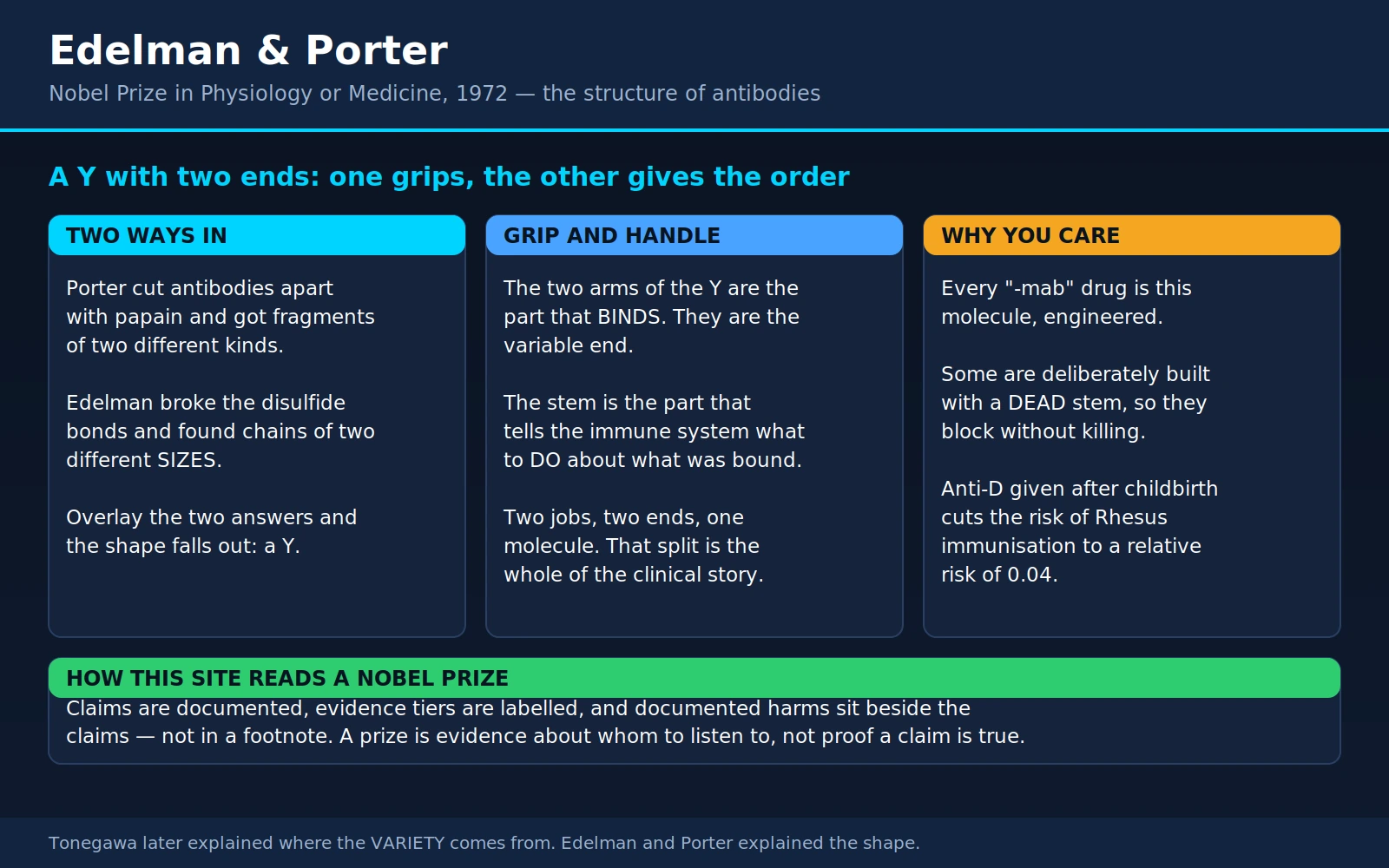
Table of Contents
- Overview
- Porter's Knife: Papain and the Three Fragments
- Edelman's Solvent: Two Different Sizes of Chain
- The Y: How the Two Halves of the Answer Fit Together
- Fab and Fc: The Grip and the Handle
- The Whole Molecule, Spelled Out
- The Question They Could Not Answer
- The 1972 Nobel Prize
- Every "-mab" Is This Molecule, Engineered
- Rh Immunoglobulin: An Antibody Given to Prevent Immunisation
- IVIG: Pooled Human Antibody Doing Two Different Jobs
- Why Antibody Drugs Have to Be Injected
- The End of "-mab": What These Drugs Are Called Now
- Edelman's Other Career: Neural Darwinism
- Key Research Papers
- Connections
- Featured Videos
1. Overview
In 1972 the Nobel Prize in Physiology or Medicine went jointly to two men who had never worked in the same laboratory and who competed with each other for a decade: Gerald M. Edelman (1929–2014), a young American physician-turned-chemist at the Rockefeller Institute in New York, and Rodney R. Porter (1917–1985), an English biochemist who had trained under Frederick Sanger at Cambridge and who did his decisive work at the National Institute for Medical Research in London before moving to Oxford. The citation was for their discoveries concerning the chemical structure of antibodies.
To see why that mattered, picture where medicine stood in the mid-1950s. Doctors had known for sixty years that blood contained something that neutralised diphtheria toxin and killed bacteria. They could measure it, transfer it from one person to another, and use it to save lives. They called it antitoxin, or immune serum, or gamma globulin. What nobody could say was what it was. It was a protein, it was big, and it came in an apparently limitless number of specificities — one for tetanus, one for measles, one for ragweed pollen. That combination looked impossible. Proteins are built from a fixed genetic blueprint. How can one blueprint produce a molecule that comes in millions of different shapes?
Edelman and Porter solved the first half of that puzzle: not where the variety comes from, but what the molecule looks like. They did it with two completely different tricks, and each trick alone would have been ambiguous. Porter took a protein-cutting enzyme from papaya and chopped the antibody into pieces, then asked which pieces still grabbed their target. Edelman used a harsh solvent plus a reducing agent to snap the chemical staples holding the molecule together, then asked how many separate strings fell out and how long each one was. Put the two answers side by side and a shape appears: a Y, built from four separate protein chains — two long "heavy" chains and two short "light" chains — held together by sulfur-to-sulfur bonds.
The Y has two ends and they do different jobs. The two tips of the Y vary enormously from one antibody to the next; they are the part that recognises and grips a specific target. The stem is nearly identical in every antibody of a given class; it is the part that other immune cells read, the handle by which the immune system picks up a flagged target and decides what to do with it. That single distinction — variable tips, constant stem — is the reason this page belongs on a health site rather than only in a history book. It is the design principle behind roughly every antibody drug on the market, behind the injection that has all but eliminated Rh disease of the newborn, and behind the pooled-antibody infusions given to people whose own immune systems cannot make enough.
2. Porter's Knife: Papain and the Three Fragments
Porter's approach was the one a butcher would recognise: if you cannot see the animal, cut it up and look at the joints. His knife was papain, the protein-digesting enzyme in papaya fruit — the same enzyme sold in supermarkets as meat tenderiser. Papain cuts proteins, but not randomly; a folded protein exposes only certain stretches of its chain to attack, so a limited digestion tends to sever a molecule at its flexible, accessible joints and leave the compact regions intact.
Porter had been picking at this since the 1940s, and the version that worked used crystalline papain plus a reducing agent, applied gently. Rabbit gamma globulin, treated this way, came apart into three pieces, which he separated by column chromatography. Two of them were essentially identical to each other. The third was different — and it did something no one expected of a fragment of a blood protein: it crystallised.
Then came the test that made the experiment famous. Porter asked which fragments still recognised the antibody's target. The two identical fragments did. The crystallising fragment did not — it had no detectable ability to grab anything. Each of the two binding fragments could attach to its target but, unlike the whole antibody, could no longer clump targets together into visible aggregates, which meant each fragment carried one binding site and the intact molecule carried two.
Those observations gave the fragments their permanent names, still on every drug label and immunology exam today: Fab, for fragment, antigen binding, and Fc, for fragment, crystallisable. It is worth pausing on how strange the second name is. Fc is named not for what it does, but for the laboratory accident that it forms crystals — and it forms crystals precisely because it is the same in every antibody molecule in the preparation. Identical molecules stack into a lattice; a mixture of millions of different shapes cannot. The crystallisation was itself the first clue that one end of the antibody is constant.
3. Edelman's Solvent: Two Different Sizes of Chain
Edelman came at it from the opposite direction, and from a much less senior position. He was a graduate student at the Rockefeller Institute, working in Henry Kunkel's laboratory, when in 1959 he published a short communication in the Journal of the American Chemical Society with a title of four words: "Dissociation of gamma-globulin."
His trick was chemical rather than enzymatic. Proteins are often stapled together by disulfide bonds — a chemical bridge between two sulfur atoms on the amino acid cysteine. A disulfide bond is a covalent link, strong enough that ordinary handling will not break it, which is why gamma globulin behaved for decades as if it were one single object. Edelman treated it with a reducing agent to snap those sulfur bridges, in a strong urea solution to unfold the protein so the bridges were reachable and so the pieces would not immediately re-associate.
The molecule fell apart. Working with M. D. Poulik, Edelman published the full account in the Journal of Experimental Medicine in 1961: after reduction in strong urea, the apparent molecular weight of human and rabbit gamma globulin fell to roughly one third of the starting value, new free sulfhydryl groups appeared exactly as breaking disulfide bonds predicts, and the products could be partially separated by chromatography and starch-gel electrophoresis. Crucially, the separated products were not all the same. One isolated component had an amino acid composition different from the starting material and different from the remaining components. Edelman and Poulik concluded that gamma globulin was built of multiple polypeptide chains linked by disulfide bonds — and that the chains were not all alike.
This is the observation the prize turned on. Porter's cut said the molecule had a two-armed grip and a distinct tail. Edelman's reduction said the molecule was not one chain at all, but several, of at least two different sizes. Neither result made full sense without the other.
4. The Y: How the Two Halves of the Answer Fit Together
Porter's own group closed the circle. Working with J. B. Fleischman and E. M. Press, he applied Edelman's reduction chemistry to his own papain fragments — first in a 1962 report and then, definitively, in a 1963 paper in the Biochemical Journal titled "The arrangement of the peptide chains in gamma-globulin." By labelling chains and asking which chain ended up in which fragment, they could work out the topology rather than just the parts list.
The answer, in modern units, is this. A typical antibody of the IgG class weighs about 150,000 daltons and is built from four chains: two identical heavy chains of roughly 50,000 daltons each and two identical light chains of roughly 25,000 daltons each. Each light chain is disulfide-bonded to a heavy chain; the two heavy chains are disulfide-bonded to each other in the middle. Draw that and you get a Y.
Now overlay Porter's cut. Papain severs the two heavy chains at the flexible hinge just above where they join. That releases the two arms of the Y — each one a light chain plus the top half of a heavy chain, which is a Fab — and leaves the bottom of the Y, the paired lower halves of the two heavy chains, as the Fc. Two Fabs and one Fc: exactly the three fragments Porter had counted. The arithmetic works, the binding activity sits where it should, and the crystallising fragment turns out to be the one made of nothing but constant material.
A useful mental image, and one that survives contact with the real chemistry: an antibody is a pair of tweezers with a handle. The two tips are shaped to grip one particular thing and nothing else. The handle is standard-issue, the same on every pair in the drawer, and it exists so that something else can pick the tweezers up.
5. Fab and Fc: The Grip and the Handle
Everything clinically useful on this page follows from that division of labour, so it is worth stating slowly.
The Fab tips answer the question "what?" Their amino acid sequence differs wildly from one antibody to another, and it is that difference that lets one antibody recognise tetanus toxin while another recognises cat dander. The variable stretches at the very tips fold into a surface that is complementary in shape and charge to a small patch on the target — a lock cut to fit one key.
The Fc stem answers the question "so what?" Binding a target, on its own, accomplishes remarkably little. Sticking a label on a bacterium does not kill it. What kills it is that immune cells carry receptors on their surface that read the Fc stem, and that a set of blood proteins called complement can latch onto clustered Fc stems and punch holes. When a natural killer cell, a macrophage or a neutrophil grips the Fc stem of an antibody that is already stuck to a target, it destroys what the antibody is holding. The stem is not decoration; it is the instruction.
The same stem does several other jobs that matter to patients. It binds a specialised receptor called FcRn, the neonatal Fc receptor, which continually rescues IgG molecules from being degraded and recycles them back into circulation. That recycling is why IgG survives in the blood for around three weeks rather than a few hours, and it is why an antibody drug can be given every few weeks instead of daily. The same receptor ferries IgG across the placenta, which is how a newborn arrives already carrying its mother's antibody repertoire — borrowed protection through the first months of life, delivered entirely by the constant end of the molecule.
6. The Whole Molecule, Spelled Out
Edelman did not stop at the outline. Between 1965 and 1969 his laboratory at Rockefeller sequenced an entire human antibody — every amino acid of a myeloma protein designated Eu — and mapped where every disulfide bond went. The paper appeared in the Proceedings of the National Academy of Sciences in 1969 under the flatly descriptive title "The covalent structure of an entire gammaG immunoglobulin molecule." It was the first antibody ever read end to end, at a time when sequencing a protein meant years of chemical degradation and paper chromatography rather than a machine.
Two ideas came straight out of that sequence, and both were right.
The first is the domain hypothesis. Edelman noticed that the sequence was not one continuous novelty but a series of repeats: the constant part of the heavy chain contained three stretches that resembled one another and resembled the constant region of the light chain, and each such stretch carried exactly one internal disulfide bond. He proposed that the antibody is built of repeating compact modules — domains — each folding independently and each contributing its own function. That is now the standard description of the molecule, and it is why an engineer can swap one domain for another without collapsing the rest.
The second is the translocation hypothesis. Comparing his sequence with another myeloma protein, Edelman saw that the variable regions of heavy and light chains were similar in length and homologous to each other, and he proposed something that sounded outrageous: that each antibody chain is specified by two genes, a V gene that encodes the antigen-binding half and a C gene that encodes the rest, fused into a single gene. Genes, in 1969, were supposed to sit still. Suggesting that a cell assembles a working gene from separate pieces was a serious departure from the textbook.
7. The Question They Could Not Answer
Structure told you the shape of the answer, not the source of the variety. If the tips of the Y differ between antibodies, and the difference is written in the amino acid sequence, then the difference must be written in DNA — and a human genome is nowhere near large enough to store a separate gene for every antibody a person can make. The number of distinct antibody specificities a healthy immune system can generate runs into the billions. There are not billions of genes.
Edelman's translocation hypothesis pointed at the answer without proving it. The proof came from Susumu Tonegawa, who showed in the mid-1970s that the DNA in an antibody-producing cell is physically different from the DNA in an embryonic cell — that the cell cuts and rejoins its own genome to build each antibody gene from a small library of interchangeable parts. Tonegawa received the 1987 Nobel Prize for it, and his 1983 Nature review "Somatic generation of antibody diversity" is still the standard summary.
The two prizes are one story told in the right order: Edelman and Porter established the shape; Tonegawa explained where the variety in that shape comes from. Our page on Susumu Tonegawa takes up exactly where this one leaves off, and is the natural next read.
The other essential sequel is technological. In 1975 Georges Köhler and César Milstein fused an antibody-producing cell to a tumour cell and got a hybrid that grew forever while making one single antibody of a chosen specificity — the hybridoma. Their Nature paper is one page long. Without Edelman and Porter's structure, a monoclonal antibody would have been a curiosity of unknown composition; with it, the hybridoma became a manufacturing platform, and the first therapeutic monoclonal was approved in 1986. That story is told on our Jerne, Köhler & Milstein page.
8. The 1972 Nobel Prize
The prize was divided equally between the two men. Both delivered their Nobel lectures in December 1972 and both were published in Science the following year, in consecutive issues: Porter's "Structural studies of immunoglobulins" and Edelman's "Antibody structure and molecular immunology." Reading them side by side is instructive, because their temperaments were opposite. Porter is spare, experimental and cautious. Edelman is expansive and theoretical, already reaching past the molecule toward general principles of biological recognition.
Their rivalry had been real and occasionally sharp — two laboratories racing on the same molecule with incompatible methods, each initially sceptical of the other's interpretation. The award is one of the cleaner examples of a Nobel committee recognising that a discovery genuinely required both approaches. The official record is at nobelprize.org — 1972 Prize in Physiology or Medicine. Our Nobel Prize in Physiology or Medicine page places it among the other immunology awards, of which there are a striking number: von Behring in 1901 for serum therapy, Ehrlich in 1908 for the theory of antibodies, Landsteiner in 1930 for blood groups, Burnet and Medawar in 1960 for immune tolerance, and Doherty and Zinkernagel in 1996 for how T cells see infected cells.
Porter did not live to see most of what his molecule became. He continued at Oxford on the complement system — the blood proteins that antibodies recruit, the other half of the killing mechanism — and died in a road accident in September 1985, aged 67, days before he was due to retire.
9. Every "-mab" Is This Molecule, Engineered
Here is the practical payoff. A monoclonal antibody drug is not a new kind of molecule. It is the molecule Edelman and Porter described, with the tips redesigned to grip a chosen human target and the stem chosen deliberately for the effect the designer wants. Once you know the Y has two independently adjustable halves, the whole drug class stops looking like a jumble of unpronounceable names and starts looking like a small number of design decisions.
When the stem is the point: rituximab
Rituximab grips CD20, a protein sitting on the surface of B lymphocytes and present on more than 90 per cent of B-cell lymphomas. Gripping CD20 does not, by itself, do much. What kills the cell is the Fc stem: immune cells latch onto it and destroy what it is attached to. In the pivotal 1998 trial, 166 patients with relapsed low-grade or follicular lymphoma received four weekly infusions, and 48 per cent responded — a response rate comparable to single-agent chemotherapy, with mostly mild toxicity.
The neatest human evidence that the stem is doing the work came in 2002. The receptor that immune cells use to read the Fc stem, FcgammaRIIIa, comes in two common genetic versions in people; one version grips IgG more tightly than the other. In 49 patients with previously untreated follicular lymphoma, those who carried two copies of the tight-binding version responded at 100 per cent at two months and 90 per cent at one year, against 67 per cent and 51 per cent in carriers of the weaker version. The tips were identical in every patient — the drug was the same drug. What differed was how well each patient's own cells could grab the handle.
When both ends work: trastuzumab
Trastuzumab grips HER2, a growth-signal receptor that is amplified and overexpressed in roughly 25 to 30 per cent of breast cancers, making those tumours more aggressive. Here the tips do real work on their own — blocking the receptor interferes with the growth signal — and the stem adds immune destruction on top. In the trial that changed the treatment of HER2-positive metastatic breast cancer, 469 women were randomised to chemotherapy alone or chemotherapy plus trastuzumab. Adding the antibody raised the response rate from 32 to 50 per cent, extended time to disease progression from a median of 4.6 to 7.4 months, cut deaths at one year from 33 to 22 per cent, and lengthened median survival from 20.3 to 25.1 months.
That trial also recorded the drug's characteristic harm, and it is worth stating plainly rather than burying: heart muscle dysfunction severe enough to limit ordinary activity or occur at rest developed in 27 per cent of women who received trastuzumab together with an anthracycline and cyclophosphamide, against 8 per cent on that chemotherapy alone. This is why HER2 therapy is now given with scheduled echocardiograms and generally not simultaneously with an anthracycline. Our Breast Cancer page covers current HER2-directed treatment.
When the stem must be switched off
Sometimes killing the target cell is precisely the wrong outcome. Checkpoint-inhibitor antibodies work by binding a brake on the surface of a patient's own T cells so that those T cells become active against a tumour. An antibody that flagged those T cells for destruction would defeat itself. So these drugs are deliberately built on backbones whose stems are poor at recruiting killing — the IgG4 class, usually with an engineered hinge — and protein engineers have gone further, designing Fc regions with a small number of targeted amino acid changes that abolish binding to Fc receptors and to complement completely, while leaving FcRn recycling intact so the drug still lasts weeks in the blood.
A "dead Fc" antibody is a pair of tweezers with the handle sanded off. It grips and blocks; nothing comes to collect what it is holding. Whether a given antibody drug has a live or a dead stem is one of the most consequential decisions in its design, and it is invisible from the name.
10. Rh Immunoglobulin: An Antibody Given to Prevent Immunisation
The strangest and arguably the finest use of a purified antibody is not to attack anything. It is to stop the recipient's immune system from ever learning a target in the first place.
The problem is blood type. About 15 per cent of people of European descent lack the RhD protein on their red cells and are RhD-negative. If an RhD-negative woman carries an RhD-positive baby, a small number of fetal red cells cross into her circulation — especially at delivery, but also with miscarriage, termination, amniocentesis or abdominal trauma. Her immune system treats RhD as foreign and makes anti-RhD antibody. That antibody is IgG. And IgG, as section 5 explained, is carried across the placenta by FcRn.
In her next pregnancy with an RhD-positive baby, the mother's anti-D crosses into the fetus and destroys fetal red cells. This is haemolytic disease of the fetus and newborn, and before the 1970s it was a major cause of stillbirth, severe newborn jaundice, kernicterus (permanent brain injury from bilirubin) and death. Treatment meant intrauterine transfusion and exchange transfusion after birth. Our pages on Neonatal Jaundice and Anemia cover the consequences.
The prevention is counter-intuitive and elegant: give the mother ready-made anti-D antibody at the moment fetal cells are most likely to have entered her blood. The passive antibody finds the fetal cells and clears them before her own immune system has time to catalogue the target and build lasting memory against it. You inject the answer so the immune system never sits the exam. The mechanism of this antibody-mediated immune suppression is still not fully settled, but the clinical effect is among the best documented in obstetrics. British and American groups established it experimentally in the early 1960s — Clarke and colleagues published their key experimental series in the British Medical Journal in 1963 — and the product was approved in 1968.
The effect size is extraordinary. A Cochrane systematic review pooled six randomised trials involving more than 10,000 women who had given birth to an RhD-positive baby. Anti-D given within 72 hours of birth cut the incidence of RhD alloimmunisation at six months to a relative risk of 0.04 (95 per cent confidence interval 0.02 to 0.06) — that is, roughly a 96 per cent reduction — and cut alloimmunisation detected in a subsequent pregnancy to a relative risk of 0.12 (0.07 to 0.23). Combining antenatal with postpartum prophylaxis, as is now standard, is about 99 per cent effective at preventing maternal sensitisation.
The remaining problem is not scientific but logistical, and it is large. A 2020 analysis estimated that roughly half of the women worldwide who need this injection do not receive it, through lack of awareness, availability or affordability — leaving hundreds of thousands of fetuses and newborns at risk each year from a disease that has been preventable since 1968.
11. IVIG: Pooled Human Antibody Doing Two Different Jobs
Intravenous immunoglobulin is exactly what its name says: IgG purified from the pooled plasma of thousands of donors and infused into a vein. There is no engineering involved. It is the natural molecule, in bulk, and it is used for two purposes that have almost nothing in common.
Job one: replacement, and the evidence is straightforward
Some people cannot make enough antibody. In X-linked agammaglobulinemia the B cells never mature; in common variable immunodeficiency they are present but fail to produce functional immunoglobulin. Untreated, these conditions bring repeated bacterial pneumonias and, over years, permanent lung damage. Replacement immunoglobulin simply supplies what the body cannot make, and it is measured by the trough level — the concentration of IgG in the blood just before the next dose.
A meta-analysis of 17 studies covering 676 patients and 2,127 patient-years found that pneumonia incidence fell by 27 per cent for every additional 100 mg/dL of trough IgG (incidence rate ratio 0.726, 95 per cent confidence interval 0.658 to 0.801). In absolute terms: patients maintained at a trough of 500 mg/dL averaged 0.113 pneumonias per patient-year, which works out at roughly one pneumonia every nine years; at a trough of 1000 mg/dL the rate was 0.023, or roughly one every forty years. That is close to a fivefold difference in how often a person with this condition ends up with a chest infection serious enough to be counted. It is a large, dose-dependent, mechanistically obvious benefit, and it is why trough levels are monitored. Our Immunoglobulins lab-test page explains what those numbers mean on a report.
Job two: immune modulation, where the mechanism is genuinely contested
The second use is stranger. At doses roughly ten times higher than replacement, immunoglobulin does not merely top up a deficit — it actively damps down an overactive immune system. This was discovered by accident. In 1981 Paul Imbach and colleagues gave high-dose intravenous immunoglobulin to 13 children with immune thrombocytopenic purpura, a condition in which the immune system destroys the patient's own platelets. In every child the platelet count rose sharply within five days. Nobody had predicted that adding antibody would stop antibody-mediated destruction. High-dose immunoglobulin is now used in Kawasaki disease, Guillain-Barré syndrome, chronic inflammatory demyelinating polyneuropathy, myasthenia gravis and a long list of other autoimmune and inflammatory conditions.
Two practical points. Immunoglobulin is a pooled human blood product, so supply is finite and shortages are recurrent and real. And it can also be given under the skin rather than into a vein — subcutaneous immunoglobulin, self-administered at home — which many people on long-term replacement prefer, because it gives steadier levels and avoids infusion-centre days.
12. Why Antibody Drugs Have to Be Injected
Patients ask this constantly, and the answer is a direct consequence of the structure. An antibody is a large protein, and the digestive tract is a machine for destroying large proteins.
Swallow an antibody and it meets stomach acid, which unfolds it, then pepsin, then in the small intestine trypsin, chymotrypsin and a battery of other protein-cutting enzymes. Notice that pepsin and papain are the same category of tool Porter used deliberately in the laboratory to dismantle this exact molecule; your gut does it faster and does not stop at three fragments. Whatever survives faces a second obstacle: the intestinal lining is built to absorb small molecules, and a folded 150,000-dalton protein does not cross it in useful quantity. Both barriers have to be defeated, and no oral antibody formulation has yet defeated them for systemic therapy.
Injection bypasses both. Intravenous delivery puts the antibody straight into the blood; subcutaneous injection puts it under the skin, from which large proteins are taken up through lymphatic vessels over hours to days. Once in the circulation, FcRn recycling keeps it there for weeks — which is why an antibody given every few weeks can hold a steady level even though nothing is being absorbed in between.
There is one genuine exception, and it proves the rule. Antibody that is supposed to work inside the gut can be swallowed. Secretory IgA in breast milk is built to survive there and protects the infant's intestinal surface locally. Oral immunoglobulin preparations act in the same way — in the gut lumen, on gut contents. They do not deliver antibody into your bloodstream. Any supplement marketed as providing "immune antibodies" by mouth for a systemic condition is making a claim that the anatomy of the digestive tract does not support, whatever the label says. If a treatment genuinely needs antibody in your blood, it will be an injection.
13. The End of "-mab": What These Drugs Are Called Now
For thirty years, one syllable told you what a drug was. The World Health Organization's International Nonproprietary Names programme introduced its first naming scheme for monoclonal antibodies in 1991, and everything containing an antibody variable domain got the ending -mab. Infliximab, rituximab, trastuzumab, adalimumab, pembrolizumab: the ending was a promise that you were looking at an antibody.
The scheme collapsed under its own success. By 2021 there were 879 approved names ending in -mab, the syllables in front of them were being stretched into ever longer and less distinguishable strings, and the ending itself had been redefined so many times that it no longer told you much. Prescribing safety depends on drug names being hard to confuse with one another, and -mab had stopped delivering that.
So at its 73rd consultation in October 2021, the WHO INN Expert Group did something drastic: it retired -mab for new names and replaced it with four endings. Existing drugs keep the names they have — rituximab is still rituximab — but nothing new will be called -mab again.
What makes this worth a section on this page is that the four new endings are organised almost exactly along the lines Edelman and Porter drew:
- -tug — "unmodified immunoglobulins." A full-length antibody with a natural, unengineered constant region. Tips redesigned, stem left alone.
- -bart — "artificial immunoglobulins." A full-length antibody whose constant region has been deliberately engineered: altered complement binding, altered Fc receptor binding, altered FcRn binding, a stabilised hinge. The WHO's own worked example is an IgG4 hinge carrying a serine-to-proline change — which is precisely the modification used in the checkpoint inhibitors described in section 9.
- -ment — "immunoglobulin fragments." Constructs with at least one variable domain but only part of a constant region, or none at all. This is Porter's Fab, industrialised.
- -mig — "multi-specific immunoglobulins." Bispecific and multispecific molecules, whose tips grip two different things at once — a cancer cell with one arm and a T cell with the other, for instance.
Read that list again with the Y in mind. The new drug names classify antibodies by whether the stem is natural, engineered, or missing — and by whether the two tips point at the same target or at different ones. Fifty years after the prize, the international naming system for an entire class of medicines is built on the anatomy these two men worked out with papaya enzyme and urea.
14. Edelman's Other Career: Neural Darwinism
Edelman lived until 2014, and he spent most of his post-Nobel career on something else entirely. First came genuine and durable work on cell adhesion molecules — the surface proteins by which cells recognise and stick to one another during development, shaping tissues and wiring nervous systems. That research produced a striking evolutionary observation: the gene family behind the neural cell adhesion molecule is ancestral to the immunoglobulin system itself. The molecular machinery of immune recognition appears to have been built from a toolkit that first existed for cells to recognise each other. That is Edelman-scale thinking, and it is well supported.
Then he turned to consciousness. From 1978 onward Edelman developed a theory he called neural Darwinism, or the theory of neuronal group selection, set out in a 1993 article in Neuron and in several books. The claim is that the brain is not a computer executing instructions but a selectional system operating on the same logic as the immune system he had spent his early career on: an initially over-produced and highly variable population of neuronal groups, pruned and strengthened by experience, coordinated by massive recursive "reentrant" signalling between brain regions — and that consciousness emerges from that process rather than from computation.
15. Key Research Papers
The structure itself
- Porter RR. The hydrolysis of rabbit y-globulin and antibodies with crystalline papain. Biochem J 1959;73(1):119-26 — the papain cut; no abstract available.
- Edelman GM. Dissociation of gamma-globulin. J Am Chem Soc 1959;81(12):3155-6 — the two-page communication that started it; not indexed in PubMed, no abstract available.
- Edelman GM, Poulik MD. Studies on structural units of the gamma-globulins. J Exp Med 1961;113(5):861-84 — the full account of reduction in urea, and the conclusion that the subunits are held by disulfide bonds.
- Fleischman JB, Porter RR, Press EM. The arrangement of the peptide chains in gamma-globulin. Biochem J 1963;88(2):220-8 — the four-chain topology; no abstract available.
- Edelman GM, Cunningham BA, Gall WE, Gottlieb PD, Rutishauser U, Waxdal MJ. The covalent structure of an entire gammaG immunoglobulin molecule. Proc Natl Acad Sci U S A 1969;63(1):78-85 — the complete sequence, the domain hypothesis and the translocation hypothesis.
- Porter RR. Structural studies of immunoglobulins (Nobel Lecture). Science 1973;180(4087):713-6 — no abstract available.
- Edelman GM. Antibody structure and molecular immunology (Nobel Lecture). Science 1973;180(4088):830-40 — no abstract available.
What came next
- Köhler G, Milstein C. Continuous cultures of fused cells secreting antibody of predefined specificity. Nature 1975;256(5517):495-7 — the hybridoma, and the beginning of monoclonal antibody manufacture.
- Tonegawa S. Somatic generation of antibody diversity. Nature 1983;302(5909):575-81 — where the variety in the variable tips comes from.
Antibodies as medicines
- Clarke CA, Donohoe WT, McConnell RB, et al. Further experimental studies on the prevention of Rh haemolytic disease. Br Med J 1963;1(5336):979-84 — the experimental basis of anti-D prophylaxis; no abstract available.
- Crowther C, Middleton P. Anti-D administration after childbirth for preventing Rhesus alloimmunisation. Cochrane Database Syst Rev 2000;(2):CD000021 — six trials, more than 10,000 women; relative risk 0.04 (0.02-0.06) at six months.
- Pegoraro V, Urbinati D, Visser GHA, et al. Hemolytic disease of the fetus and newborn due to Rh(D) incompatibility: a preventable disease that still produces significant morbidity and mortality in children. PLoS One 2020;15(7):e0235807 — about 99 per cent effective, and about half the women who need it do not get it.
- Imbach P, Barandun S, d'Apuzzo V, et al. High-dose intravenous gammaglobulin for idiopathic thrombocytopenic purpura in childhood. Lancet 1981;1(8232):1228-31 — the accidental discovery that high-dose IgG is anti-inflammatory.
- Orange JS, Grossman WJ, Navickis RJ, Wilkes MM. Impact of trough IgG on pneumonia incidence in primary immunodeficiency: a meta-analysis of clinical studies. Clin Immunol 2010;137(1):21-30 — 27 per cent fewer pneumonias per 100 mg/dL of trough IgG.
- Nimmerjahn F, Ravetch JV. Anti-inflammatory actions of intravenous immunoglobulin. Annu Rev Immunol 2008;26:513-33 — the review that states plainly how much of the high-dose mechanism remains unexplained.
- McLaughlin P, Grillo-López AJ, Link BK, et al. Rituximab chimeric anti-CD20 monoclonal antibody therapy for relapsed indolent lymphoma. J Clin Oncol 1998;16(8):2825-33 — 166 patients, 48 per cent response to four weekly infusions.
- Cartron G, Dacheux L, Salles G, et al. Therapeutic activity of humanized anti-CD20 monoclonal antibody and polymorphism in IgG Fc receptor FcgammaRIIIa gene. Blood 2002;99(3):754-8 — the human evidence that the Fc stem determines the outcome.
- Slamon DJ, Leyland-Jones B, Shak S, et al. Use of chemotherapy plus a monoclonal antibody against HER2 for metastatic breast cancer that overexpresses HER2. N Engl J Med 2001;344(11):783-92 — median survival 25.1 versus 20.3 months, and the cardiac toxicity signal.
- Schlothauer T, Herter S, Koller CF, et al. Novel human IgG1 and IgG4 Fc-engineered antibodies with completely abolished immune effector functions. Protein Eng Des Sel 2016;29(10):457-66 — how a stem is deliberately switched off while leaving FcRn recycling intact.
- Guimaraes Koch SS, Thorpe R, Kawasaki N, et al. International nonproprietary names for monoclonal antibodies: an evolving nomenclature system. MAbs 2022;14(1):2075078 — the WHO's own account of retiring -mab and defining -tug, -bart, -ment and -mig.
Edelman's later work
- Edelman GM. Neural Darwinism: selection and reentrant signaling in higher brain function. Neuron 1993;10(2):115-25 — a theoretical framework, not an experimental result; see the caveat in section 14.
Live PubMed Searches
- Immunoglobulin G structure, Fab and Fc
- Fc effector function in therapeutic antibodies
- Anti-D immunoglobulin prophylaxis
- Intravenous immunoglobulin: mechanism of action
- FcRn and antibody half-life
- Bispecific antibodies and T-cell engagers
Connections
- All Notable Doctors
- Susumu Tonegawa — the direct sequel: Edelman and Porter found the shape, Tonegawa found where the variety in that shape comes from
- Jerne, Köhler & Milstein — the hybridoma, which turned this molecule into a manufacturable drug
- Paul Ehrlich — the side-chain theory that predicted specific receptors decades before anyone could see one
- Emil von Behring — serum therapy: using antibody as medicine sixty years before anyone knew what it was
- Karl Landsteiner — blood groups, without which the Rh story in section 10 could not exist
- Burnet & Medawar — clonal selection and immune tolerance, the theory this structure had to fit
- Doherty & Zinkernagel — how T cells recognise targets, the other arm of adaptive immunity
- Nobel Prize in Physiology or Medicine — every laureate, 1901 onward
- Immunoglobulins (Lab Test) — what IgG, IgA, IgM and IgE levels mean on a blood report
- Blood Type — ABO and RhD, and why RhD status matters in pregnancy
- Immunology — the full section on immune-system disorders
- Common Variable Immunodeficiency — a leading reason for lifelong immunoglobulin replacement
- X-Linked Agammaglobulinemia — when B cells never mature and no antibody is made at all
- IgA Deficiency — the commonest antibody deficiency, and why it usually needs no treatment
- Breast Cancer — HER2-positive disease and trastuzumab
- Biologics in Lupus — antibody drugs used against an autoimmune disease
- Rheumatoid Arthritis — where antibody drugs became routine rheumatology
- Granulomatosis with Polyangiitis — B-cell depletion as treatment for a vasculitis
- Multiple Sclerosis — another disease reshaped by antibody therapy
- Neonatal Jaundice — what haemolytic disease of the newborn does, and why preventing it matters
- Anemia — including immune destruction of red blood cells
- Vaccine Immunity (interactive) — watch antibody responses build and fade
- Blood Typing (interactive) — antibodies agglutinating red cells, on screen