Acanthamoeba Keratitis — The Contact Lens Eye Infection
If you wear contact lenses and you have a red, painful eye that is getting worse instead of better — especially after any contact with water — this page is for you. Acanthamoeba keratitis is rare, but it is one of the few eye infections that can take your sight, and it is far more treatable when it is caught in the first days than after it has been missed for weeks. The single most important thing to know is simple, and it can protect you for the rest of your life: keep water away from your contact lenses. No swimming, showering, or sleeping in them. No rinsing or storing them in tap water. That one habit prevents the great majority of cases.
Table of Contents
- What Is Acanthamoeba Keratitis?
- How a Contact Lens Opens the Door
- Symptoms — and the One That Matters Most
- Why It Is Missed at First
- Who Gets It — Risk Factors
- The Real Stories Behind the Warnings
- Diagnosis
- Outcomes and Why Early Treatment Is Everything
- Treatment in Brief
- Prevention — Keep Water Away From Lenses
- Key Research Papers
- Featured Videos
1. What Is Acanthamoeba Keratitis?
Acanthamoeba keratitis is a sight-threatening infection of the cornea — the clear, curved front window of the eye, the dome you look out through and the surface a contact lens sits on. The infection is caused by Acanthamoeba, a microscopic, free-living amoeba (a single-celled organism) that lives almost everywhere in the environment: in soil, dust, ocean and lake water, hot tubs, swimming pools, and ordinary tap water, including the water in your bathroom pipes. Acanthamoeba is not a parasite that needs a host to survive — it lives perfectly well on its own, grazing on bacteria. It only becomes dangerous when it gets trapped against the human cornea and starts to feed on it instead.
The word "keratitis" simply means inflammation of the cornea. Acanthamoeba keratitis is the version of that inflammation caused by this amoeba. It is uncommon, but it is not exotic: it happens to ordinary people in ordinary places, and the overwhelming majority of those people have one thing in common. The great majority of cases occur in contact lens wearers. In countries where contact lenses are widely used, well over 8 in 10 people who develop Acanthamoeba keratitis wear them. That single fact is the most useful clue in this whole story: if you do not wear contacts, your risk is very low; if you do, the way you handle your lenses and water is what matters.
Acanthamoeba has two life stages, and understanding both explains why this infection is so stubborn to treat. In its active, feeding form it is called a trophozoite — this is the stage that crawls, multiplies, and digests corneal tissue. When conditions turn hostile (cold, dryness, lack of food, or anti-amoeba medication), it withdraws into a tough, dormant shell called a cyst. The cyst is remarkably hardy: it shrugs off drying out, freezing, chlorine at swimming-pool levels, and many disinfectants, and it can survive for years, then re-awaken into an active amoeba when things improve. Curing the eye means killing both forms — and the cyst is the hard part.
2. How a Contact Lens Opens the Door
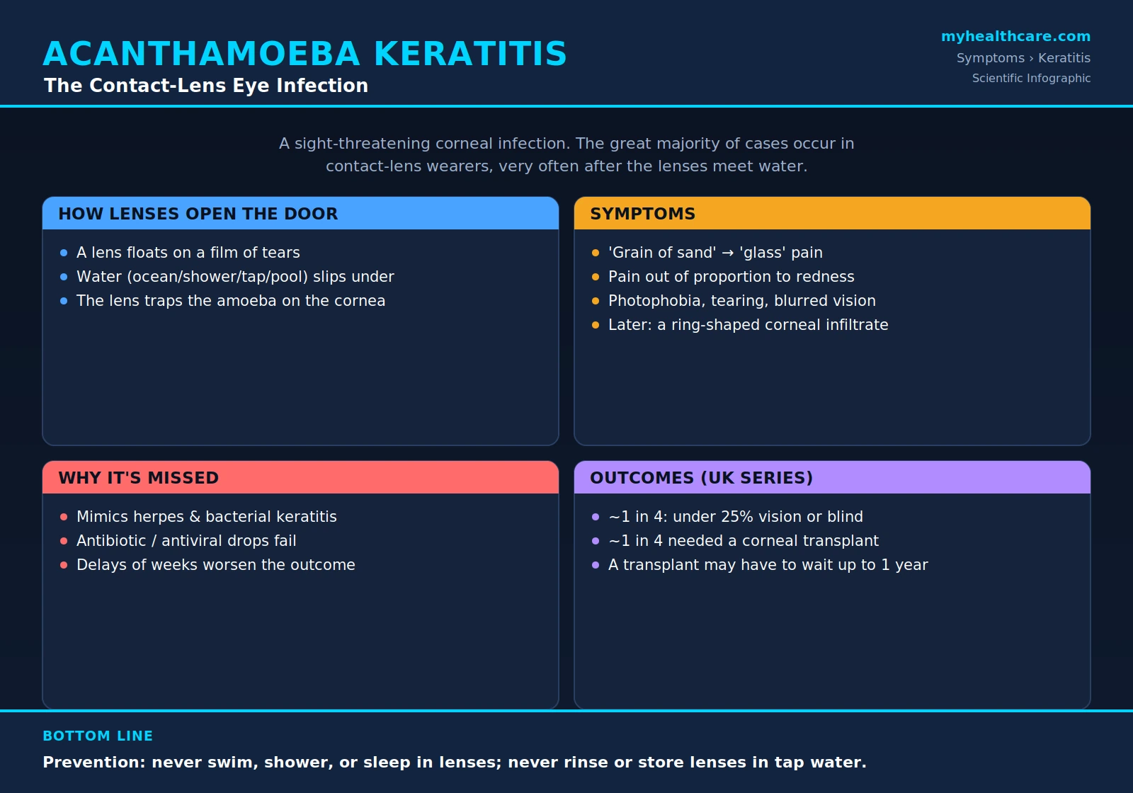
Here is the part that surprises most people: a contact lens does not seal tightly against your eye. It floats. Beneath every soft lens there is a paper-thin film of tears, and that means there is a microscopic gap between the back of the lens and the surface of your cornea. Tiny things can get into that gap. Most of the time that is harmless. But when the water you expose your eye to is carrying Acanthamoeba — ocean, lake, river, shower, tap water, a hot tub, a swimming pool — some of those amoebae can slip under the lens.
Now the lens turns from a victim into an accomplice. Instead of letting your blinking and your tears flush the amoeba away, the lens does the opposite: it holds the amoeba pressed against the warm, dark, moist cornea for hours at a time, exactly the conditions Acanthamoeba likes. It is a snug, undisturbed pocket with a steady food supply. The amoeba settles in, attaches to the surface cells, and begins to feed. A microorganism that would otherwise have been rinsed harmlessly out of your eye in seconds is instead given a sheltered place to live and grow.
There is a second, deeper reason the cornea is so vulnerable, and it is a kind of beautiful, cruel trade-off in human anatomy. The cornea has no blood vessels. It is one of the only tissues in your body with no blood supply at all, and that is on purpose: blood vessels are not perfectly transparent, so if the cornea had them, you would be looking out through a faint red mesh. To stay crystal clear, it does without blood. But your immune system travels in the blood. White blood cells, antibodies, and the body's whole infection-fighting army arrive at threatened tissue through the bloodstream. Because the cornea has no vessels, the immune system can barely reach it. An infection that your body would crush in minutes almost anywhere else gets, in the cornea, a quiet refuge where it can dig in before your defenses ever arrive. The same feature that keeps your vision clear is the feature that lets this infection take hold.
3. Symptoms — and the One That Matters Most
The earliest sign is easy to dismiss. It usually starts as a foreign-body sensation — the persistent feeling that there is a grain of sand, an eyelash, or grit in your eye — that will not rinse away no matter what you do. You blink, you flush it with drops, you take the lens out and clean it, and the feeling stays. That refusal to wash out is itself a warning. As the days pass, the symptoms build:
- Severe, deep eye pain — patients very often describe it as feeling "like glass in the eye," a bottle cap, or a hot needle. It is not a mild scratchy discomfort; it is genuine, intense pain.
- Redness of the white of the eye that keeps getting worse.
- Extreme sensitivity to light (photophobia) — ordinary daylight or a lit room becomes hard or impossible to tolerate, and you find yourself wanting to keep the eye shut.
- Heavy watering and tearing, with the eye streaming on its own.
- Blurred or declining vision in the affected eye.
- Later, doctors may see a ring-shaped infiltrate — a circular, cloudy band in the cornea — which is a classic (though late) sign of this particular infection.
But here is the single most important clue, the one that should put Acanthamoeba on the table for any contact lens wearer: the hallmark is pain that is out of all proportion to how the eye looks. Early on, an examining doctor may see only a modestly irritated cornea — nothing that should hurt much — while the patient is in agony. That mismatch is the tell. The reason is specific and grim: Acanthamoeba attacks the corneal nerves, and the cornea is the most densely nerve-packed tissue in the entire human body, far more sensitive per square millimeter than your fingertip. When the amoeba damages and inflames those nerves, the pain is extraordinary even before there is much visible to see. If you are a contact lens wearer and your eye hurts far more than it looks like it should, say exactly that to whoever examines you, and ask directly: "Could this be Acanthamoeba?"
4. Why It Is Missed at First
This infection is frequently missed in its early weeks, and it is worth understanding why, because knowing the trap is how you avoid falling into it. Two things work against an early diagnosis. First, it is rare — a busy eye doctor or emergency department may see a painful red eye in a contact lens wearer many times before they ever see a true case of Acanthamoeba, so it is simply not the first thing that comes to mind. Second, and more dangerously, in its early stages it looks almost exactly like the common eye infections it is not.
In particular, early Acanthamoeba keratitis mimics bacterial keratitis and herpes simplex keratitis (a cold-sore-family virus infection of the cornea). All three can produce a red, painful, light-sensitive eye in a contact lens wearer. So the natural first move is to prescribe what those common infections need: antibiotic eye drops for suspected bacteria, or antiviral drops for suspected herpes. Against Acanthamoeba, those treatments do nothing — the amoeba is neither a bacterium nor a virus, and the drugs that kill those do not kill it. The eye fails to improve, or briefly seems calmer and then flares again. Sometimes a course of steroid drops is added, which can actually let the amoeba grow faster. Meanwhile the real organism keeps feeding.
The result is that diagnostic delays of several weeks are common, and delay is not harmless. The longer the amoeba feeds before the right treatment starts, the deeper it burrows and the worse the final outcome tends to be. This is exactly why your own awareness matters so much. You may be the person in the room who knows to push: a contact lens wearer with a worsening, intensely painful eye and a history of any water exposure should specifically raise Acanthamoeba with their eye doctor and, if things are not getting better, ask for referral to a corneal specialist. Being politely persistent is reasonable here. It can be the difference between a saved eye and a lost one.
5. Who Gets It — Risk Factors
Almost every avoidable risk factor comes down to one theme: letting your contact lenses meet water, or skipping the steps that keep them clean. The strongest risk factors, drawn from careful case-control studies of real patients, include:
- Swimming, showering, or using a hot tub while wearing lenses. This is the classic exposure — the moment water carrying the amoeba can get under the lens.
- Sleeping in your lenses (overnight wear), which gives any trapped amoeba many uninterrupted hours against a closed, warm eye.
- Rinsing or storing lenses in tap water instead of fresh disinfecting solution — tap water is not sterile and can contain Acanthamoeba.
- "Topping off" old solution — pouring a little fresh solution onto yesterday's used solution instead of emptying the case and refilling it. Old solution loses its disinfecting power and lets organisms build up.
- Poor lens-case hygiene — not emptying, rinsing, drying, and regularly replacing the case. The case itself can grow a biofilm that shelters amoebae.
Now the part that matters most for how you feel about this, and it is genuinely important: this is not simply a punishment for carelessness, and no contact lens wearer is fully immune. People who follow textbook hygiene to the letter have still developed Acanthamoeba keratitis. People who use fresh daily disposable lenses — a new sterile pair every day, thrown away each night, the cleanest way to wear contacts — have still gotten it. If it has happened to you and you did everything "right," you did not bring this on yourself through some failure, and you do not deserve to carry that guilt. The reason perfect hygiene is not perfect protection is exactly why the water rule is the heart of prevention: hygiene reduces how many organisms reach the lens, but the decisive event is water getting under the lens in the first place. You cannot disinfect your way out of a swim or a shower in your contacts. That is why "keep water away from your lenses" is the rule that protects everyone, the careful and the casual alike.
6. The Real Stories Behind the Warnings
It is one thing to read that an infection "can take your sight." It is another to understand that this happens to real, ordinary people, doing ordinary things, who never imagined a swim or a shower could cost them an eye. Documented public cases — reported in news coverage and patient-advocacy campaigns — make the danger concrete in a way statistics cannot. (The medical facts below are accurate; we deliberately leave out personal names, because automatically generated video captions and second-hand retellings frequently garble the spelling of people's names, and we will not risk attaching the wrong name to anyone's story.)
- A young woman in her early twenties from Texas, who worked as a barista, was left effectively blind in one eye after swimming in the Gulf of Mexico while wearing her monthly contact lenses. A short swim, lenses left in, ocean water under the lens — and a sight-threatening infection followed.
- A woman in the United Kingdom lost an eye after the everyday habit of showering while wearing her contact lenses allowed the amoeba into her eye.
- Another wearer was repeatedly misdiagnosed with a herpes infection and saw multiple specialists, with the wrong treatment failing each time, before anyone identified Acanthamoeba as the true cause — a direct, painful illustration of the misdiagnosis trap described above.
- A patient in the United Kingdom who went blind in the affected eye despite practicing perfect hygiene with daily disposable lenses — the cleanest way there is to wear contacts — went on to found a public campaign pressing for a simple, plain warning to be printed on contact-lens packaging: keep water away from your lenses. Their case is the clearest possible proof that this is not only about carelessness, and that a single, clear warning could prevent a great deal of suffering.
The thread running through all of these is the same as the science: it was water and a worn lens, and in several cases a delay before the right diagnosis. None of these people did anything unusual. They swam, they showered, they wore the lenses they were sold. That is precisely why the prevention message is worth taking to heart even though the infection is rare — because the people it happens to almost never see it coming.
7. Diagnosis
Because Acanthamoeba keratitis is rare and easily mistaken for other infections, getting a firm diagnosis usually means going beyond a routine eye exam to tests that can actually find the amoeba. A corneal specialist (an ophthalmologist who focuses on the cornea) has three main tools, often used together:
- In vivo confocal microscopy. This is a specialized, non-invasive microscope that scans through the living cornea in fine optical slices and, in skilled hands, can reveal the round, bright Acanthamoeba cysts directly in the tissue — a result available in minutes rather than days. In experienced hands it is a sensitive way to spot the infection, which is why it is so valuable for getting treatment started early.
- Corneal scraping with culture. The specialist gently scrapes a small sample of cells from the affected cornea and grows (cultures) it in the laboratory. Acanthamoeba is grown by a clever, classic method: the sample is placed on a "non-nutrient" agar plate that has been seeded with a lawn of E. coli bacteria. The plate offers the amoeba no food of its own — except the bacteria. Any living Acanthamoeba wakes up and crawls across the plate eating the bacteria, leaving visible tracks that confirm its presence.
- PCR (polymerase chain reaction). This molecular test detects Acanthamoeba's DNA in a corneal sample. It is fast and highly specific — if the amoeba's genetic signature is there, PCR finds it.
For a fuller walk-through of how the diagnosis is made and what each test is like to undergo, see the Symptoms & Diagnosis hub. The practical takeaway for patients is simple: these are specialist tests. If you are a contact lens wearer with a worsening painful eye and the standard treatments are not working, the goal is to reach a corneal specialist who can perform them — the sooner the better.
8. Outcomes and Why Early Treatment Is Everything
The good news first, because it is real and it is the whole reason this page urges speed: when Acanthamoeba keratitis is caught early and treated promptly, many eyes are saved with good vision. Early disease, while the amoeba is still confined to the surface layers of the cornea, responds far better to treatment. Time is genuinely on your side at the start.
The hard news is what happens when it is not caught early. Given weeks of delay, the amoeba burrows deep into the cornea, dissolving the structural tissue as it goes, and the damage at that depth can be permanent and devastating. The numbers from real patient series are sobering. In a major United Kingdom hospital series, roughly 1 in 4 patients ended up with severe visual loss — less than a quarter of normal vision in the affected eye, or outright blindness — and about 1 in 4 needed a corneal transplant (a graft of donor corneal tissue) to try to restore the eye. These are not the outcomes of early disease caught fast; they are heavily weighted by cases that were severe or that were diagnosed late.
And even a corneal transplant is not a quick rescue. There is a cruel catch built into this infection: doctors often cannot even attempt a transplant for up to a year. The reason is the cyst. A single surviving Acanthamoeba cyst, hidden and dormant in the eye, can wake up and infect the brand-new donor cornea, destroying the graft and putting the patient right back where they started. So before transplanting, surgeons generally have to be confident the amoeba is truly dead — which means months of anti-amoeba treatment and waiting and watching first, to prove the infection is gone. The whole arc — the months-long medication, the long wait, the staged surgery — is why everything depends on the beginning. The earlier the right treatment starts, the more likely none of this is ever needed.
9. Treatment in Brief
Treating Acanthamoeba keratitis is less like taking a course of antibiotics and more like laying a months-long siege. The goal is to kill not only the active, feeding amoebae but also the tough dormant cysts — and the cysts are what make the campaign so long and so demanding. The backbone of treatment is a class of antiseptic eye drops called biguanides, most commonly PHMB (polyhexamethylene biguanide / polihexanide) or chlorhexidine. Their great advantage is that they kill both the trophozoites and the cysts, which is exactly what is needed. They are frequently paired with a second type of drop called a diamidine — usually propamidine (sold as Brolene) — to attack the amoeba on two fronts at once.
What makes the early phase so grueling is the schedule. To beat the infection back, these drops may be given as often as every 30 to 60 minutes around the clock, day and night, in the first days — an exhausting regimen that is then slowly tapered over weeks and months as the eye responds. In severe or stubborn cases, doctors may add an oral medication called miltefosine, taken as a pill, to help bring the infection under control. Treatment commonly continues for six months to a year or more, and is tapered only carefully, because stopping too soon can let a surviving cyst reignite the whole infection.
This is a brief overview, not a treatment manual — the specifics (drug concentrations, dosing schedules, how the taper is managed, and what to do if it relapses) belong with your corneal specialist. For the full picture, see the Treatments hub and the detailed Conventional Medical Treatment page.
10. Prevention — Keep Water Away From Lenses
This is the most important section on the page, and it is short on purpose, because the rules are few and they work. The overwhelming majority of Acanthamoeba keratitis is preventable, and prevention costs nothing. If you wear contact lenses:
- Never swim in your lenses — not in the ocean, a lake, a river, a pool, or a hot tub. If you must wear vision correction in the water, use prescription goggles, or take the lenses out.
- Never shower or wash your face in your lenses, and keep your eyes shut if water hits your face with lenses still in. Shower water comes straight from the tap and is not sterile.
- Never sleep in your lenses unless a doctor has specifically prescribed an overnight lens and told you it is safe.
- Never rinse or store your lenses (or your lens case) in tap water. Use only fresh contact lens disinfecting solution — every single time.
- Never "top off" old solution. Empty the case completely and refill it with fresh solution at each use.
- Replace your lens case regularly (every one to three months), and after each use empty it, rinse it with fresh solution, wipe it, and let it air-dry face down.
- Consider daily disposable lenses, which start each day sterile and remove the case and storage steps entirely — just remember that even daily disposables must never go near water.
- See an eye doctor promptly for any painful, red eye, and if you are a lens wearer with worsening pain — especially pain out of proportion to how the eye looks — ask specifically about Acanthamoeba and request a corneal specialist if you are not improving.
That is the whole of it. The science of this infection is intricate, but the protection is one plain sentence you can keep for life: keep water away from your contact lenses.
Key Research Papers
Peer-reviewed studies on Acanthamoeba keratitis — spanning its biology and pathogenesis, the diagnostic tests, the contact-lens risk factors confirmed in real patients, treatment trials of the biguanide drops, and the visual outcomes that make early diagnosis so urgent. Each citation links to the full record via DOI. Author names, titles, and journals are shown as plain text; only the year, volume, and pages link out.
- Lorenzo-Morales J, Khan NA, Walochnik J. An Update on Acanthamoeba Keratitis: Diagnosis, Pathogenesis and Treatment. Parasite. 2015;22:10.
- Carnt N, Hoffman JJ, Verma S, Hau S, et al. Acanthamoeba Keratitis: Confirmation of the UK Outbreak and a Prospective Case-Control Study Identifying Contributing Risk Factors. British Journal of Ophthalmology. 2018;102(12):1621–1628.
- Marciano-Cabral F, Cabral G. Acanthamoeba spp. as Agents of Disease in Humans. Clinical Microbiology Reviews. 2003;16(2):273–307.
- Clarke DW, Niederkorn JY. The Pathophysiology of Acanthamoeba Keratitis. Trends in Parasitology. 2006;22(4):175–180.
- Clarke DW, Niederkorn JY. The Immunobiology of Acanthamoeba Keratitis. Microbes and Infection. 2006;8(5):1400–1405.
- Dart JKG, Papa V, Rama P, et al. The Orphan Drug for Acanthamoeba Keratitis (ODAK) Trial: PHMB 0.08% (Polihexanide) and Placebo versus PHMB 0.02% and Propamidine 0.1%. Ophthalmology. 2024;131(3):277–287.
- Lim N, Goh D, Bunce C, et al. Comparison of Polyhexamethylene Biguanide and Chlorhexidine as Monotherapy Agents in the Treatment of Acanthamoeba Keratitis. American Journal of Ophthalmology. 2008;145(1):130–135.
- Robaei D, Carnt N, Minassian DC, Dart JKG. Therapeutic and Optical Keratoplasty in the Management of Acanthamoeba Keratitis: Risk Factors, Outcomes, and Summary of the Literature. Ophthalmology. 2015;122(1):17–24.
- Ficker LA, Kirkness C, Wright P. Prognosis for Keratoplasty in Acanthamoeba Keratitis. Ophthalmology. 1993;100(1):105–110.
- Jasim H, Grzeda MT, Foot B, Tole D, et al. Incidence of Acanthamoeba Keratitis in the United Kingdom in 2015: A Prospective National Survey. Cornea. 2024;43(3):269–276.
- Moshtaghion SM, Abolhosseini M, Yaseri M, et al. Diagnostic Accuracy of Confocal Scan in Detecting Acanthamoeba Keratitis and Fungal Keratitis: A Systematic Review and Meta-Analysis. International Ophthalmology. 2023;43(8):3011–3022.
- Seal DV, Hay J, Kirkness CM. Chlorhexidine or Polyhexamethylene Biguanide for Acanthamoeba Keratitis. The Lancet. 1995;345(8942):136.
Live PubMed Searches
Each link opens a live PubMed query so results stay current as new papers are indexed.
- Acanthamoeba keratitis contact lens
- Acanthamoeba keratitis risk factors
- Acanthamoeba keratitis diagnosis confocal microscopy
- Acanthamoeba keratitis PHMB / polihexanide treatment
- Acanthamoeba keratitis chlorhexidine
- Acanthamoeba keratitis miltefosine
- Acanthamoeba keratitis keratoplasty outcomes
- Acanthamoeba keratitis misdiagnosis (herpes)
- Acanthamoeba keratitis incidence and epidemiology
- Acanthamoeba cyst resistance to disinfection
Connections
- Acanthamoeba (Overview)
- Symptoms & Diagnosis Hub
- Granulomatous Amebic Encephalitis (GAE)
- Acanthamoeba Treatments Hub
- Conventional Medical Treatment
- All Parasites
- Ophthalmology
- Dry Eye Disease
- Escherichia coli
- Infectious Disease
- Meningitis
- All Conditions
- Remedies