Ascariasis Symptoms and Diagnosis
Pulmonary Ascariasis & Löffler Syndrome
When migrating larvae pass through the lungs — cough, wheeze, and eosinophilic pneumonitis.
Intestinal Obstruction & Biliary Complications
The dangerous complications — a worm bolus blocking the gut, and worms in the bile and pancreatic ducts.
Malnutrition & Childhood Growth
How chronic infection saps nutrition and stunts growth and learning in children.
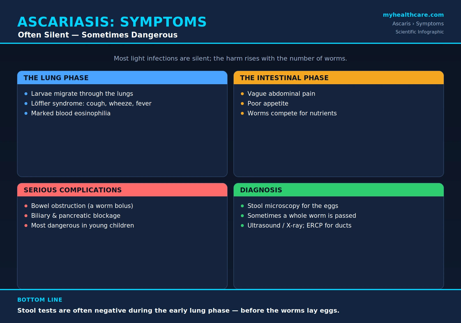
Ascariasis is the infection caused by the giant intestinal roundworm Ascaris lumbricoides — one of the most common parasitic infections of human beings, carried by hundreds of millions of people, mostly in warm regions with limited sanitation. The single most important thing to understand about its symptoms is that they depend almost entirely on how many worms a person is carrying. Most infected people harbor only a handful of worms and feel nothing at all; a smaller number carry heavy worm burdens and can develop anything from vague belly pain and poor growth to a life-threatening blockage of the bowel. The worm also has a curious life cycle in which its larvae travel through the lungs before settling in the intestine, so the very same infection can briefly cause a cough and wheeze weeks before it ever causes a stomach complaint. This page walks through that whole spectrum — from silent infection to severe disease — and explains how doctors confirm the diagnosis, including the important reason why a stool test can come back negative even in someone who is truly infected.
Table of Contents
- A Spectrum That Depends on Worm Burden
- The Lung-Migration Phase (Löffler Syndrome)
- The Intestinal Phase
- Malnutrition and Impaired Growth in Children
- Serious Mechanical Complications
- How Ascariasis Is Diagnosed
- Why Stool Tests Can Be Negative
- When to Suspect and Test
- Key Research Papers
- Featured Videos
1. A Spectrum That Depends on Worm Burden
The defining feature of ascariasis is that the illness it causes is burden-dependent: the more worms a person carries, the more likely they are to have symptoms and complications. This is not a minor detail — it is the key that makes the whole disease make sense. A person with two or three worms may go their entire life without a single symptom, while a child packed with dozens or even hundreds of worms can become seriously, even dangerously, ill.
Worms are not spread evenly across an infected population. In any community, a small fraction of people carry the great majority of the worms — a pattern parasitologists call overdispersion or aggregation. Most infected individuals have light infections and few or no symptoms; a minority carry heavy burdens and account for most of the disease, most of the complications, and most of the eggs that contaminate the environment and keep the cycle going. Children, especially preschool and early-school-age children, tend to carry the heaviest burdens, which is why severe ascariasis is overwhelmingly a childhood problem.
Because of this, the same infection can look completely different from one person to the next:
- Light infection — usually silent. No symptoms at all, or only vague, occasional discomfort. The infection may only ever be discovered if a worm is passed in stool or an egg is found incidentally on a stool test.
- Moderate infection — vague abdominal symptoms: intermittent belly pain, poor appetite, nausea, bloating, and in children a contribution to poor nutrition and growth.
- Heavy infection — the source of the serious disease: significant malnutrition and growth failure in children, and a real risk of mechanical complications such as the bowel being blocked by a tangled mass of worms, or worms migrating into the bile or pancreatic ducts.
Holding this spectrum in mind keeps everything else in perspective. Ascariasis is enormously common, and in most people it is harmless; its medical importance comes from the heavily-infected minority — usually children — in whom it can do real and lasting harm.
2. The Lung-Migration Phase (Löffler Syndrome)
One of the most distinctive things about Ascaris is that it does not go straight to the intestine. After a person swallows infective eggs (from soil, hands, or food contaminated with human feces), the eggs hatch in the small intestine and release tiny larvae — but those larvae then take a remarkable detour. They burrow through the gut wall, enter the bloodstream, are carried to the lungs, break out into the tiny air sacs, climb up the airways, and are swallowed again before finally maturing in the intestine. This whole journey takes roughly one to two weeks.
While the larvae are passing through the lungs, they can provoke a temporary but striking allergic-type inflammation. This appears, usually a couple of weeks after a heavy exposure, as a dry cough, wheeze, shortness of breath, and sometimes fever, frequently accompanied by a sharp rise in eosinophils (a type of allergy-associated white blood cell) and fleeting shadows on a chest X-ray. This pattern — transient lung infiltrates with a high eosinophil count — is known as Löffler syndrome (also spelled Loeffler), and ascariasis is one of its classic causes.
Two practical points matter here. First, this lung phase is self-limiting: it typically settles on its own within one to two weeks as the larvae move on to the gut. Second, and critically for diagnosis, it happens weeks before the worms are mature and laying eggs — which is exactly why a stool test will usually be negative during the lung phase (see Why Stool Tests Can Be Negative below). A patient can have a textbook cough-and-eosinophilia picture from Ascaris while every stool sample comes back clear.
The lung phase, including how it is recognized, why eosinophils rise, and how it differs from asthma and other causes of eosinophilic pneumonia, is covered in depth on the dedicated page: Pulmonary Ascariasis and Löffler Syndrome.
3. The Intestinal Phase
Once the larvae complete their lung journey and return to the small intestine, they grow into the long adult worms that give the parasite its reputation — adult females can reach 20 to 35 centimeters in length. It is in this intestinal phase, which begins roughly two to three months after the eggs were swallowed and can last a year or two, that most of the familiar symptoms of ascariasis arise — though, again, only when the worm burden is more than trivial.
In light or moderate infection, the intestinal phase tends to produce symptoms that are vague and easy to dismiss:
- Intermittent abdominal pain — often a dull, crampy discomfort around the navel that comes and goes.
- Poor appetite and a feeling of fullness or early satiety.
- Nausea, occasional vomiting, and bloating.
- Irregular bowel habits, including occasional diarrhea.
These complaints overlap with countless ordinary digestive problems, so ascariasis is rarely the first thing suspected on symptoms alone. Often the most telling clue is not a symptom at all but an event: a person — or a worried parent — sees a large, pinkish-tan, earthworm-like worm passed in the stool, or, less pleasantly, vomited or coughed up. The sight of an actual adult worm is, understandably, alarming, but it is also one of the most specific signs of the infection and frequently the thing that finally brings a patient to the clinic. Adult worms can also wander, occasionally emerging from the nose or mouth, which is distressing but again points clearly to the diagnosis.
The reassuring counterweight is that for most people with light intestinal infection, these symptoms are mild, and the infection responds quickly and completely to a short course of deworming medication (see Anthelmintic Treatment). The serious face of the intestinal phase — obstruction and biliary disease — is reserved for heavy worm burdens and is discussed below.
4. Malnutrition and Impaired Growth in Children
Beyond any single dramatic complication, the quieter but more widespread harm of ascariasis is its toll on the nutrition, growth, and development of children. A child living in an endemic area is rarely infected with just one kind of worm and rarely just briefly — infection is often heavy, repeated, and sustained over the years when the body is growing fastest and the brain is developing. Over time, a substantial worm burden can act as a chronic drain on a child already living close to nutritional margins.
The mechanisms are several and overlapping: worms can interfere with appetite and food intake, they can impair the digestion and absorption of nutrients (including fat, protein, and key vitamins), and the low-grade ongoing inflammation of chronic infection diverts energy that a growing child needs. The downstream consequences that researchers have repeatedly linked to heavy or chronic infection include poor weight gain, stunted growth (being short for one's age), and impaired physical fitness and learning. Because these effects unfold slowly and silently, they are easy to miss in any individual child, yet across a community they represent a meaningful and entirely preventable burden — which is much of the rationale for school-based mass deworming.
This is an overview; the nutritional and developmental consequences — how worms steal nutrition, what the evidence on growth and cognition actually shows, and how treatment can help recovery — are explored in detail on the dedicated page: Malnutrition and Childhood Growth.
5. Serious Mechanical Complications
The dangerous, occasionally life-threatening side of ascariasis comes not from any toxin but from the worms' sheer physical presence. In a heavy infection — especially in a young child with a narrow bowel — the worms can cause mechanical problems by physically blocking or invading parts of the digestive system. These complications are uncommon relative to the vast number of light infections, but where ascariasis is common they are an important cause of surgical emergencies, particularly in children.
Intestinal obstruction is the best-known of these. When many worms are present, they can tangle into a dense bolus that physically plugs the small bowel, most often near the lower end of the small intestine. This produces the picture of a bowel blockage: severe crampy abdominal pain, a swollen (distended) belly, vomiting, and an inability to pass stool or gas. It is a genuine emergency — if not relieved, the bowel can become damaged, perforate, or die — and it falls disproportionately on young children, in whom the smaller intestinal diameter is more easily blocked.
Biliary and pancreatic complications are the other major group. Adult worms are restless and have a tendency to migrate, and they are drawn to openings. A worm can wriggle into the bile ducts, the gallbladder, or the pancreatic duct, causing intense pain and a range of serious problems — blockage of bile flow (jaundice), infection of the bile ducts (cholangitis), inflammation of the gallbladder, and inflammation of the pancreas (pancreatitis). This is a particularly important cause of acute biliary and pancreatic disease in regions where the parasite is common, and it often requires endoscopic or surgical intervention to remove the worm.
Because these complications are mechanical, they are also the situations in which imaging — ultrasound, X-ray, and endoscopic procedures — becomes central to both diagnosis and treatment, as described in the next section. The full clinical picture, including how obstruction and biliary ascariasis are managed (from conservative care to ERCP and surgery), is covered on the dedicated page: Intestinal Obstruction and Biliary Complications.
6. How Ascariasis Is Diagnosed
Diagnosing ascariasis brings together a few complementary approaches, and which one is used depends a great deal on which phase of the infection is in question — the early lung phase, the established intestinal phase, or a mechanical complication.
Stool microscopy is the mainstay. The standard, everyday way to confirm an intestinal Ascaris infection is to examine a sample of stool under the microscope for the parasite's characteristic eggs. Ascaris eggs are distinctive — oval, with a thick, often knobbly outer shell — and are usually present in large numbers when adult female worms are established and laying, because each female produces an enormous quantity of eggs every day. Because the worms are so prolific, even a single direct smear is often enough; concentration techniques and egg-counting methods (such as the Kato–Katz method) can increase sensitivity and, importantly, estimate how heavy the worm burden is, which matters for gauging the risk of complications and for monitoring deworming programs.
Passing an adult worm is itself diagnostic. When a person passes, vomits, or coughs up a recognizable adult roundworm, that single observation confirms the infection without any laboratory test — and it is frequently the event that prompts testing in the first place. Sometimes a worm is also seen unexpectedly during an endoscopy or a surgical procedure.
Imaging comes into its own for the complicated and the migrating worm. Ultrasound is especially useful and is the workhorse for suspected biliary ascariasis: an experienced operator can actually see worms inside the bile ducts or gallbladder, classically as long, tubular, moving structures. Plain abdominal X-ray can show signs of intestinal obstruction and sometimes a tangled mass of worms, and contrast studies may outline worms within the bowel. When worms are lodged in the bile or pancreatic ducts, ERCP (endoscopic retrograde cholangiopancreatography) and other forms of endoscopy serve a double purpose — confirming the diagnosis by direct visualization and allowing the worm to be extracted, which is often the treatment itself.
Blood tests play a supporting role. A raised eosinophil count is a useful clue, particularly during the larval lung-migration phase when eosinophilia tends to be most marked; it points toward a parasitic process but is not specific to Ascaris. Routine blood tests are otherwise used mainly to assess the consequences of heavy infection (such as anemia or nutritional deficits) and the complications (such as the inflammation of cholangitis or pancreatitis), rather than to make the diagnosis directly.
7. Why Stool Tests Can Be Negative
One of the most important and easily misunderstood points in diagnosing ascariasis is that a negative stool test does not rule the infection out — and the reason traces straight back to the worm's life cycle.
Stool microscopy detects eggs, and eggs only appear once mature adult female worms are present and laying. But it takes roughly two to three months from swallowing the infective eggs for the larvae to complete their migration and grow into egg-producing adults. During that entire pre-patent window — including the early lung-migration phase, when a patient may be coughing, wheezing, and showing a high eosinophil count — there are no adult worms laying eggs in the gut, so there is simply nothing for the microscope to find. This is precisely why someone can have classic Löffler-syndrome symptoms from Ascaris while every stool sample comes back clean. The diagnosis in that early window often rests on the clinical picture (recent exposure, cough, eosinophilia, fleeting chest-X-ray shadows) rather than on stool testing.
A few other situations can also yield a falsely reassuring stool result. An infection consisting only of male worms produces no eggs at all (only females lay), so a single-sex infection will be egg-negative. A very light infection may shed too few eggs to be caught on a single examination, which is one reason repeat or concentrated samples improve detection. And if a worm has migrated out of the intestine — into the bile ducts, for example — the clinical problem can be dominated by that displaced worm even when intestinal egg output is low.
The practical lesson is to match the test to the phase of disease: stool microscopy is the right tool for an established intestinal infection, but in the early lung phase, in suspected biliary disease, or in any situation where the picture strongly suggests Ascaris despite a negative stool, clinicians lean on eosinophilia, imaging (especially ultrasound), the history of exposure, and repeat testing rather than accepting a single negative stool as the final word.
8. When to Suspect and Test
Because most ascariasis is silent and self-limiting, testing is guided less by minor symptoms in the general public than by particular situations in which the parasite is genuinely likely or potentially dangerous. Ascariasis is worth actively considering and testing for when:
- A worm has been seen. Anyone — child or adult — who passes, vomits, or coughs up a recognizable roundworm should be evaluated and treated; the sighting is essentially diagnostic, and other family members in the same household often warrant attention too.
- A child in an endemic area has poor growth or vague abdominal symptoms. In regions where the parasite is common, a child with unexplained poor appetite, recurrent belly pain, bloating, or faltering growth is a natural candidate for stool examination, given how often heavy worm burdens contribute to exactly this picture.
- There is a cough with eosinophilia after likely exposure. A dry cough, wheeze, and transient chest-X-ray shadows accompanied by a high eosinophil count — especially in someone from or recently traveled to an endemic area — should raise the possibility of the larval lung phase, even though the stool may still be negative at that stage.
- Acute abdominal, biliary, or pancreatic disease arises in an endemic setting. In areas where ascariasis is common, intestinal obstruction in a child, or unexplained biliary colic, cholangitis, or pancreatitis at any age, should prompt thought of a worm — with ultrasound a particularly valuable first step for the biliary forms.
Outside these situations — a healthy person in a low-prevalence area with vague, transient digestive symptoms and no exposure history — routine testing for Ascaris is usually unnecessary. The skill of diagnosis lies in recognizing the settings and the people for whom this extraordinarily common, usually harmless worm becomes something that genuinely needs to be found and treated.
Key Research Papers
Peer-reviewed reviews, cohort and surgical studies, and global-burden analyses covering the clinical spectrum, complications, nutritional consequences, and diagnosis of Ascaris lumbricoides infection. Journal names appear as plain text; the year/volume/pages link opens the full citation via DOI.
- Dold C, Holland CV. Ascaris and Ascariasis. Microbes and Infection. 2011;13(7):632–637.
- Crompton DWT. Ascaris and Ascariasis. Advances in Parasitology. 2001;48:285–375.
- O'Lorcain P, Holland CV. The Public Health Importance of Ascaris lumbricoides. Parasitology. 2000;121(S1):S51–S71.
- Bethony J, Brooker S, Albonico M, et al. Soil-Transmitted Helminth Infections: Ascariasis, Trichuriasis, and Hookworm. The Lancet. 2006;367(9521):1521–1532.
- Jourdan PM, Lamberton PHL, Fenwick A, Addiss DG. Soil-Transmitted Helminth Infections. The Lancet. 2018;391(10117):252–265.
- Pullan RL, Smith JL, Jasrasaria R, Brooker SJ. Global Numbers of Infection and Disease Burden of Soil Transmitted Helminth Infections in 2010. Parasites & Vectors. 2014;7:37.
- Holland CV, Behnke JM, Dold C. Larval Ascariasis: Impact, Significance, and Model Organisms. In: Ascaris: The Neglected Parasite. 2013:107–125.
- de Silva NR, Guyatt HL, Bundy DAP. Morbidity and Mortality Due to Ascaris-Induced Intestinal Obstruction. Transactions of the Royal Society of Tropical Medicine and Hygiene. 1997;91(1):31–36.
- Khuroo MS, Zargar SA, Mahajan R. Hepatobiliary and Pancreatic Ascariasis in India. The Lancet. 1990;335(8704):1503–1506.
- Khuroo MS, Zargar SA, Yattoo GN, et al. Worm Extraction and Biliary Drainage in Hepatobiliary and Pancreatic Ascariasis. Gastrointestinal Endoscopy. 1993;39(5):680–685.
- Crompton DWT. Ascariasis and Childhood Malnutrition. Transactions of the Royal Society of Tropical Medicine and Hygiene. 1992;86(6):577–579.
- Stephenson LS, Latham MC, Ottesen EA. Malnutrition and Parasitic Helminth Infections. Parasitology. 2000;121(S1):S23–S38.
Live PubMed Searches
Each link opens a live PubMed query so results stay current as new papers are indexed.
- Ascaris lumbricoides clinical manifestations
- Ascariasis intestinal obstruction in children
- Hepatobiliary and pancreatic ascariasis
- Pulmonary ascariasis and Löffler syndrome
- Ascaris, nutrition, and child growth
- Ascaris stool microscopy and Kato–Katz diagnosis
- Ascariasis ultrasound and biliary diagnosis
- Ascaris eosinophilia and larval migration
Connections
- Ascaris Overview
- Pulmonary Ascariasis & Löffler Syndrome
- Intestinal Obstruction & Biliary Complications
- Malnutrition & Childhood Growth
- Treatment & Prevention
- Anthelmintic Treatment (Albendazole & Mebendazole)
- Prevention: Sanitation & Hygiene
- Mass Deworming Programs
- All Parasites
- Gastroenterology
- Infectious Disease
- All Conditions