Intestinal Obstruction and Biliary Complications of Ascariasis
Most people who carry Ascaris lumbricoides — the large human roundworm — never feel a thing. But when the worms are big, many, and on the move, they can cause some of the most dramatic mechanical emergencies in all of medicine. A tangled ball of worms can plug a child's small intestine like a wad of wet rope in a drainpipe. A single restless worm can wriggle up into the tube that drains the liver and pancreas and trigger searing pain, jaundice, infection, or a pancreatitis attack. This page is about those dangerous complications — why worm size and number are what make ascariasis turn from a silent passenger into a surgical emergency, how the obstruction and the biliary problems unfold, how doctors find the worms (ultrasound is the hero here), and how most cases are treated — often without an operation at all. These complications fall heaviest on young children in places where heavy infection is common, which is exactly why prevention and deworming matter so much.
Table of Contents
- Why Worm Size and Number Matter
- Intestinal (Bowel) Obstruction
- When Obstruction Turns Dangerous
- Hepatobiliary and Pancreatic Ascariasis
- Appendicular Ascariasis
- What Makes Worms Migrate
- Diagnosis: Ultrasound Is Key
- Management and Treatment
- A True Emergency in Children
- Key Research Papers
- Featured Videos
1. Why Worm Size and Number Matter
To understand why Ascaris causes mechanical trouble, picture the worm itself. An adult female roundworm is large — roughly the thickness of a pencil and commonly 20 to 35 centimeters (8–14 inches) long. This is not a microscopic parasite. It is a substantial physical object living loose in the small intestine.
A person harboring just one or two of these worms usually has no symptoms at all. The problem is worm burden — the total number of worms a person carries. In communities where contaminated soil leads to repeated swallowing of eggs, a single child can accumulate dozens, even hundreds, of adult worms. The record cases in the medical literature describe hundreds of worms recovered from one small child. When that many pencil-thick worms occupy a child's narrow gut, simple arithmetic becomes a medical emergency: there is only so much room.
Two facts combine to make heavy infection dangerous:
- The worms are bulky. Many of them together form a real, space-occupying mass.
- The worms are active. They are not stationary. They writhe, knot together, and migrate — and when they wander out of the small intestine into places they do not belong (the bile duct, the pancreatic duct, the appendix), they cause focused, localized disasters.
So the two great families of complication on this page both trace back to the same root: too many large, mobile worms. A high burden leads to bulk problems (obstruction); even a low burden can lead to migration problems (biliary and pancreatic disease) because it only takes one worm in the wrong tube. Worldwide, ascariasis remains one of the most common human infections, and although the great majority of carriers are healthy, the sheer number of infected children means these serious complications add up to a substantial burden of illness and a real number of deaths each year, concentrated in low-resource settings.
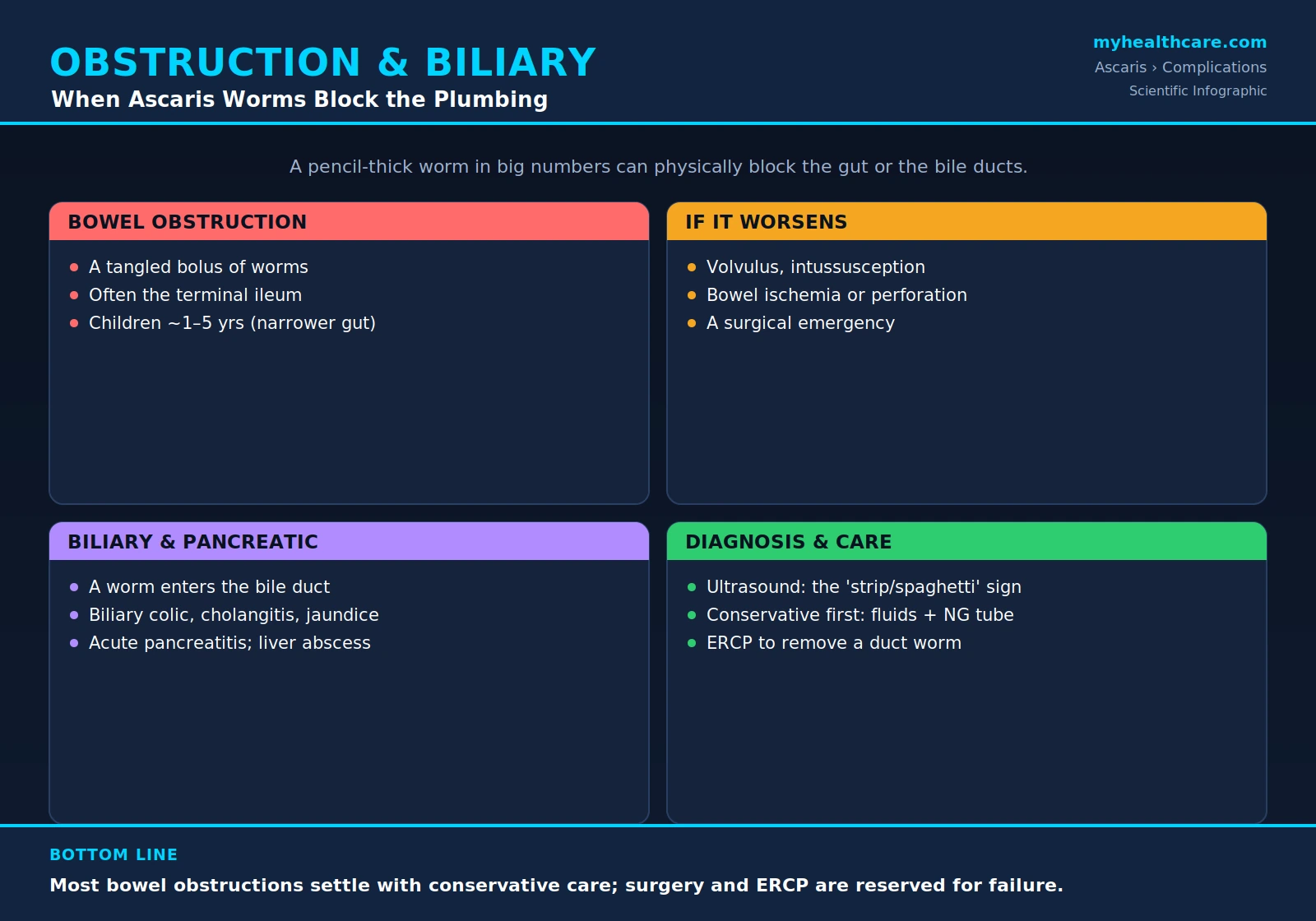
2. Intestinal (Bowel) Obstruction
The most common life-threatening complication of heavy ascariasis is intestinal obstruction — a physical blockage of the small bowel. It happens when a large number of worms tangle together into a firm, ropey bolus (a mass) that the intestine simply cannot push past. The worms knot around one another, sometimes provoked into clumping by fever, illness, or a dose of deworming medicine, and the resulting wad jams the channel shut.
This bolus classically lodges in the terminal ileum — the last and narrowest segment of the small intestine, just before it joins the large bowel. It is the natural choke point: the gut is at its slimmest there, so a mass that traveled freely higher up gets stuck at this narrowing.
Crucially, this is overwhelmingly a disease of young children, most often between roughly 1 and 5 years of age. There are two reasons. First, young children in endemic areas tend to carry the heaviest worm burdens. Second — and just as important — a small child's intestine is narrower, so it takes far fewer worms to plug it than it would in an adult. The same handful of worms that an adult would pass without noticing can completely obstruct a toddler's gut.
The symptoms of bowel obstruction are the classic ones, and parents should know them:
- Crampy, colicky abdominal pain — coming in waves as the bowel strains against the blockage.
- Vomiting — which may become bile-stained (greenish) as the obstruction holds.
- Abdominal distension — a swollen, bloated belly as gas and fluid back up behind the blockage.
- A palpable mass — sometimes a doughy, movable lump of worms can actually be felt through the child's abdominal wall.
- Constipation / failure to pass gas or stool — the downstream sign that the bowel is truly blocked. Occasionally a child vomits or passes a worm, which makes the diagnosis obvious.
A child with vomiting, a distended belly, and crampy pain in a region where ascariasis is common should be evaluated urgently — this is one of the leading causes of childhood bowel obstruction in the developing world.
3. When Obstruction Turns Dangerous: Volvulus, Intussusception, Ischemia, Perforation
A worm bolus that simply blocks the bowel is serious but often manageable. The real danger is what can happen next if it is not relieved. A heavy mass of worms can drag and distort the bowel, setting off a cascade of more severe surgical complications:
- Volvulus — the weighted loop of bowel twists on itself, like a kinked garden hose. This not only blocks the contents completely but pinches off the loop's blood supply.
- Intussusception — one segment of bowel telescopes inside the segment ahead of it, the worm mass acting as the "lead point" that drags it in. This too blocks the gut and threatens its blood flow.
- Bowel ischemia — once blood supply is cut off (by twisting, telescoping, or sheer pressure from a tense, over-distended loop), the affected stretch of intestine begins to starve of oxygen. The wall swells and is injured.
- Perforation — the final, catastrophic step. Ischemic, dying bowel wall can rupture, or worms can occasionally bore through a weakened or inflamed wall. The contents of the gut — and the worms — spill into the sterile abdominal cavity, causing peritonitis, a severe, rapidly spreading infection that is frequently fatal without emergency surgery.
This progression — from a simple worm bolus, to a strangled or twisted loop, to dead bowel, to perforation and peritonitis — is what converts ascariasis from a treatable infection into a life-threatening surgical crisis. It is also why a child with bowel obstruction needs close monitoring: the picture can change from "settling with conservative care" to "needs the operating room now" within hours. Warning signs that the obstruction has turned dangerous include constant (rather than wave-like) pain, a rigid or exquisitely tender abdomen, fever, a fast heart rate, and signs of shock.
4. Hepatobiliary and Pancreatic Ascariasis
The second great family of complications comes not from a crowd of worms but from a single wandering one. The small intestine connects to the liver and pancreas through the ampulla of Vater — a small opening that is the shared drainage outlet for the common bile duct (carrying bile from the liver and gallbladder) and the pancreatic duct. A restless adult Ascaris can find this opening, push its head through, and climb up into the biliary tree or pancreatic duct. This is hepatobiliary and pancreatic ascariasis, a syndrome studied and described in detail by Khuroo and colleagues in regions of the world (notably the Kashmir valley) where it is common.
Once a worm has invaded the bile duct, it can cause a whole spectrum of disease, depending on how long it stays, whether it dies there, and whether it provokes infection:
- Biliary colic — sudden, intense pain in the upper-right or upper-middle abdomen, often coming in severe waves, as the worm irritates and partially blocks the duct. Patients frequently describe it as the worst pain of their lives.
- Acute cholangitis — infection of the obstructed bile duct, a dangerous combination of jaundice, fever with chills, and abdominal pain. Cholangitis can become life-threatening if the infected, blocked bile is not drained.
- Obstructive jaundice — the worm physically blocks bile flow, so bile pigment backs up into the blood, turning the skin and the whites of the eyes yellow and the urine dark.
- Acute pancreatitis — if the worm blocks the pancreatic duct, digestive enzymes back up and inflame the pancreas, causing severe upper-abdominal pain radiating to the back, nausea, and vomiting. In endemic areas, ascariasis is a recognized and important cause of acute pancreatitis.
- Liver abscess — a worm that migrates deep into the smaller bile ducts within the liver can carry bacteria with it and seed a pocket of pus in the liver tissue.
- Intrahepatic stones and recurrent cholangitis — a worm that dies inside the bile ducts can leave behind eggs and fragments that act as a nidus (a seed) around which pigment stones form over time. These stones can cause repeated bouts of duct infection (recurrent pyogenic cholangitis) for years afterward — a long-term consequence that outlasts the original worm.
The key insight here is that the biliary complications do not require a heavy worm burden. It only takes one worm in the wrong place. This is why hepatobiliary ascariasis can strike adults — including adult women, in whom it is notably common in endemic regions — not just the heavily-infected young children who get bowel obstruction.
5. Appendicular Ascariasis
The appendix is a small dead-end pouch hanging off the start of the large intestine, and its narrow opening sits close to where worms congregate. A wandering Ascaris can crawl into the appendix and block its opening — and a blocked appendix is exactly the recipe for appendicitis.
When a worm plugs the appendiceal lumen, the appendix can become inflamed, swollen, infected, and (if not treated) can perforate, just as in ordinary appendicitis. Appendicular ascariasis presents like any other appendicitis — pain that starts around the navel and settles in the lower-right abdomen, loss of appetite, nausea, and fever — and is usually only discovered to be worm-related at operation, when the surgeon finds a roundworm in or protruding from the appendix. It is an uncommon but well-documented cause of appendicitis in regions where ascariasis is endemic, and a reminder that these worms can turn up almost anywhere they can physically reach.
6. What Makes Worms Migrate
For most of their life, adult Ascaris worms sit quietly in the small intestine. The serious complications described above almost all happen when something provokes them to move. Understanding these triggers is genuinely useful, because some of them are medical interventions, and because a sudden bout of pain in someone known to carry worms often has a recent trigger behind it. Worms are known to become restless and migrate in response to:
- Fever and illness — a high temperature or an unrelated infection seems to disturb the worms and drive them to wander. A child who develops biliary or obstructive symptoms in the middle of a feverish illness is a classic scenario.
- Anthelmintic (deworming) drugs — this is a critical and somewhat paradoxical point. Some deworming treatments, by irritating or partially paralyzing the worms, can provoke a mass of them to clump and tangle (worsening or precipitating obstruction) or drive an individual worm to bolt into the bile duct. This is one reason the timing of deworming matters during an acute episode (discussed below) — you generally do not want to stir up a fragile situation. For the drug details, see Anthelmintic Treatment: Albendazole and Mebendazole.
- Anesthesia and surgery — general anesthesia and abdominal operations can disturb the gut and prompt worms to migrate, sometimes appearing unexpectedly during or after an operation.
- Hunger, pregnancy, and other physiological stresses — various changes in the gut environment have been associated with worm restlessness.
The practical takeaway: in someone known to carry roundworms, a recent fever, a recent dose of deworming medicine, or a recent operation can be the spark that turns a quiet infection into an acute abdominal emergency.
7. Diagnosis: Ultrasound Is Key
The single most valuable tool for diagnosing these complications — especially the biliary ones — is the abdominal ultrasound. It is cheap, widely available, involves no radiation (safe for children and pregnant women), and, remarkably, can show the living worm directly. Because Ascaris is large and has a distinctive body wall, it produces classic, almost diagnostic patterns on ultrasound that Khuroo and colleagues first carefully described:
- The "strip" or "spaghetti" sign — viewed along its length, a worm in the bile duct appears as a long, echogenic (bright) strip or noodle-like band running inside the duct.
- The "target" or "bull's-eye" sign — viewed in cross-section, the worm appears as a small round structure with a central core (its own gut), looking like a target or a bull's-eye inside the duct.
- Visible movement — perhaps the most striking finding of all: because the worm is alive, a real-time ultrasound can actually show it moving — coiling, shifting, or curling within the bile duct. A moving worm on ultrasound is essentially diagnostic.
Other imaging plays supporting roles:
- Plain abdominal X-ray — in bowel obstruction, a dense mass of worms can sometimes be seen as a swirled, tangled bundle — described as a "whirlpool" or "medusa" appearance — against the gas in the intestine. Plain films also show the dilated, fluid-filled loops and air-fluid levels typical of any obstruction.
- MRCP (magnetic resonance cholangiopancreatography) — a non-invasive MRI technique that produces a detailed map of the bile and pancreatic ducts and can clearly outline a worm sitting inside them, useful when ultrasound is unclear.
- ERCP (endoscopic retrograde cholangiopancreatography) — a procedure in which an endoscope is passed down to the ampulla of Vater. It is both diagnostic (the duct can be imaged and the worm seen at the opening) and, importantly, therapeutic — the same scope can be used to grab and pull the worm out (see below).
Because ultrasound is so good at this, it has also become the tool of choice for following a worm over the days of treatment — watching to see whether it retreats back out of the duct on its own (which most do) or stays put and needs to be removed.
8. Management and Treatment
A reassuring and important theme runs through the modern management of these complications: most do not need immediate surgery. Both the bowel obstruction and the biliary worm tend to respond to a measured, stepwise approach, with the operating room held in reserve.
Bowel obstruction — conservative care first
The great majority of worm-related bowel obstructions are partial, not complete, and a large proportion settle with conservative (non-surgical) treatment. The standard approach is:
- Stop oral intake and rest the bowel. The child is given nothing to eat or drink by mouth.
- Intravenous (IV) fluids. Fluids and salts are given through a vein to correct dehydration from vomiting and to keep the child stable.
- Nasogastric (NG) decompression. A thin tube passed through the nose into the stomach drains away the backed-up fluid and gas, relieving the distension and vomiting and "taking the pressure off" the obstructed bowel.
- Then — once the bowel is moving again — an anthelmintic. Crucially, the deworming drug is given after the situation has begun to settle, not at the height of a tense, tightly-packed obstruction (because a drug that stirs up the worm mass at the wrong moment could make things worse). Historically, piperazine has been favored in this setting because it paralyzes the worms in a relaxed, limp state, helping the bolus loosen and pass; albendazole is also used. As the worms are paralyzed and the bowel resumes its normal squeezing, the loosened mass is moved along and passed.
Surgery is reserved for failure or for the dangerous turn. An operation becomes necessary when conservative care does not work (the obstruction does not resolve), or when there are signs of the serious complications described earlier — suspected strangulation, volvulus, intussusception, dead bowel, or perforation. At operation the surgeon may be able to "milk" the worm mass along the bowel and break it up without opening the intestine, or may need to open the bowel (enterotomy) to remove the worms directly; dead or perforated segments must be removed.
Biliary and pancreatic worms — from conservative care to ERCP
For a worm that has invaded the bile or pancreatic duct, the encouraging fact is that most worms migrate back out of the duct into the intestine on their own within days, especially with supportive care (pain control, fluids, antibiotics if there is infection) plus an anthelmintic to address the rest of the worm burden. Serial ultrasound is used to watch the worm and confirm it leaves.
When the worm does not leave — if it stays lodged in the duct, if it dies inside the duct, or if there is severe cholangitis, worsening pancreatitis, or persistent obstructive jaundice — the definitive treatment is endoscopic extraction by ERCP. Khuroo and colleagues showed that the worm can be grasped with a basket or forceps at the ampulla and pulled out endoscopically, draining the duct and resolving the crisis without open surgery. ERCP can also be used to clear any stones or debris a dead worm has left behind. Open surgery on the bile duct is now needed only in the minority of cases that endoscopy cannot resolve (for example, complicated intrahepatic disease, abscess, or stones not retrievable endoscopically).
9. A True Surgical Emergency in Children
It is worth stating plainly, because it can be lost amid the reassurance that "most cases settle": in young children, complicated ascariasis is a genuine surgical emergency and a real cause of childhood death in the parts of the world where heavy infection is common. Bowel obstruction from worms is one of the leading causes of acute abdomen in children in endemic regions, and the complications — strangulation, perforation, peritonitis — can kill quickly.
For families and frontline health workers, the message is this: a young child in a high-prevalence area who develops persistent vomiting, a swollen belly, and crampy pain — or who develops jaundice with severe upper-abdominal pain — needs prompt medical assessment, not watchful waiting at home. Early evaluation (with that all-important ultrasound), early IV fluids and bowel rest, and timely deworming relieve the great majority of cases. The small fraction that progress are exactly the ones for whom early recognition and access to surgery make the difference between life and death.
And because every one of these complications begins with swallowed eggs from contaminated soil, the truest "treatment" is to stop the infection before it starts: clean water and sanitation, hand and food hygiene, and periodic community deworming. See Prevention: Sanitation and Hygiene and Mass Deworming Programs.
Key Research Papers
Peer-reviewed reviews, classic case series, and imaging studies on the mechanical and hepatobiliary complications of ascariasis. Journal names and authors appear as plain text; the year/volume/pages link opens the full citation via DOI.
- Khuroo MS, Zargar SA, Mahajan R. Hepatobiliary and Pancreatic Ascariasis in India. The Lancet. 1990;335(8704):1503–1506.
- Khuroo MS, Rather AA, Khuroo NS, Khuroo MS. Hepatobiliary and Pancreatic Ascariasis. World Journal of Gastroenterology. 2016;22(33):7507–7517.
- Khuroo MS, Zargar SA, Yattoo GN, Javid G, Dar MY, Boda MI. Worm Extraction and Biliary Drainage in Hepatobiliary and Pancreatic Ascariasis. Gastrointestinal Endoscopy. 1993;39(5):680–685.
- Khuroo MS, Zargar SA, Mahajan R, Bhat RL, Javid G. Sonographic Appearances in Biliary Ascariasis. Gastroenterology. 1987;93(2):267–272.
- Desai S, Tobin K. Biliary Ascariasis: Sonographic Findings. American Journal of Roentgenology. 1995;164(3):767–768.
- López L, Cáceres R, Servin J, Esquivel J, Chirico M, Rodriguez-Morales AJ. Surgical Diagnosis and Management of Intestinal Obstruction Due to Ascaris lumbricoides. Surgical Infections. 2010;11(2):183–185.
- Blumenthal DS, Schultz MG. Incidence of Intestinal Obstruction in Children Infected with Ascaris lumbricoides. The American Journal of Tropical Medicine and Hygiene. 1975;24(5):801–805.
- Bethony J, Brooker S, Albonico M, et al. Soil-Transmitted Helminth Infections: Ascariasis, Trichuriasis, and Hookworm. The Lancet. 2006;367(9521):1521–1532.
- Jourdan PM, Lamberton PHL, Fenwick A, Addiss DG. Soil-Transmitted Helminth Infections. The Lancet. 2018;391(10117):252–265.
- Chan MS. The Global Burden of Intestinal Nematode Infections — Fifty Years On. Parasitology Today. 1997;13(11):438–443.
- de Silva NR, Brooker S, Hotez PJ, Montresor A, Engels D. Soil-Transmitted Helminth Infections: Updating the Global Picture. Trends in Parasitology. 2003;19(12):547–551.
Live PubMed Searches
Each link opens a live PubMed query so results stay current as new papers are indexed.
- Ascaris intestinal obstruction in children
- Hepatobiliary and pancreatic ascariasis
- Biliary ascariasis ultrasonography
- Ascaris-induced pancreatitis
- ERCP worm extraction for biliary ascariasis
- Appendicular ascariasis and appendicitis
- Ascaris perforation and volvulus
- Conservative management of ascariasis obstruction
Connections
- Ascaris Overview
- Symptoms & Diagnosis
- Pulmonary Ascariasis & Löffler's Syndrome
- Malnutrition & Childhood Growth
- Treatment & Prevention
- Anthelmintic Treatment: Albendazole & Mebendazole
- Prevention: Sanitation & Hygiene
- Mass Deworming Programs
- All Parasites
- Gastroenterology
- Infectious Disease
- All Conditions