Pulmonary Ascariasis and Löffler Syndrome
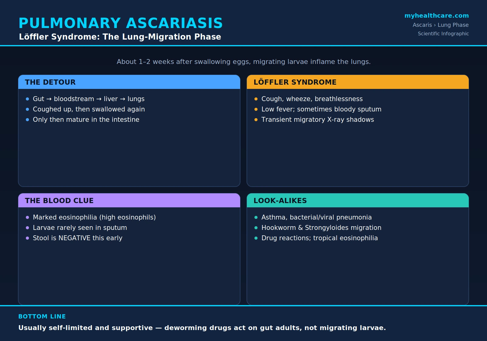
When most people picture a roundworm infection, they imagine worms living in the gut. But before Ascaris lumbricoides ever becomes an adult worm in the intestine, its tiny larvae take a remarkable detour through the lungs. For a week or two during that journey, the lungs — not the gut — are where the action is. This brief but distinctive phase can cause a cough, wheeze, and breathlessness with a striking rise in a particular white blood cell (the eosinophil), a picture doctors call Löffler syndrome. It is usually mild and passes on its own, but it is easy to mistake for asthma or pneumonia, and recognizing it for what it is can spare a patient unnecessary tests and treatments. This page explains the lung-migration phase of ascariasis: what is happening, why the timing makes the diagnosis tricky, how it is told apart from look-alike conditions, and when it actually needs treatment.
Table of Contents
- What Pulmonary Ascariasis Is
- The Larval Detour Through the Body
- Löffler Syndrome: The Symptoms
- Blood Eosinophilia and the Chest X-Ray
- The Timing Trap: Why Stool Tests Are Negative
- Seasonal "Ascaris Pneumonia"
- Telling It Apart: The Differential Diagnosis
- How It Is Diagnosed
- Management and Treatment
- When It Becomes Serious
- Key Research Papers
- Featured Videos
1. What Pulmonary Ascariasis Is
Pulmonary ascariasis is the lung phase of a roundworm infection — the stage when immature larvae are physically passing through the lungs, not the later disease caused by adult worms in the intestine. This distinction matters enormously, because the two phases happen at different times, cause different symptoms, are found by different tests, and are not even well treated by the same drugs.
The disease that most people associate with Ascaris — the foot-long adult worms, the abdominal discomfort, the rare but dangerous intestinal blockages — is the intestinal phase, and it comes later, once worms have matured in the gut. (That phase is covered on the Intestinal Obstruction and Biliary Complications page.) The pulmonary phase, by contrast, happens within the first couple of weeks after a person swallows infective eggs, while the parasite is still a microscopic larva on the move.
Think of it as two separate chapters of the same infection. In the lung chapter, there are no worms in the intestine yet, no eggs being produced, and often nothing abnormal to find in the stool. What there is — for a short window — is an inflamed, irritated set of lungs reacting to thousands of larvae squeezing through the delicate air sacs on their way to the throat. Understanding that this is a transient migration event, and not an established lung infection, is the key to making sense of everything else about it.
2. The Larval Detour Through the Body
To understand why the lungs are involved at all, it helps to follow the parasite's strange journey. Ascaris is a gut worm as an adult, yet its larvae take a long, looping route through the body before they ever settle in the intestine. The path is sometimes called the tracheal migration or the Löffler-Wakana cycle, and it goes like this:
- Swallowed eggs reach the gut. A person ingests microscopic Ascaris eggs — usually from soil, hands, or food and water contaminated with human feces. The eggs travel to the small intestine.
- Larvae hatch and burrow into the bloodstream. In the intestine, the eggs hatch and release tiny larvae. Rather than staying put, these larvae penetrate the intestinal wall and enter the bloodstream (and lymphatic vessels).
- The blood carries them to the liver, then the lungs. Riding the circulation, the larvae pass first through the liver and then are swept to the lungs, where they become lodged in the tiny capillaries surrounding the air sacs.
- They break into the airways. The larvae break out of the capillaries into the alveoli (the lungs' microscopic air pockets) and crawl up the bronchial tubes toward the windpipe and throat. This is the moment that produces the cough and the lung symptoms.
- They are coughed up and swallowed. Reaching the back of the throat, the larvae are coughed up and then swallowed back down — a second trip through the esophagus and stomach.
- They mature in the intestine. Back in the small intestine, the now larger larvae finally mature into adult worms, and weeks later begin producing eggs.
The whole detour exists because the larvae must grow and develop in the lung tissue before they are mature enough to survive as adults; they cannot simply stay in the gut from the start. The practical consequence is that the lung symptoms arrive early — during the migration, roughly one to two weeks after the eggs were swallowed — and well before any worm is laying eggs in the intestine. The general thread of the parasite's life cycle is described on the Ascaris Overview, but the lung phase is where that life cycle becomes briefly, and sometimes dramatically, visible.
3. Löffler Syndrome: The Symptoms
The cluster of lung symptoms produced by migrating Ascaris larvae is the classic example of Löffler syndrome (also written Löffler's syndrome or eosinophilic pneumonitis). The Swiss physician Wilhelm Löffler described, in the early 1930s, patients with fleeting lung shadows on X-ray and a high eosinophil count who recovered on their own — and migrating worm larvae, including Ascaris, are among the best-known causes.
The symptoms come from the body's inflammatory reaction as larvae rupture into the air sacs and irritate the airways. They typically include:
- Cough — often dry at first, sometimes becoming productive; this is the most consistent feature.
- Wheezing — a whistling sound and chest tightness, because the irritated airways narrow, much as they do in asthma.
- Breathlessness — a feeling of shortness of breath, usually mild but occasionally pronounced.
- Low-grade fever — a modest temperature rise rather than a high, spiking fever.
- Blood-tinged sputum — some people cough up phlegm streaked with a little blood, reflecting tiny areas of bleeding where larvae crossed from capillary into air sac. Frank, heavy bleeding is uncommon.
- Chest discomfort, malaise, and sometimes hives or an itchy rash — reflecting a more general allergic-type reaction to the parasite.
The single most reassuring feature of Löffler syndrome from Ascaris is that it is almost always self-limited and transient. The larvae do not stay in the lungs; within days to a week or two they have moved on to the throat and been swallowed, and the lung inflammation settles as they leave. Most people recover fully without any specific treatment. The challenge is rarely the danger of the illness itself — it is recognizing what it is, because a cough, wheeze, and low fever look like a great many ordinary chest complaints.
4. Blood Eosinophilia and the Chest X-Ray
Two findings, more than the symptoms themselves, point a clinician toward a parasitic cause: a markedly raised eosinophil count in the blood, and fleeting shadows on the chest X-ray.
Marked blood eosinophilia. The eosinophil is a type of white blood cell that the immune system deploys especially against parasites and in allergic reactions. During larval migration, eosinophils surge, and a blood test (the complete blood count, or CBC, with differential) often shows a strikingly high eosinophil percentage and count. This is a major clue: an unexplained cough or wheeze accompanied by a high eosinophil count should always raise the question of a migrating parasite, among other causes. The eosinophilia of Ascaris migration tends to be most pronounced during the lung phase and then declines.
Transient, migratory infiltrates on chest X-ray. The chest X-ray during Löffler syndrome typically shows patchy, hazy areas of shadowing (called infiltrates) that represent inflamed lung tissue around the migrating larvae. Their hallmark is that they are fleeting and shifting: a shadow seen in one part of the lung today may have faded by next week, while a new one appears elsewhere. This wandering, here-today-gone-tomorrow pattern — sometimes called “fleeting” or “migratory” pulmonary infiltrates — is very different from the fixed, solid consolidation of an ordinary bacterial pneumonia, and is one of the most characteristic signs of the whole eosinophilic-lung family of diseases. The combination of cough + wheeze + high eosinophils + shifting X-ray shadows is, in effect, the fingerprint of Löffler syndrome.
5. The Timing Trap: Why Stool Tests Are Negative
Here is the single most important and most counter-intuitive point about diagnosing pulmonary ascariasis, and it trips up patients and clinicians alike: at the moment the lungs are most symptomatic, the standard test for ascariasis — finding eggs in the stool — is usually negative.
The reason is pure timing. The lung symptoms appear during larval migration, roughly one to two weeks after the eggs were swallowed. But at that point the worms have not yet matured into egg-laying adults in the intestine. Egg production does not begin until the larvae have completed their lung journey, been swallowed, grown up in the gut, and reached sexual maturity — a process that takes on the order of two to three months from the original infection. So during the lung phase there are simply no eggs in the stool to find, because no adult worm is producing them yet.
This creates a genuine diagnostic trap. A patient may have an obvious eosinophilic pneumonitis driven by Ascaris, yet a stool examination sent at that very time comes back clean — falsely reassuring everyone that worms are not the cause. The resolution is to understand the sequence:
- During the lung phase: diagnosis rests on the pattern (cough/wheeze + high eosinophils + fleeting X-ray infiltrates + relevant exposure), not on stool eggs. Stool is typically negative.
- Weeks to months later: once the worms have matured, stool examination becomes positive for the characteristic Ascaris eggs, often after the lung symptoms have completely resolved. A stool test repeated some weeks down the line can therefore confirm in retrospect what the lung phase suggested.
Recognizing this lag — lung symptoms early, stool eggs late — is what separates a confident diagnosis from a missed one. It is also why a careful history and the eosinophil count carry so much weight during the acute illness.
6. Seasonal "Ascaris Pneumonia"
In some parts of the world where Ascaris transmission follows a seasonal rhythm — tied to rainfall, soil moisture, and patterns of human contact with contaminated earth — large numbers of people swallow infective eggs at roughly the same time of year. A few weeks later, those simultaneous infections produce a wave of lung-phase illness, sometimes described as seasonal “Ascaris pneumonia” or seasonal pneumonitis with eosinophilia.
A classic and well-documented example came from Saudi Arabia, where investigators in the 1960s described an annual outbreak of cough, wheeze, fever, fleeting lung infiltrates, and high eosinophil counts that swept through a community each year at a predictable time — and was traced to seasonal Ascaris larval migration. The lesson from such outbreaks is twofold. First, it underscores that the lung phase is a migration event: it appears in clusters precisely because exposure occurred in clusters a couple of weeks earlier. Second, it reminds clinicians in endemic regions to think of ascariasis when they see a seasonal spike of eosinophilic respiratory illness, rather than assuming a run of ordinary viral or bacterial chest infections. The seasonality is, in a sense, the epidemiological echo of the parasite's life cycle written across a whole population.
7. Telling It Apart: The Differential Diagnosis
Because the lung phase produces such common-sounding symptoms, the real diagnostic work is distinguishing it from the conditions it imitates. The differential diagnosis falls into a few groups.
Common chest conditions it mimics:
- Asthma — the wheeze, cough, and breathlessness can look just like an asthma flare. The clues that point away from simple asthma are the very high eosinophil count, the shifting X-ray infiltrates, and a relevant exposure history.
- Bacterial or viral pneumonia — an ordinary chest infection also causes cough, fever, and shadows on the X-ray. But typical pneumonia produces fixed consolidation rather than fleeting, migratory infiltrates, and it does not usually drive a dramatic eosinophilia.
Other parasites whose larvae also migrate through the lungs. Ascaris is not the only worm that takes a tracheal detour, so other helminths can produce an essentially identical Löffler picture:
- Hookworm (Ancylostoma duodenale, Necator americanus) — its larvae also pass through the lungs en route to the gut and can cause the same transient cough and eosinophilia.
- Strongyloides (Strongyloides stercoralis) — another soil-transmitted worm with a lung-migration phase; importantly, it can persist for years and, in people with weakened immunity, cause severe disease, so it is an essential one to consider and exclude.
Non-parasitic causes of Löffler / eosinophilic lung disease. The same syndrome of pulmonary infiltrates with eosinophilia can arise without any worm at all:
- Drug reactions — many medications can trigger a drug-induced eosinophilic pneumonitis that looks like Löffler syndrome; a careful medication history is essential.
- Tropical pulmonary eosinophilia — a distinct, more sustained eosinophilic lung disease caused by an immune reaction to lymphatic filariae (the worms behind filariasis), seen in endemic tropical regions, with extremely high eosinophil counts; unlike the brief migration of Ascaris, it tends to be persistent and to respond specifically to antifilarial treatment.
- The wider eosinophilic-pneumonia family — conditions such as acute and chronic eosinophilic pneumonia and allergic bronchopulmonary aspergillosis, which clinicians weigh when no parasite or drug is found.
In practice, sorting through this list relies on the exposure history (travel, soil contact, sanitation, medications), the time course, the degree and persistence of eosinophilia, and targeted testing for the specific parasites in question.
8. How It Is Diagnosed
Diagnosis of pulmonary ascariasis is, more than anything, a matter of putting the picture together, because no single quick test confirms it during the lung phase. The pieces include:
- History. The starting point: a relevant exposure (living in or travel to an area with poor sanitation, contact with contaminated soil, a setting where ascariasis is common), the timing of symptoms, and the absence of a better explanation.
- Blood eosinophilia. A complete blood count showing a markedly raised eosinophil count strongly supports a migrating-parasite cause, especially alongside the right symptoms and X-ray.
- Chest imaging. A chest X-ray (or CT) showing the characteristic transient, migratory infiltrates fits Löffler syndrome and argues against fixed bacterial consolidation.
- Larvae in sputum or gastric aspirate. Occasionally, the migrating larvae themselves can be found under the microscope in coughed-up sputum or in fluid aspirated from the stomach (where swallowed larvae collect). This is not commonly done, but when larvae are seen it provides direct confirmation during the acute phase.
- Stool examination — later. As explained above, stool is typically negative during the lung phase. A stool test repeated weeks to a few months later, once the worms have matured, can reveal the characteristic Ascaris eggs and confirm the diagnosis after the fact.
Because of this, a clinician will often make a presumptive (working) diagnosis of pulmonary ascariasis during the acute illness — based on the eosinophilia, the X-ray pattern, and the exposure — and then look for confirmatory stool eggs at a follow-up visit later on. The general approach to confirming intestinal ascariasis by stool microscopy is covered on the Symptoms & Diagnosis hub.
9. Management and Treatment
For the great majority of people, the lung phase of ascariasis needs only supportive care, because it is self-limited — the larvae move on within days to a couple of weeks, and the inflammation subsides as they go. The aims of treatment are simply to ease symptoms while the body clears the migration.
Supportive care for symptoms:
- Bronchodilators (inhaled airway-opening medicines, the same kind used in asthma) can relieve wheeze and breathlessness when those are troublesome.
- Corticosteroids may be used in more severe cases to dampen the inflammatory reaction in the lungs, reserved for patients whose symptoms are marked rather than mild.
- General measures — rest, fluids, and time — suffice for many people, since the syndrome resolves on its own.
A genuine nuance about antiparasitic (deworming) drugs. It is natural to assume that the cure for a worm infection is a deworming pill. But here the timing creates a real subtlety. The standard anthelmintic drugs — albendazole and mebendazole — act primarily on the adult worms in the intestine, not on the larvae that are migrating through the lungs. So giving a deworming drug during the acute lung phase does not reliably target the very larvae causing the symptoms; the lung disease is driven by parasites the gut-acting drug does not effectively reach. For this reason, treatment of the lung phase is mainly supportive, and definitive antiparasitic therapy is generally directed at the later intestinal stage, once the worms have matured into the adults that the drugs do clear. (There is also a theoretical concern that killing larvae mid-migration could intensify the inflammatory reaction, which is a further reason the lung phase is usually managed supportively rather than with immediate larvicidal intent.) The mechanics and timing of albendazole and mebendazole are covered on the Anthelmintic Treatment page, and the broader strategy on the Treatment & Prevention hub.
The practical upshot: support the patient through the brief lung phase, then treat the intestinal infection that follows — and, most importantly, prevent the next infection through sanitation and hygiene, since the entire cycle begins with swallowing eggs from a contaminated environment (see Prevention: Sanitation and Hygiene).
10. When It Becomes Serious
Although pulmonary ascariasis is usually mild, it is worth knowing the circumstances in which it deserves more attention or can turn serious:
- A heavy larval load. When a person swallows a very large number of eggs, the simultaneous migration of huge numbers of larvae can produce a more intense pneumonitis, with significant breathlessness and more extensive lung involvement than the usual mild, transient picture.
- Severe wheeze or respiratory distress. Marked airway narrowing — especially in children or in people with underlying asthma — can cause genuine difficulty breathing that needs prompt bronchodilator and sometimes steroid treatment.
- Significant coughing of blood. While blood-streaked sputum is common and minor, heavier bleeding is unusual and warrants evaluation.
- Diagnostic confusion that delays the right care. Perhaps the most common “harm” is not from the worm but from the mix-up: being treated repeatedly for asthma or bacterial pneumonia while the true cause is missed, or, conversely, being put through unnecessary investigations. Recognizing the eosinophilia-plus-fleeting-infiltrate pattern prevents both.
- The look-alikes that are more dangerous. Because Strongyloides can cause life-threatening disease in immunocompromised people, and because tropical pulmonary eosinophilia and drug reactions need their own specific management, the bigger risk is sometimes mistaking one of these for simple Ascaris migration — another reason a careful differential matters.
Reassuringly, serious outcomes from the Ascaris lung phase itself are uncommon in otherwise healthy people; the migration is typically a brief, self-resolving episode. The more lasting concerns of ascariasis come from the intestinal worms that follow — nutritional effects in children (see Malnutrition and Childhood Growth) and, occasionally, blockage of the bowel or bile ducts (see Intestinal Obstruction and Biliary Complications). The lung phase is the body's brief, noisy announcement that those gut worms are on their way.
Key Research Papers
Peer-reviewed reviews and studies on ascariasis, larval lung migration, Löffler syndrome, and the broader family of eosinophilic and parasitic lung diseases. Journal names appear as plain text; the year/volume/pages link opens the full citation via DOI.
- Löffler W. Transient Lung Infiltrations with Blood Eosinophilia. International Archives of Allergy and Applied Immunology. 1956;8:54–59.
- Dold C, Holland CV. Ascaris and Ascariasis. Microbes and Infection. 2011;13(7):632–637.
- Bethony J, Brooker S, Albonico M, et al. Soil-Transmitted Helminth Infections: Ascariasis, Trichuriasis, and Hookworm. The Lancet. 2006;367(9521):1521–1532.
- Jourdan PM, Lamberton PHL, Fenwick A, Addiss DG. Soil-Transmitted Helminth Infections. The Lancet. 2018;391(10117):252–265.
- Chitkara RK, Krishna G. Parasitic Pulmonary Eosinophilia. Seminars in Respiratory and Critical Care Medicine. 2006;27(2):171–184.
- Akuthota P, Weller PF. Eosinophilic Pneumonias. Clinical Microbiology Reviews. 2012;25(4):649–660.
- Allen JN, Davis WB. Eosinophilic Lung Diseases. American Journal of Respiratory and Critical Care Medicine. 1994;150(5):1423–1438.
- Cottin V, Cordier JF. Eosinophilic Pneumonias. Allergy. 2005;60(7):841–857.
- Gelpi AP, Mustafa A. Seasonal Pneumonitis with Eosinophilia: A Study of Larval Ascariasis in Saudi Arabs. The American Journal of Tropical Medicine and Hygiene. 1967;16(5):646–657.
- Ong RKC, Doyle RL. Tropical Pulmonary Eosinophilia. Chest. 1998;113(6):1673–1679.
- Vijayan VK. Tropical Pulmonary Eosinophilia: Pathogenesis, Diagnosis and Management. Current Opinion in Pulmonary Medicine. 2007;13(5):428–433.
Live PubMed Searches
Each link opens a live PubMed query so results stay current as new papers are indexed.
- Pulmonary ascariasis larval migration
- Löffler syndrome eosinophilic pneumonia
- Ascaris lumbricoides lung eosinophilia
- Transient pulmonary infiltrates with eosinophilia
- Tropical pulmonary eosinophilia (filariasis)
- Parasitic pulmonary eosinophilia (helminths)
- Larvae in sputum / gastric aspirate
- Seasonal ascariasis pneumonitis
Connections
- Symptoms & Diagnosis
- Intestinal Obstruction & Biliary Complications
- Malnutrition & Childhood Growth
- Treatment & Prevention
- Anthelmintic Treatment (Albendazole & Mebendazole)
- Prevention: Sanitation & Hygiene
- Mass Deworming Programs
- Ascaris Overview
- All Parasites
- Pulmonology
- Infectious Disease
- All Conditions