Rothman, Schekman and Südhof: The Cell's Delivery Service, and What Happens When It Misfires
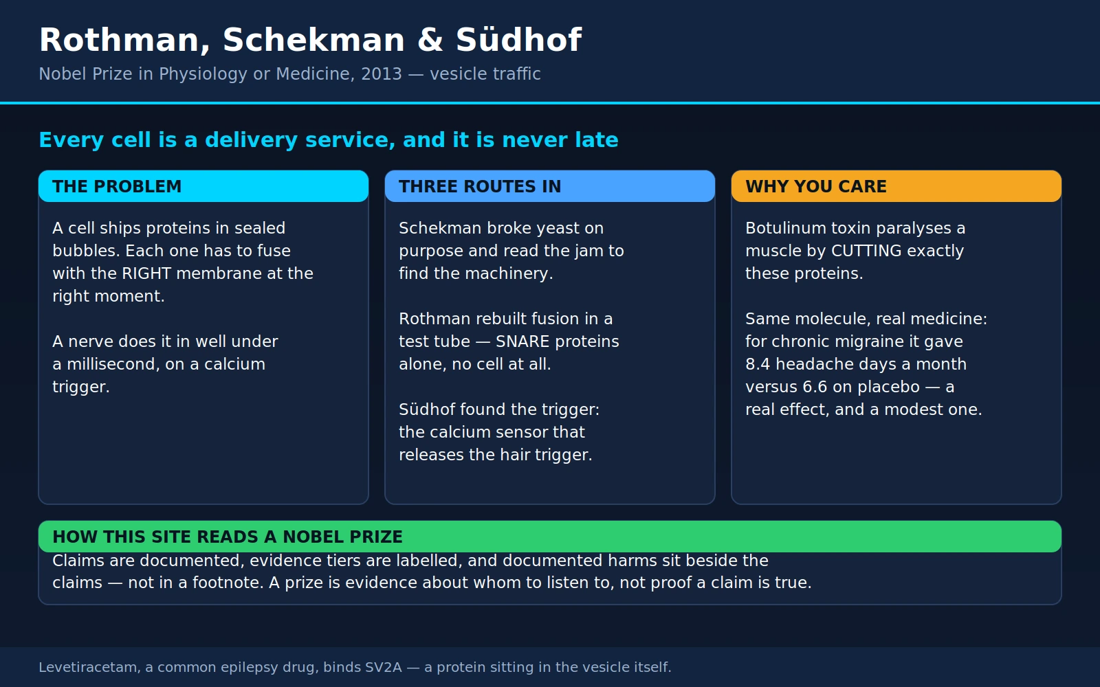
The 2013 Nobel Prize in Physiology or Medicine was awarded jointly to James E. Rothman (Yale University), Randy W. Schekman (University of California, Berkeley) and Thomas C. Südhof (Stanford University) — in the committee's words, "for their discoveries of machinery regulating vesicle traffic, a major transport system in our cells." Three laboratories, three completely different methods, one machine.
It is one of the least glamorous-sounding prizes on this site and one of the most useful. The machinery they described is why insulin reaches your bloodstream, why a nerve can speak to a muscle in under a millisecond, why Botox works, why tetanus kills, and why a common epilepsy drug binds a protein nobody had connected to seizures. If you want a single page that explains what a cell actually does all day, this is it.
Table of Contents
- The Logistics Problem
- Schekman: The Jam That Tells You Where the Pipeline Broke
- Rothman: The Address System, Built in a Test Tube
- Südhof: The Trigger, and Why Speed Is the Hard Part
- Where This Shows Up in an Ordinary Day
- Botulinum and Tetanus: One Cut, Two Opposite Diseases — and a Medicine
- Diabetes and the Insulin Granule
- Epilepsy, Neurodevelopment, and a Thread to Parkinson's
- Levetiracetam: An Epilepsy Drug That Binds a Vesicle
- The 2013 Prize and the Credit Question
- The Same Story, Told Across Five Prizes on This Site
- Where Mainstream Medicine Agrees — and What Remains Debated
- Key Research Papers
- Connections
- Featured Videos
1. The Logistics Problem
A cell is a warehouse with no aisles. It manufactures proteins at one address — ribosomes on the endoplasmic reticulum, deep inside — and needs an enormous fraction of them somewhere else entirely: embedded in the outer membrane, dropped into a lysosome, or pushed clean out of the cell into the bloodstream. Proteins cannot simply drift there. Most of them are far too big to cross a membrane, and the compartments they must reach are sealed off from one another by those same membranes.
The cell's solution is a delivery van. It pinches off a small bubble of membrane — a vesicle — loads the cargo inside, buds it away from one compartment, moves it, and fuses it into the wall of the next. Fusion is the delivery: when the vesicle's membrane merges with the target's, the contents are simply there, on the other side, without anything ever having crossed a barrier.
Building the van turns out not to be the hard part. The hard parts are the two that any real courier company would recognise:
- Addressing. A cell has dozens of destinations and is running thousands of vesicles at once. A van carrying digestive enzymes must not empty itself into the nucleus. Something has to guarantee that a vesicle fuses with the right membrane and no other.
- Timing. Some deliveries are constant and unsupervised. Others must be held at the loading dock, fully packed, for hours — and then released in a burst, on a signal, faster than almost any other biological event. Nothing about a bubble of fat naturally waits.
The 2013 prize covers exactly that division of labour. Schekman found the parts list. Rothman found how the parts do the addressing. Südhof found how the machine is held back and then triggered. Three independent attacks on one problem, using genetics, biochemistry and electrophysiology — and, remarkably, they converged on the same set of molecules.
2. Schekman: The Jam That Tells You Where the Pipeline Broke
Randy Schekman's move, beginning in the late 1970s, was to stop looking at the machinery and start breaking it. He worked in baker's yeast, Saccharomyces cerevisiae — a single-celled organism that secretes proteins the same way our cells do, but which can be mutated and screened by the million.
The trick was to make the defect conditional. He hunted for temperature-sensitive mutants: yeast that grow and secrete normally at 25 °C but whose secretion fails when shifted to 37 °C. That way a lethal defect can be switched on for study and switched off again.
The first one, reported with Peter Novick in 1979, was named sec1. At the restrictive temperature the mutant cells kept manufacturing their secreted enzymes — protein synthesis carried on for hours — but the enzymes stopped coming out. The internal pool of the secreted enzymes invertase and acid phosphatase rose up to eightfold. Under the electron microscope the reason was visible and unmistakable: the cells had filled up with membrane-bound vesicles, and staining showed the trapped enzyme was inside them. Return the cells to the permissive temperature and the vesicles disappeared as the backed-up cargo was finally secreted.
Why a jam is so informative. This is the logic that makes the whole approach work, and it is worth stating plainly. A secretory pathway is an assembly line. If you disable one station, everything upstream of it keeps arriving and nothing moves on — so cargo piles up immediately before the broken step. The shape of the pile-up tells you which step the gene performs. Cargo stuck in the endoplasmic reticulum means the gene works at ER export. Cargo stuck in the Golgi means it works in the Golgi. Cargo stuck in finished vesicles sitting at the cell surface — as in sec1 — means the gene is needed at the very last step, fusion with the plasma membrane. You never have to see the machine. You infer its parts, and their order, from where the traffic stops.
In 1980, Novick, Charles Field and Schekman turned that into an industrial-scale screen. They had noticed something useful about the mutants: a cell that keeps making cargo it cannot release becomes physically denser. So they mutagenised yeast, shifted them to the restrictive temperature and spun them on a density gradient, harvesting the heavy cells. From those they recovered 188 mutant clones that accumulated an abnormally large internal pool of invertase, and complementation testing sorted them into 23 distinct genes — the sec genes, sec1 through sec23. Electron microscopy showed that nearly every one of them accumulated a specific membrane-bound structure. The authors were explicit that they had not exhausted the list; the distribution of alleles suggested more genes were out there, and more were duly found.
That is what a parts list looks like when you have no idea what the parts are. Schekman had named, ordered and assigned rough jobs to two dozen components of the secretory pathway before anyone knew what a single one of the proteins looked like.
3. Rothman: The Address System, Built in a Test Tube
James Rothman came at the identical problem from the opposite end. Rather than break the machine inside a living cell, he set out to rebuild it outside one — on the principle that if you can reconstitute a process from purified components in a tube, you necessarily know what the components are.
This was not an obviously reasonable ambition. Membrane fusion in a living cell is fast, directional and exquisitely regulated; a bag of broken cell parts should just produce mush. Rothman's laboratory nonetheless built a working cell-free transport system, described in 1984 with William Balch, William Dunphy and William Braell. They used a viral glycoprotein as trackable cargo and mixed Golgi membranes from two different cell lines — a "donor" population missing a particular sugar-adding enzyme and an "acceptor" population that had it. If, and only if, the cargo was genuinely transported from one compartment into the next, it picked up a radioactive sugar tag. It did. Transport in the tube was almost as efficient as inside the cell, and it required ATP and cytosol — meaning there were soluble factors to be purified.
Purifying them is exactly what the lab then did, and one result deserves its own paragraph. The soluble fusion factor they isolated from mammalian cells — NSF, named for its sensitivity to the chemical N-ethylmaleimide — was cloned in 1989 and turned out to be the product of the yeast SEC18 gene. One of Schekman's sec genes, found by breaking yeast, and Rothman's fusion protein, found by rebuilding transport in hamster-cell extracts, were the same thing. As the paper put it, the mechanism of vesicular fusion is highly conserved, both between species and between different stages of transport. Yeast and human beings run the same delivery service. It is one of the cleanest examples in modern biology of two utterly different experimental strategies arriving at one molecule.
The address system itself came in 1993. Thomas Söllner and colleagues in Rothman's lab used the natural stickiness of NSF and its adaptor proteins as bait to fish their binding partners out of cow brain. The catch was named SNAREs (SNAP receptors), and its arrangement was the answer to the addressing problem: the four principal proteins recovered were all synaptic, and they came in two flavours — one type sitting in the synaptic vesicle membrane, another type sitting in the target plasma membrane. Vesicle SNAREs and target SNAREs. Further work showed there were many SNARE-family members, each apparently associated with one particular kind of vesicle or one particular target membrane.
The image that has stuck — and it is a fair one — is a zip. Half the zip is on the van, half is on the loading dock, and the two halves interlock along their length, pulling the membranes together until they merge. A zip closes only with its own matching half; a vesicle carrying one v-SNARE finds the membrane carrying the complementary t-SNAREs and no other. Addressing and fusion are the same event.
In 1998, Thomas Weber and colleagues in Rothman's lab supplied the decisive test. They put purified, recombinant v-SNAREs into one set of artificial lipid vesicles and t-SNAREs into another, mixed them, and watched the two sets of membranes fuse spontaneously at body temperature — no cell, no cytoplasm, nothing else. SNAREs alone were, in their phrase, the minimal machinery for membrane fusion.
4. Südhof: The Trigger, and Why Speed Is the Hard Part
Thomas Südhof took the machine to the place where it is pushed hardest: the synapse. And at the synapse, the SNARE zip alone is not a sufficient explanation — because it explains fusion, and what a synapse needs is fusion that does not happen until it is told, and then happens almost instantly.
The numbers are the point. When an action potential arrives at a nerve terminal, calcium channels open, calcium floods in, and neurotransmitter is released into the synaptic cleft in well under a millisecond. Vesicles have to sit at the membrane fully assembled, held on a hair trigger, doing nothing at all — and then dump their contents in a fraction of the time it takes a nerve impulse to travel a centimetre. A spontaneously fusing SNARE complex would be a catastrophe. What the synapse requires is a clamp and a switch.
Südhof's laboratory identified the switch: synaptotagmin, a protein sitting in the synaptic vesicle membrane that binds calcium and, in doing so, drives the waiting SNARE complex to complete fusion. It is the calcium sensor — the component that converts "calcium has entered" into "release now."
The decisive experiment came in 1994, with Markus Geppert and colleagues. They deleted the synaptotagmin I gene in mice. The animals were born but died within 48 hours, and recordings from their cultured hippocampal neurons showed a very specific failure: the fast, synchronous, calcium-triggered component of release was severely impaired, while spontaneous release and slower asynchronous release carried on. That selectivity is what makes the result a sensor result rather than a general "the synapse is broken" result. Take away synaptotagmin I and the synapse does not stop releasing — it stops releasing on time.
How this sits beside Katz. Our page on Katz, von Euler and Axelrod covers the 1970 prize, where Bernard Katz showed that neurotransmitter is not released as a smooth stream but in discrete, uniform packets — quanta — and that their release is triggered by calcium entering the nerve terminal. Katz established, from electrical recordings alone, that the packets and the calcium trigger existed. He could not say what a packet physically was, or what calcium bound to.
This prize is the answer to both. The quantum is a synaptic vesicle. The calcium trigger is synaptotagmin. Katz described the behaviour; Rothman, Schekman and Südhof described the machine producing it. Read the two pages together and you have roughly a century of work on a single question, from the first electrical hint to the named proteins.
5. Where This Shows Up in an Ordinary Day
Regulated secretion — cargo packed in advance, held, and released on a signal — is not a specialist neuronal trick. It is how much of the body's minute-to-minute chemistry is delivered. A partial list of things you did today using this exact machinery:
- Released insulin after eating. Beta cells in the pancreas keep insulin pre-packed in granules. Rising blood glucose changes the cell's electrical behaviour, calcium enters, and docked granules fuse with the cell surface and empty insulin into the blood. The granule is the van; the trigger is calcium; the fusion is SNARE-mediated. See Diabetes.
- Digested a meal. Pancreatic acinar cells hold digestive enzymes — amylase, lipase, proteases — in zymogen granules and release them into the gut on hormonal and neural cues. Stomach parietal cells traffic their acid pumps to the surface the same way.
- Ran your nervous system. Every thought, movement and sensation is neurotransmitter released from vesicles: acetylcholine at the neuromuscular junction, glutamate and GABA in the brain, dopamine, serotonin and noradrenaline in the pathways that mood and attention run on.
- Made antibodies. Plasma cells are secretory factories, exporting immunoglobulin continuously through the same ER–Golgi–vesicle route Schekman's sec mutants mapped.
- Had an allergic twinge. Mast cells hold histamine pre-packed in granules; cross-linking of IgE on their surface triggers calcium entry and mass fusion. The itch, the wheal and the runny nose of an allergic reaction are a regulated exocytosis event — which is why antihistamines block the receptor for the released cargo rather than the release itself.
- Clotted a small cut. Platelets degranulate on activation, releasing their contents to recruit more platelets.
The unifying observation is that a cell wanting to say something to the rest of the body almost always says it by fusing a vesicle. Which means a single molecular machine is a shared dependency for endocrinology, neurology, immunology and haematology at once — and a single point of failure.
6. Botulinum and Tetanus: One Cut, Two Opposite Diseases — and a Medicine
If you want proof that the SNARE machine is real and not a diagram, the clostridial neurotoxins supply it. They are the clearest clinical demonstration of the whole prize, and they arrived at the answer at almost the same moment the biochemists did.
The mechanism. In 1992, Giampietro Schiavo, Cesare Montecucco and colleagues in Padua showed that tetanus toxin and botulinum toxin type B are zinc-dependent proteases — molecular scissors — and that their substrate is synaptobrevin, the vesicle SNARE. They cut it at one specific peptide bond. The specificity is startling: the rat synaptobrevin-2 isoform is cleaved, while synaptobrevin-1, which has a different single amino acid at the equivalent position, is not. The following year, José Blasi, Reinhard Jahn and colleagues showed that botulinum toxin type A cleaves SNAP-25 instead — a different SNARE, near its far end. Different serotypes, different scissors, different SNARE targets, same consequence.
That consequence is total. Cut the zip and the vesicle can no longer fuse. The nerve terminal is intact, full of neurotransmitter, electrically excitable, and completely silent. This is why botulinum toxin is the most poisonous substance known by weight: it does not damage the neuron, it disconnects one specific step, and that step happens to be the only way a neuron has of speaking.
Why the same class of mechanism produces opposite-looking diseases. Botulism causes floppy paralysis; tetanus causes rigid paralysis, with clenched jaw and arched back. Two toxins from closely related bacteria, cleaving the same family of proteins by the same chemistry, produce clinical pictures that look like each other's mirror image. The explanation is not in the chemistry at all. It is in the address.
- Botulinum toxin stays peripheral. It binds motor nerve terminals at the neuromuscular junction and stops there. The command "contract" never reaches the muscle. The muscle goes slack — flaccid paralysis. Death, when it comes, is from the diaphragm going slack too.
- Tetanus toxin travels. After entering peripheral motor and sensory neurons it is carried backwards along the axon into the spinal cord, where it moves into the inhibitory interneurons and blocks their release of glycine and GABA. Those interneurons exist to restrain motor neurons. Silence them and the motor neurons lose their brakes and fire continuously — spastic paralysis.
Same lesion, opposite outcome, because one toxin silences the accelerator and the other silences the brake. It is the neatest available illustration of a general principle in neurology: knowing what a drug or toxin does molecularly tells you nothing about the clinical picture until you also know which cells it reaches. Our page on Tetanus covers that disease and its vaccine.
The therapeutic turn
The idea of injecting the most potent toxin in nature into people on purpose sounds unhinged until you state it correctly: in tiny, localised doses, botulinum toxin is a way to switch off one named muscle or one named gland, reversibly, for a few months. That is a genuinely unusual capability, and medicine has found a lot of uses for it.
Approved uses for onabotulinumtoxinA (Botox) on the current US label include cervical dystonia; blepharospasm and strabismus (age 12 and up); spasticity in upper and lower limbs (age 2 and up); severe primary axillary hyperhidrosis (excessive underarm sweating); overactive bladder with urge incontinence in adults who have not tolerated or responded to an anticholinergic; urinary incontinence from neurogenic detrusor overactivity (age 5 and up); and prophylaxis of chronic migraine in adults — defined as 15 or more headache days a month, with headaches lasting four hours or more. The cosmetic product is separately approved for moderate-to-severe glabellar lines, lateral canthal lines (crow's feet), forehead lines and platysma bands. (DailyMed — BOTOX label; DailyMed — BOTOX Cosmetic label.)
Two of those deserve their actual numbers rather than a wave.
Post-stroke spasticity. A randomised, double-blind, placebo-controlled trial published in the New England Journal of Medicine in 2002 injected 126 people who had wrist and finger spasticity after a stroke with a single dose of botulinum toxin A or placebo. Muscle tone improved more in the treated group at every follow-up through 12 weeks. On the outcome that matters more — self-reported disability in the one area each patient had chosen as their treatment target, such as hygiene or dressing — 62% of the treated group reported meaningful improvement at six weeks versus 27% on placebo.
Chronic migraine. The two PREEMPT trials, pooled and published in 2010, randomised 1,384 adults with chronic migraine to onabotulinumtoxinA (155–195 units, injected across a fixed set of head and neck sites every 12 weeks) or placebo. At 24 weeks, headache days fell by 8.4 per month on active treatment versus 6.6 on placebo — a statistically robust difference, and one worth reading carefully. The placebo group improved a great deal. The genuine drug effect is the roughly two extra headache-free days a month, which for someone living with 20 headache days a month can be meaningful, and is also a good deal less than the marketing implies. Adverse events occurred in 62.4% of treated patients versus 51.7% on placebo; discontinuation for adverse events was uncommon (3.8% versus 1.2%).
The honest caveats
- It always wears off. The nerve terminal is not killed; the cleaved SNARE is eventually replaced and sprouted terminals re-establish contact. For glabellar lines the label puts the duration at roughly three to four months, and the labelled re-treatment interval for the medical indications is generally no sooner than 12 weeks. Every benefit described above is a benefit that must be bought again, indefinitely.
- Repeat dosing can provoke antibodies. Because the toxin is a foreign protein, the immune system can learn to neutralise it — and then the injections simply stop working. This is not rare. A 2019 study of 596 patients on long-term botulinum toxin A for neurological indications found neutralising antibodies in 83 of them (13.9%). The probability rose with both the single dose and the cumulative dose and varied by formulation, while treatment duration by itself added nothing. The practical implication, which the label also gives, is to use the lowest effective dose at the longest workable interval — the opposite of the instinct to top up early when the effect fades.
- The effect can spread beyond the injection site. Every botulinum toxin product carries a boxed warning that the effect may spread from the injection area hours to weeks afterwards, and that swallowing and breathing difficulties can be life-threatening. Reported cases have clustered around the higher-dose indications such as spasticity, and around unapproved uses. This is a prescription procedure with a real risk profile, not a cosmetic accessory.
- Units are not interchangeable between brands. Onabotulinumtoxin, abobotulinumtoxin, incobotulinumtoxin and the rest are dosed on different unit scales and cannot be substituted one-for-one.
None of this is treatment advice, and nothing here should be used to choose a dose. The point of the section is different and, we think, more interesting: a paralytic toxin became one of the most widely used drugs in medicine because three laboratories worked out what it cuts.
7. Diabetes and the Insulin Granule
Insulin release is a textbook case of regulated exocytosis, and every stage of it is the machinery this prize describes. A beta cell manufactures insulin, packs it into dense-core granules, and stores them. When blood glucose rises, glucose metabolism inside the cell closes potassium channels, the membrane depolarises, voltage-gated calcium channels open, and calcium entry triggers granules that are already docked at the plasma membrane to fuse and empty. The insulin in your blood after lunch arrived by exactly the route Südhof's synapse work describes, on a timescale of seconds rather than microseconds.
The natural question is whether the secretion machinery itself goes wrong in type 2 diabetes. Here we have to be careful, because this is exactly the kind of place where a plausible mechanism gets over-sold.
What is well established. Type 2 diabetes involves both insulin resistance in the tissues and a genuine defect in insulin secretion by the beta cell — the two together, not resistance alone. One of the most consistent findings is the loss of first-phase insulin release: in a healthy person, a glucose load produces a sharp initial burst within minutes (the already-docked granules going) followed by a slower sustained phase (granules recruited and prepared). In type 2 diabetes that first burst is blunted or gone, often years before diagnosis.
What is well-supported mechanism, at the level of human tissue. A 2018 study in Cell Metabolism examined insulin granules in human beta cells directly and found that secretion depends critically on how many granules are docked at the membrane and ready to go — and that in islets from donors with type 2 diabetes, granule docking was strongly reduced. Glucose normally accelerates docking; in the diabetic cells that acceleration was absent. Gene-expression analysis showed key docking proteins were downregulated, and putting them back increased docking. That is a coherent molecular account of where the missing first-phase burst goes.
What is not established. Whether a docking defect is a cause of type 2 diabetes or a consequence of the metabolic environment beta cells endure in it — chronic high glucose, high fatty acids, years of overwork — is not settled by studies like this, which compare tissue from people who already have the disease. Nor is there any therapy that targets the exocytosis machinery. Every drug currently used to increase insulin secretion works upstream of it: sulfonylureas close the potassium channel, and GLP-1 receptor agonists amplify the response to glucose through signalling pathways. Nothing on the market fixes a SNARE.
8. Epilepsy, Neurodevelopment, and a Thread to Parkinson's
If a machine is required for every synapse in the brain, mutations in its parts should cause brain disease. They do, and the pattern is consistent: severe, early, and neurodevelopmental rather than degenerative.
STXBP1 is the clearest example. The gene encodes syntaxin-binding protein 1, also called Munc18-1 — a partner protein essential for assembling the SNARE complex. It is the same gene family Schekman found in yeast as sec1. De novo mutations in it cause a severe childhood epilepsy and encephalopathy, first reported in 2008. A 2016 international study assembled the largest picture of the condition, describing 147 patients: all had intellectual disability, severe to profound in 88%; 95% had epilepsy. About a fifth presented as Ohtahara syndrome and around a tenth as West syndrome, but the majority had a non-syndromic early-onset epilepsy. Strikingly, the authors found no correlation between how severe the seizures were and how severe the intellectual disability was, which led them to reframe the condition: STXBP1 encephalopathy is best understood as a neurodevelopmental disorder that includes epilepsy, rather than an epilepsy that damages the brain through seizures. That distinction matters enormously to families, because it means controlling the seizures — worth doing — is not expected on its own to change the developmental trajectory.
STXBP1 is not alone. Mutations in other synaptic vesicle and release genes — including STX1B, SYT1, DNM1 and SNAP25 — are recognised causes of developmental and epileptic encephalopathies, and this whole class of gene is now part of routine epilepsy gene panels. For most of them there is no targeted treatment yet; the value of a diagnosis is prognostic and practical — ending the diagnostic odyssey, guiding which antiseizure medicines to try, and connecting families to gene-specific registries.
The thread to Parkinson's is real but thinner, and should be labelled as such. Alpha-synuclein is the protein that aggregates into the Lewy bodies that define Parkinson's disease pathologically, and duplications and mutations of its gene cause inherited Parkinson's. What was not clear for a long time was what the protein normally does. In 2010 Südhof's laboratory reported an answer squarely inside this prize's territory: alpha-synuclein binds directly to the vesicle SNARE synaptobrevin-2 and promotes SNARE-complex assembly, acting as a kind of chaperone that keeps a nerve terminal capable of assembling fusion machinery over and over across a lifetime of firing. Mice lacking all three synucleins developed age-dependent neurological impairment, showed reduced SNARE-complex assembly, and died early.
9. Levetiracetam: An Epilepsy Drug That Binds a Vesicle
Levetiracetam (Keppra) deserves its own short section, because it is one of the most widely prescribed antiseizure medicines in the world and its story runs backwards through this entire page.
It was found the old-fashioned way — it protected animals against seizures — and it had an unusual profile: effective in animal models where the standard drugs were not, well tolerated, and remarkably free of drug–drug interactions. What nobody could say was how it worked. It did not fit the known mechanisms. It was not blocking sodium channels, not enhancing GABA receptors, not doing any of the things antiepileptics were supposed to do.
In 2004, Berkley Lynch and colleagues answered it, and the answer was a surprise. The brain binding site for levetiracetam is SV2A — synaptic vesicle glycoprotein 2A, a protein sitting in the membrane of synaptic vesicles. The evidence was tight: the binding site was enriched in purified synaptic vesicles; brain membranes from mice lacking SV2A did not bind the drug at all; SV2A expressed in fibroblasts was sufficient to confer binding; the closely related isoforms SV2B and SV2C did not bind; and across a series of chemical analogues, how tightly a compound bound SV2A predicted how well it protected animals against seizures.
So a drug that works in humans binds a synaptic vesicle protein. That finding validated an entirely new target class, and two successor drugs — brivaracetam and padsevonil — were designed against the same site.
What is still not settled is the step in between. Binding SV2A is established. What SV2A normally does, and how occupying it translates into fewer seizures, is not. SV2A is thought to modulate vesicle availability and release probability, and one attractive idea is that the drug damps down excessive release specifically during the high-frequency firing that characterises a seizure while leaving normal transmission largely alone — which would fit the clinical profile. But a 2024 review of the SV2A drug class puts the state of knowledge bluntly: although it is widely accepted that levetiracetam acts through SV2A, the molecular basis of its action remains unknown.
That gap is worth sitting with, because it is a fair picture of how much of pharmacology actually works. We have a common, effective, well-tolerated drug; we know precisely what molecule it grabs; we know that molecule lives on a synaptic vesicle; and the mechanism connecting the two is still being argued about two decades later. See Epilepsy for the condition itself.
10. The 2013 Prize and the Credit Question
The prize was announced on 7 October 2013 and awarded jointly, with the three laureates' contributions summarised as complementary: Schekman discovered the genes required for vesicle traffic, Rothman worked out the protein machinery that lets vesicles fuse with their targets, and Südhof showed how signals instruct vesicles to release their cargo with precision.
Nobody disputes that the work deserved a prize. The one point worth recording is a structural feature of the Nobel rules rather than a controversy: a prize may be shared by at most three people, and this field had more than three central figures.
The documented illustration is that in the very same year, the Albert Lasker Basic Medical Research Award — often a Nobel forerunner — went to Richard Scheller and Thomas Südhof for the molecular machinery underlying neurotransmitter release. Scheller, then at Stanford and later at Genentech, had worked in parallel with Südhof through the 1980s and 1990s and identified syntaxin, one of the target-membrane SNAREs at the heart of the whole model. He was honoured by the Lasker jury for that work and was not among the three Nobel laureates. Reinhard Jahn, whose laboratory co-authored the discovery that botulinum toxin A cleaves SNAP-25 and who contributed extensively to the identification and characterisation of the synaptic SNAREs, is another name that recurs in accounts of the field.
We state that as a fact about how prizes are allocated, not as a grievance. Discovery here was genuinely collective and genuinely international: yeast geneticists in Berkeley, biochemists in New York, neuroscientists in Texas, and toxin chemists in Padua all converged on one machine within about fifteen years, each group's result making the next one interpretable.
11. The Same Story, Told Across Five Prizes on This Site
This page is not a standalone. It is the middle of a chain that runs through several other Nobel pages here, and naming the chain is the most useful thing this section can do.
- 1974 — the map. Albert Claude, Christian de Duve and George Palade took the cell apart by centrifuge and electron microscope and described its compartments. Palade in particular traced the secretory pathway itself: he followed labelled protein from the endoplasmic reticulum through the Golgi into secretory granules and out of the cell, establishing that there is a route and what its stations are. De Duve found the lysosome, one of the destinations.
- 1999 — the labels. Günter Blobel won the prize for the signal hypothesis: proteins carry short address tags built into their own sequence that direct them into the ER and on to their destination. Palade had the route; Blobel had the labels stuck to the parcels.
- 1970 — the behaviour. Bernard Katz showed that neurotransmitter release is quantal and calcium-triggered — discrete packets, on a calcium signal — without being able to say what a packet was made of.
- 2013 — the machine. This page. Schekman's parts list, Rothman's SNARE address system, Südhof's calcium trigger. The vehicles that run Palade's route, the mechanism that reads Blobel's labels to their final destination, and the physical identity of Katz's quantum.
- 1991 and 2016 — the instruments and the reverse gear. Erwin Neher and Bert Sakmann built the patch clamp, which made it possible to record the currents and, with capacitance measurements, to watch single vesicles fuse in real time. Yoshinori Ohsumi used Schekman's exact strategy — yeast genetics, conditional mutants, a screen — to find the ATG genes of autophagy, membrane traffic running inward instead of outward.
Read in order, these are not six separate discoveries. They are one question — how does a cell move things? — answered in layers over roughly seventy years, each layer only askable because the one before it existed. That is a better description of how biology actually progresses than any single-genius story, and it is why this site keeps a complete list of the laureates rather than a highlights reel.
12. Where Mainstream Medicine Agrees — and What Remains Debated
Settled, and not seriously contested by anyone
- Proteins destined for secretion travel through the ER and Golgi in membrane vesicles, and the pathway's components were identified genetically in yeast as the sec genes.
- SNARE proteins on the vesicle and on the target membrane pair with each other, and that pairing both specifies the destination and drives fusion. Purified SNAREs in artificial membranes are sufficient to fuse them.
- The machinery is conserved from yeast to humans — the same gene, found twice by opposite methods.
- Synaptotagmin is the calcium sensor for fast synchronous neurotransmitter release.
- Botulinum and tetanus toxins are proteases that cleave SNARE proteins; that is why they block transmitter release. Botulinum toxin is an effective, approved treatment for cervical dystonia, spasticity, blepharospasm, axillary hyperhidrosis, certain bladder conditions, and chronic migraine prophylaxis.
- Levetiracetam binds SV2A, a synaptic vesicle protein.
- De novo mutations in STXBP1 cause a severe neurodevelopmental disorder with epilepsy.
Genuinely open
- How SV2A binding produces an anticonvulsant effect. The target is certain; the mechanism is not, twenty years on.
- How much of the beta-cell secretory defect in type 2 diabetes is a primary machinery problem versus a consequence of chronic metabolic stress. Human islet data show reduced granule docking; the direction of causation is unresolved.
- How central presynaptic failure is to Parkinson's disease. Alpha-synuclein has a demonstrated normal role in SNARE assembly, and that is a real clue; whether presynaptic dysfunction initiates the disease, or aggregation elsewhere does, is actively argued.
- Botulinum toxin for chronic pain beyond migraine. Trials in various pain conditions have produced mixed results and this remains an area of ongoing study rather than settled practice.
- How to prevent neutralising antibodies to botulinum toxin, and how to manage patients who develop them. Lower doses at longer intervals is the current best advice, not a solution.
Where this page declines to go
There is no supplement, diet, herb or lifestyle intervention with evidence that it improves vesicle fusion, SNARE assembly or exocytosis in human beings, and this page makes no such suggestion. Calcium's role here is as an intracellular signal at concentrations the cell controls with great precision; it has nothing to do with dietary calcium intake, and taking more of it does not make synapses faster. Anyone selling you otherwise on the strength of a Nobel Prize is using the prize as decoration.
13. Key Research Papers
- Novick P, Schekman R. Secretion and cell-surface growth are blocked in a temperature-sensitive mutant of Saccharomyces cerevisiae. Proc Natl Acad Sci U S A 1979;76(4):1858-62
- Novick P, Field C, Schekman R. Identification of 23 complementation groups required for post-translational events in the yeast secretory pathway. Cell 1980;21(1):205-15
- Balch WE, Dunphy WG, Braell WA, Rothman JE. Reconstitution of the transport of protein between successive compartments of the Golgi measured by the coupled incorporation of N-acetylglucosamine. Cell 1984;39(2 Pt 1):405-16
- Wilson DW, Wilcox CA, Flynn GC, et al. A fusion protein required for vesicle-mediated transport in both mammalian cells and yeast. Nature 1989;339(6223):355-9
- Schiavo G, Benfenati F, Poulain B, et al. Tetanus and botulinum-B neurotoxins block neurotransmitter release by proteolytic cleavage of synaptobrevin. Nature 1992;359(6398):832-5
- Söllner T, Whiteheart SW, Brunner M, et al. SNAP receptors implicated in vesicle targeting and fusion. Nature 1993;362(6418):318-24
- Blasi J, Chapman ER, Link E, et al. Botulinum neurotoxin A selectively cleaves the synaptic protein SNAP-25. Nature 1993;365(6442):160-3
- Geppert M, Goda Y, Hammer RE, et al. Synaptotagmin I: a major Ca2+ sensor for transmitter release at a central synapse. Cell 1994;79(4):717-27
- Weber T, Zemelman BV, McNew JA, et al. SNAREpins: minimal machinery for membrane fusion. Cell 1998;92(6):759-72
- Brashear A, Gordon MF, Elovic E, et al. Intramuscular injection of botulinum toxin for the treatment of wrist and finger spasticity after a stroke. N Engl J Med 2002;347(6):395-400
- Lynch BA, Lambeng N, Nocka K, et al. The synaptic vesicle protein SV2A is the binding site for the antiepileptic drug levetiracetam. Proc Natl Acad Sci U S A 2004;101(26):9861-6
- Dodick DW, Turkel CC, DeGryse RE, et al. OnabotulinumtoxinA for treatment of chronic migraine: pooled results from the double-blind, randomized, placebo-controlled phases of the PREEMPT clinical program. Headache 2010;50(6):921-36
- Burré J, Sharma M, Tsetsenis T, et al. Alpha-synuclein promotes SNARE-complex assembly in vivo and in vitro. Science 2010;329(5999):1663-7
- Hurst JH. Richard Scheller and Thomas Südhof receive the 2013 Albert Lasker Basic Medical Research Award. J Clin Invest 2013;123(10):4095-101
- Brose N. All roads lead to neuroscience: the 2013 Nobel Prize in Physiology or Medicine. Neuron 2014;81(4):723-7
- Stamberger H, Nikanorova M, Willemsen MH, et al. STXBP1 encephalopathy: A neurodevelopmental disorder including epilepsy. Neurology 2016;86(10):954-62
- Pirazzini M, Rossetto O, Eleopra R, Montecucco C. Botulinum Neurotoxins: Biology, Pharmacology, and Toxicology. Pharmacol Rev 2017;69(2):200-235
- Gandasi NR, Yin P, Omar-Hmeadi M, et al. Glucose-Dependent Granule Docking Limits Insulin Secretion and Is Decreased in Human Type 2 Diabetes. Cell Metab 2018;27(2):470-478.e4
- Albrecht P, Jansen A, Lee JI, et al. High prevalence of neutralizing antibodies after long-term botulinum neurotoxin therapy. Neurology 2019;92(1):e48-e54
- Megighian A, Pirazzini M, Fabris F, Rossetto O, Montecucco C. Tetanus and tetanus neurotoxin: From peripheral uptake to central nervous tissue targets. J Neurochem 2021;158(6):1244-1253
- Wu PP, Cao BR, Tian FY, Gao ZB. Development of SV2A Ligands for Epilepsy Treatment: A Review of Levetiracetam, Brivaracetam, and Padsevonil. Neurosci Bull 2024;40(5):594-608
Live PubMed Searches
- SNARE proteins and membrane fusion
- Synaptotagmin as calcium sensor
- Botulinum toxin for chronic migraine
- STXBP1 encephalopathy
- Insulin granule exocytosis and SNAREs
Connections
- All Notable Doctors
- Nobel Prize in Physiology or Medicine — every laureate from 1901 onward, in one list
- Claude, de Duve and Palade — the 1974 prize that mapped the secretory pathway this machinery runs on
- Katz, von Euler and Axelrod — quantal, calcium-triggered release: the behaviour whose machine this page describes
- Neher and Sakmann — the patch clamp, which made single fusion events measurable
- Yoshinori Ohsumi — the same yeast-screen strategy, applied to membrane traffic running inward
- Otto Loewi and Henry Dale — the proof that nerves signal chemically at all
- Hodgkin, Huxley and Eccles — the electrical impulse that arrives at the terminal and opens the calcium channels
- Carlsson, Greengard and Kandel — what happens after the transmitter is released
- Gilman and Rodbell — G proteins, the other great intracellular switch
- Frederick Banting — the discovery of insulin, the cargo in section 7
- Type 2 Diabetes — where the insulin secretion defect shows up clinically
- Epilepsy — the condition levetiracetam and the STXBP1 genes connect to
- Migraine — including where botulinum toxin fits, and where it does not
- Parkinson's Disease — alpha-synuclein, and the presynaptic thread
- Tetanus — the rigid half of the toxin story
- Myasthenia Gravis — failure on the receiving side of the same junction
- Covered in depth on its own page: Calcium