Claude, de Duve and Palade: The Cell's Recycling System, and What Fasting Really Does
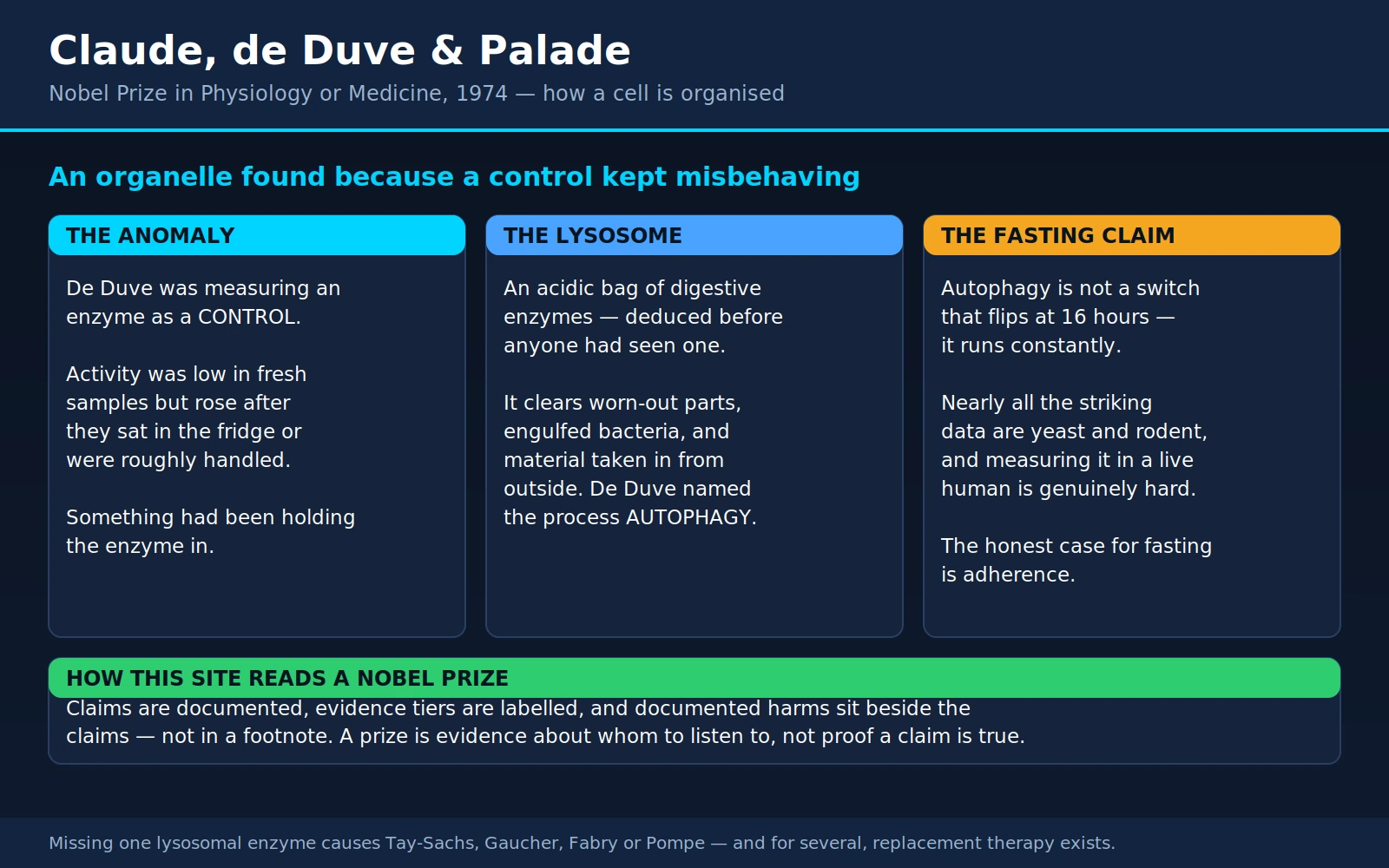
Table of Contents
- Overview: The 1974 Prize
- Three Men and a Centrifuge
- The Lysosome, Found by Accident
- What a Lysosome Actually Does
- Lysosomal Storage Diseases
- Palade and the Secretory Pathway
- Autophagy: de Duve Coined the Word
- The Fasting Question
- Chloroquine, Hydroxychloroquine and the Lysosome
- Lysosomes and Neurodegeneration
- Rapamycin, mTOR and the Longevity Claim
- Where Mainstream Medicine Agrees
- What Remains Debated
- Key Research Papers
- Connections
- Featured Videos
1. Overview: The 1974 Prize
In 1974 the Nobel Prize in Physiology or Medicine went to three men — Albert Claude, Christian de Duve and George Emil Palade — "for their discoveries concerning the structural and functional organization of the cell." It is one of the least glamorous citations in the prize's history and one of the most consequential. What those three did, between roughly 1940 and 1970, was turn the cell from a bag of chemistry into a place with rooms.
Before them, a biochemist ground up tissue, measured what enzymes were in the soup, and had almost no way of saying where in the cell any of it had been. Textbook diagrams showed a nucleus, some mitochondria and a lot of hopeful stippling. After them, the cell had a floor plan: manufacturing in the endoplasmic reticulum, packaging and address-labeling in the Golgi, power in the mitochondria, and — the discovery this page is built around — a demolition and recycling department called the lysosome.
That last one belongs to de Duve, and it is the reason this page exists on a health site rather than only in a cell-biology course. The lysosome is where more than seventy inherited diseases go wrong. It is where hydroxychloroquine does most of its work. It is the destination of the process de Duve named autophagy — the process that the entire modern fasting industry invokes, usually incorrectly. If you have ever read that skipping breakfast "switches on autophagy" and cleans out your cells, you have read a claim about de Duve's organelle. The honest version of that claim is the hardest section on this page, and the most important one: Section 8.
A note on scope. Yoshinori Ohsumi won the 2016 Nobel Prize for working out the genetics of autophagy, and this site has a full page on him: Yoshinori Ohsumi — Autophagy, Fasting, and the Cell's Recycling Program. If you want the molecular machinery — how a cell actually builds a garbage bag, gene by gene — read that page. This page covers the discovery of the compartment, the diseases that come from it, and the practical question of what fasting does and does not do.
2. Three Men and a Centrifuge: How You Study Something You Cannot See
Here is the problem these three solved. A liver cell is about 20–25 micrometres across. Its interior contains thousands of distinct structures, most of them below the resolution of a light microscope, all of them mixed together. You cannot pick one up with tweezers. You cannot ask a chemical assay where its substrate was sitting five seconds ago. So how do you find out that a cell has rooms at all?
Albert Claude (1899–1983), a Belgian working at the Rockefeller Institute in New York, answered that question with a machine and a great deal of patience. In the 1930s he was trying to isolate the infectious agent of the Rous chicken sarcoma — a tumor virus — and in the course of failing to isolate it cleanly, he built something better than what he was looking for: differential centrifugation, a way of breaking cells open gently and then spinning the resulting slurry at successively higher speeds so that the contents settle out in order of size and density. Heavy things pellet first. Lighter things stay in suspension until you spin harder. Do it carefully enough and you get a series of fractions, each one enriched in a different kind of subcellular particle.
Claude published the mature version of the method in 1946, in a two-part paper in the Journal of Experimental Medicine so procedural that it reads like a recipe: how to homogenize liver, in what medium, at what speed, for how long. It is not thrilling prose. It is one of the most productive papers of the century. It also produced a puzzle: one of Claude's fractions contained small particles that were not mitochondria and had no name. He called them microsomes, and they turned out to be fragments of the endoplasmic reticulum — a structure nobody had described yet.
Claude then did the second decisive thing. In 1945, with the physicist Ernest Fullam, he put a thin slice of guinea pig liver in front of an electron microscope — a wartime physics instrument that had never seriously been pointed at an animal cell. The images were crude by later standards and they changed everything, because for the first time you could look at a fraction from the centrifuge and look at a picture of the intact cell and try to match them up.
George Emil Palade (1912–2008), a Romanian physician who joined Claude's laboratory in 1946, spent the next decade making both halves of that method good enough to be believed. He improved the homogenization medium (sucrose rather than water, which stopped organelles from bursting), and he attacked the far harder problem of preparing tissue for electron microscopy without destroying it — better fixatives, better embedding, thinner sections. The result was that by the mid-1950s a picture of a cell was no longer an artifact you argued about; it was data.
With that in hand, Palade found the ribosome. In 1955 he described "a small particulate component of the cytoplasm" — dense granules, about 15 nanometres across, rich in RNA, studding the surface of the endoplasmic reticulum. They were called Palade granules for a while. They are the machines that build every protein in your body.
Christian de Duve (1917–2013) was the third kind of scientist in this story: not an instrument builder but a biochemist with a physician's training and a specific medical question. He was in Louvain, Belgium, trying to understand how insulin works on the liver — a question that had led him to an enzyme called glucose-6-phosphatase. He adopted Claude's centrifugation method not to survey the cell but to purify one enzyme. What he found instead is the next section.
The point worth pausing on: in this story the method was the discovery. Claude did not set out to find the endoplasmic reticulum; he set out to build a centrifugation protocol, and the organelles fell out of it. De Duve did not set out to find the lysosome; he was fractionating liver for an unrelated reason. Palade's ribosome came out of a fixation technique. This is a pattern worth recognising, because it is the opposite of how discovery is usually narrated. Nobody in 1945 could have written a grant proposal to discover the lysosome, since nobody knew there was one. What you could propose was a better way of looking — and the better way of looking then told you what to find.
3. The Lysosome, Found by Accident
This is a genuinely good story, and it survives being told accurately, so let us tell it that way.
In the late 1940s de Duve wanted to purify hepatic glucose-6-phosphatase, the liver enzyme that releases glucose into the blood. Like any careful biochemist, he ran a comparison enzyme alongside it — acid phosphatase, a well-characterised, thoroughly boring enzyme included as a control so he could tell whether his fractionation was behaving sensibly.
The control misbehaved. In freshly prepared liver homogenates, acid phosphatase activity came out far lower than it should have. That alone might have been written off as a bad batch of reagent or a failed prep. But then de Duve's group noticed something stranger: when the same preparation was left in the refrigerator for several days and re-assayed, the activity had climbed to the level everyone expected. Vigorous mechanical treatment — putting the sample through a blender, or repeated freezing and thawing — did the same thing much faster.
The enzyme was not being made. It had been there the whole time. Something had been keeping it away from its substrate, and days in the cold, or rough handling, broke that something open.
De Duve called the property latency, and he read it correctly: the enzyme was sealed inside a membrane-bounded particle. Detergents released it. Osmotic shock released it. Anything that damaged a membrane released it. And when his group looked for other enzymes with the same behaviour, they found a whole set of them — acid phosphatase, cathepsin (a protease), acid ribonuclease, acid deoxyribonuclease, β-glucuronidase — all latent, all with acidic pH optima, all sedimenting together in the same centrifuge fraction, distinct from the mitochondria.
Five different digestive enzymes, all locked in the same bag. That was the 1955 paper in the Biochemical Journal — the sixth in a numbered series called "Tissue fractionation studies," which is worth noting because the series is often cited loosely and the parts are not interchangeable. Part 6 is the one that lays out the intracellular distribution patterns and names the particle. De Duve called it the lysosome: the lytic body, the digestive body.
He had deduced an organelle nobody had ever seen. The first electron micrographs of the isolated fraction came a year or so later, from Alex Novikoff, who was sent de Duve's material to photograph. The pictures confirmed a population of dense, membrane-bounded bodies. But the reasoning came first, from a control enzyme that would not behave.
Fifty years later, de Duve wrote a short retrospective in Nature Cell Biology whose closing observation is the moral of the whole thing: the discovery "could not have happened if strict adherence to a previously set programme had been mandatory." He was looking for one enzyme and found a compartment, because he stopped to explain an anomaly in a control instead of discarding it.
That is worth saying plainly, because it is a general lesson and not a charming anecdote: the finding was in the control. The experiment de Duve designed did not work. The reagent he included only for comparison did something inexplicable, and he chased it. Most of the time a misbehaving control really is a bad reagent. Occasionally it is a Nobel Prize.
4. What a Lysosome Actually Does
Picture a small sac, roughly a quarter to a half micrometre across, wrapped in a single membrane. There are hundreds of them in a typical cell. Inside is an acidic soup: pH around 4.5 to 5.0, roughly the acidity of tomato juice, and several hundred times more acidic than the fluid surrounding it in the cell (which sits near pH 7.2). The acidity is not incidental; it is actively maintained by a proton pump in the lysosomal membrane that spends energy pushing hydrogen ions in.
Floating in that acid are around sixty different hydrolytic enzymes — proteases that cut proteins, lipases that cut fats, nucleases that cut DNA and RNA, glycosidases that cut sugars, sulfatases, phosphatases. Between them they can take essentially any biological molecule apart into its component pieces, which are then exported back into the cell to be reused.
Three kinds of material arrive:
- Things from outside. The cell engulfs droplets of fluid or specific molecules bound to surface receptors, and those vesicles mature into, and fuse with, lysosomes. This is how a cell takes up cholesterol carried on LDL particles, and how it recovers iron from transferrin.
- Things it has eaten deliberately. Immune cells — neutrophils and macrophages — swallow bacteria whole into a phagosome, which then fuses with lysosomes. The acid and the enzymes are the kill mechanism. Some pathogens, notably Mycobacterium tuberculosis and Legionella, survive by blocking exactly that fusion step.
- Its own worn-out parts. A mitochondrion that is no longer producing ATP properly, a tangle of misfolded protein, a stretch of surplus endoplasmic reticulum — these get wrapped in a membrane and delivered to a lysosome. That is autophagy, and it is the subject of the next several sections.
Why the acid is a safety feature
It is reasonable to be alarmed by the idea of hundreds of bags of protein-digesting enzymes floating around inside your cells. The design answer is elegant: lysosomal enzymes are built to work in acid and work badly outside it. A cathepsin that is fully active at pH 4.7 loses most of its activity at the pH 7.2 of the surrounding cytoplasm. So a small leak — and small leaks presumably happen constantly — is not a catastrophe. The enzymes spill into an environment where they are largely inert, and are cleaned up.
This is not an absolute guarantee. Large-scale rupture of many lysosomes at once — what the literature calls lysosomal membrane permeabilization — genuinely can kill a cell, and is a recognised mechanism of cell death in some kinds of injury. But it takes a real insult, not an ordinary leak.
"Suicide bags": a nickname that did damage
In the early 1960s the lysosome acquired a memorable nickname — the cell's "suicide bag" — and the phrase is attributed to de Duve himself. It caught on immediately, because it is a wonderful phrase, and it gave a generation of readers a picture of the organelle that was mostly wrong.
Two things are worth separating here. The hypothesis behind the nickname was a real early-1960s idea: that when a cell needs to die — in normal development, or in disease — its lysosomes rupture and digest it from within. That idea was reasonable at the time, and it did not survive. Programmed cell death in animals turned out to run mostly through a completely different system, the caspases, which won its own Nobel Prize in 2002. Lysosomal rupture contributes to some kinds of cell death but is not the general mechanism.
The picture the nickname left behind was worse: a lysosome as an ominous, dangerous, largely destructive thing that a cell keeps at arm's length. What the last few decades established is close to the opposite. The lysosome is one of the busiest and most constructive structures in the cell — the hub through which nutrients are recovered and recycled, and, as it turned out, a signalling centre. The master nutrient sensor of the cell, mTORC1, physically docks on the outer surface of the lysosome and reads the amino acid concentration inside it. A dedicated transcription factor, TFEB, sits on the lysosomal surface and, when nutrients run short, moves to the nucleus and turns on a whole gene network that builds more lysosomes. This is why one modern review of the field is simply titled "The awesome lysosome", and describes the shift in view "from a dead-end organelle to a control center of cell metabolism."
Keep that in mind for Section 8. The organelle that fasting content describes as a garbage incinerator is, in current understanding, closer to the cell's fuel gauge.
5. Lysosomal Storage Diseases: The Direct Clinical Payoff
The logic here is simple enough to state in one sentence, and it is one of the cleanest examples of basic science producing medicine.
If a lysosome is missing one of its sixty enzymes, the molecule that enzyme was supposed to break down does not get broken down. It accumulates. Year after year, in whichever cells handle the most of it, until those cells stop working.
That is a lysosomal storage disease. There are more than seventy of them, almost all inherited, most of them autosomal recessive. Individually each is rare. Collectively they affect roughly 1 in 5,000 live births — which is not rare at all. And they were essentially uninterpretable before 1955, because you cannot understand a disease of an organelle before you know the organelle exists.
Five you may have heard of
- Tay-Sachs disease — the missing enzyme is hexosaminidase A; the molecule that piles up is GM2 ganglioside, a fatty molecule found in enormous quantity in neurons. Infants develop normally for about six months and then lose the abilities they had gained. There is a characteristic cherry-red spot at the back of the eye. The infantile form is fatal, usually in early childhood. Carrier frequency is elevated in Ashkenazi Jewish, French-Canadian, Cajun and Old Order Amish populations, and voluntary carrier screening programmes begun in the 1970s reduced the number of affected births dramatically — one of the great successes of genetic counselling.
- Gaucher disease — the missing enzyme is glucocerebrosidase, encoded by the gene GBA1. Glucocerebroside accumulates mainly in macrophages, which swell into distinctive "Gaucher cells" and crowd the spleen, liver and bone marrow. The result is a hugely enlarged spleen and liver, low platelets and easy bruising, anaemia, and bone disease — painful crises, thinned bone, avascular necrosis of the hip. Type 1, the most common form, spares the brain. Types 2 and 3 do not. Remember GBA1; it reappears in Section 10 in an unexpected place.
- Fabry disease — the missing enzyme is α-galactosidase A, and the gene is on the X chromosome. Globotriaosylceramide accumulates in the cells lining small blood vessels. It typically begins in childhood with burning pain in the hands and feet, an inability to sweat normally, and clusters of small dark red skin lesions; over decades it damages the kidneys, thickens the heart muscle, and causes stroke at unusually young ages. Because of X-inactivation, women who carry one copy are frequently affected too — sometimes seriously — which was underappreciated for years.
- Pompe disease — the missing enzyme is acid α-glucosidase, which breaks down glycogen inside the lysosome. Glycogen accumulates in muscle. The infantile form causes a profoundly weak, floppy baby with a massively thickened heart, and before treatment existed it was almost uniformly fatal in the first year of life. The late-onset form can appear at any age as slowly progressive weakness of the hips, shoulders and, critically, the diaphragm — so the first sign is sometimes breathlessness lying flat, or sleep-related breathing trouble, in an adult who has been called unathletic their whole life.
- Niemann-Pick disease — and this one carries an important technical distinction. Types A and B are a true enzyme deficiency: acid sphingomyelinase, now usually called ASMD. Type A is a severe infantile neurological disease; type B is a chronic disorder of the liver, spleen and lungs that people can live with for decades. Type C is a different disease that kept the name for historical reasons. It is not an enzyme deficiency at all — it is a defect in the proteins that move cholesterol out of the lysosome. It presents with clumsiness, a distinctive paralysis of vertical eye movement, and sudden loss of muscle tone triggered by laughter. Grouping it with A and B causes real confusion, including about which treatments could possibly apply.
Enzyme replacement therapy: the genuinely uplifting part
De Duve did not only find the compartment. In the 1960s he proposed the obvious-in-hindsight therapeutic idea: if the problem is a missing enzyme, give the enzyme back.
The obstacle was delivery. An enzyme injected into the bloodstream has to get inside cells, and then into the lysosome specifically. What made it possible was the discovery of the cell's own addressing system for lysosomal enzymes — a sugar tag, mannose-6-phosphate, attached in the Golgi, which acts as a postal code that receptors recognise and route to the lysosome. (Elizabeth Neufeld's work on that recognition marker is one of the reasons this therapy exists; she is a co-author of the standard modern review of these diseases.)
The first success came in Gaucher disease. Roscoe Brady's group at the NIH took glucocerebrosidase and chemically trimmed its sugar chains to expose mannose — because the cells that most needed the enzyme were macrophages, and macrophages carry a mannose receptor that would haul it in. The 1991 New England Journal of Medicine report of that trial is the founding document of enzyme replacement therapy: patients' spleens and livers shrank, haemoglobin rose, platelet counts recovered. Children who had been transfusion-dependent got better.
Enzyme replacement now exists for Gaucher type 1, Fabry, Pompe, several of the mucopolysaccharidoses, and ASMD, among others. For some of these it is transformative. Infantile Pompe went from a disease that essentially always killed babies in their first year to one where long-term survival is routine. In Fabry, the 2001 randomised trial showed clearance of the accumulated lipid from kidney, heart and skin blood vessels — the storage material physically went away.
Two other approaches have joined it: substrate reduction therapy, which uses a small molecule to slow production of the material upstream so less of it needs clearing, and chaperone therapy, which uses a small molecule to help a partly-functional mutant enzyme fold correctly — useful only for people whose specific mutation produces an enzyme worth rescuing.
Newborn screening, and why it matters here
Several of these diseases are now on newborn dried-blood-spot screening panels in the United States and in a growing number of other countries — Pompe and MPS I most widely, with Fabry, Gaucher and Krabbe screened in some jurisdictions and large programmes elsewhere covering six lysosomal disorders at once. The reason is not curiosity. In infantile Pompe, the difference between starting enzyme replacement at three weeks of age and starting it at six months — after the heart has thickened and motor milestones have been lost — is enormous, because you cannot un-damage tissue that has already been destroyed. Screening buys the only thing that helps: time before symptoms.
Now the honest limits
Two of them, and they are severe.
Cost. Enzyme replacement is among the most expensive medicine that exists. Annual costs commonly run into the hundreds of thousands of dollars per patient, the infusions are lifelong, and there is no version of these drugs that becomes cheap when the patent expires, because they are complex proteins manufactured in living cells, not pills. Families navigate insurance appeals, lifetime caps, and coverage that can change when a job changes. Globally, most people with a treatable lysosomal storage disease do not receive treatment, and the reason is price.
The blood-brain barrier. An enzyme is a large protein. Large proteins do not cross from blood into brain. So enzyme replacement given intravenously treats the liver, spleen, kidney, heart and muscle — and does essentially nothing for the nervous system. This is why Tay-Sachs has no enzyme replacement therapy that helps, why Gaucher types 2 and 3 remain far harder than type 1, why Niemann-Pick type A is untreated while type B responds, and why the neurological forms across this whole family of diseases remain the great unsolved problem.
Work-arounds exist and are advancing: delivering enzyme directly into the cerebrospinal fluid (this is how the approved treatment for CLN2 Batten disease is given), haematopoietic stem-cell transplantation early enough that donor-derived cells populate the brain, small molecules that are small enough to cross, and gene therapy. None of this is settled. It is the frontier, and it is where the field's effort is going.
6. Palade and the Secretory Pathway
Palade's half of the 1974 prize deserves its own section, briefly, because it is the other half of the same map — and because it explains how the lysosome gets its enzymes in the first place.
Working with pancreatic cells, which do almost nothing but manufacture and export digestive enzymes, Palade and his colleagues used a technique called pulse-chase autoradiography: feed the cells radioactively labelled amino acids for a few minutes, then wash them out, then take electron micrographs at intervals and see where the radioactivity is. The label moved. First it was over the ribosomes and the rough endoplasmic reticulum. A little later, the Golgi apparatus. Later still, the secretory granules near the cell surface. Then it was gone — secreted.
That traced out the secretory pathway, one of the fundamental circuits of biology:
ribosome → endoplasmic reticulum → Golgi apparatus → transport vesicle → cell surface
It matters practically because this is the route taken by nearly everything your body sends out of a cell. Insulin travels it, from ribosome to secretory granule in the pancreatic beta cell. Antibodies travel it, in plasma cells. So do digestive enzymes, clotting factors, most hormones, collagen, and the receptors that end up embedded in your cell membranes. It is also the machinery that biotechnology hijacks: a monoclonal antibody drug is made by engineered cells running Palade's pathway in a bioreactor.
Two later Nobel Prizes finished the picture and are worth knowing as the same story. In 1999, Günter Blobel was recognised for the "signal hypothesis" — the discovery that a protein destined for export carries a short address tag in its own sequence that directs it into the endoplasmic reticulum in the first place. In 2013, James Rothman, Randy Schekman and Thomas Südhof shared the prize for working out the machinery of vesicle traffic: how a vesicle knows where to go, and how it fuses with the right target membrane at the right moment — which is, among other things, how a nerve cell releases a neurotransmitter in under a millisecond.
And here is the link back to de Duve. A lysosomal enzyme is made on the same assembly line as a secreted one. What decides whether it goes out of the cell or into a lysosome is a single sugar modification added in the Golgi — the mannose-6-phosphate tag mentioned above. When the enzyme that attaches that tag is itself defective, the consequence is a disease called mucolipidosis II, or I-cell disease: the lysosomal enzymes get made perfectly well and then are secreted into the bloodstream instead of being delivered, so a patient's lysosomes are empty of enzymes while their plasma is full of them. Palade's pathway and de Duve's organelle are not two discoveries. They are two ends of the same one.
7. Autophagy: de Duve Coined the Word
In the early 1960s, electron microscopists kept photographing something odd inside cells: lysosome-like bodies containing recognisable pieces of the cell itself — a mitochondrion, a fragment of endoplasmic reticulum — visibly being digested. De Duve gave the process its name at a 1963 symposium: autophagy, from the Greek for "self-eating." He coined the word, and his laboratory did the first biochemistry showing that lysosomes were genuinely responsible for it.
The basic form, macroautophagy, works like this. A flat sheet of membrane appears in the cytoplasm, near whatever is to be disposed of. It curves, extends around the cargo like two hands closing, and seals into a double-membraned bubble called an autophagosome. That bubble then finds a lysosome and fuses with it. The acid and the enzymes do the rest, and the resulting amino acids, fatty acids and sugars are pumped back out into the cell to be used again.
There are variants — microautophagy, where the lysosome membrane simply dimples inward and swallows something directly; chaperone-mediated autophagy, where individual proteins bearing a particular sequence are recognised and threaded across the membrane one at a time. And there are selective forms named for their cargo: mitophagy for worn-out mitochondria, lipophagy for fat droplets, xenophagy for invading bacteria.
The genetics of how a cell actually assembles that membrane — the ATG genes, discovered in yeast — is Yoshinori Ohsumi's work, and it won the 2016 Nobel Prize. It is covered in detail on our page on Ohsumi, which is where to go for the mechanism.
The single most important fact about autophagy
Everything in the next section depends on this, so it gets its own heading.
Autophagy is constitutive. It runs all the time, in every cell of your body, at this moment. It is not a program that starts when triggered and stops when the trigger is removed. It is a continuous housekeeping process with a baseline rate that is never zero in a living cell, and that baseline is modulated up and down by circumstances — nutrient availability, energy status, exercise, infection, damage, hormones.
The proof that basal autophagy is continuous and essential is genetic, and it is unambiguous. Mice engineered to lack the core autophagy genes do not develop a mild deficiency — newborns die within the first day of life, during the brief starvation between the loss of the placental nutrient supply and the establishment of feeding. Mice in which autophagy is switched off only in neurons develop progressive neurodegeneration and accumulate protein inclusions in their brains, without any external stress at all. A cell that stops recycling does not coast; it fills up with wreckage.
The regulation runs through the nutrient sensors. mTORC1 — sitting on the lysosomal surface, as described above — is active when amino acids and insulin are plentiful, and while it is active it holds the autophagy initiation machinery in check. AMPK, which senses low cellular energy, pushes the other way. Eating raises mTORC1 activity and dampens autophagy; not eating relieves that brake. This part is not controversial and it is the seed of truth inside the fasting claims.
But notice the shape of it: a dial with a floor, not a switch with an off position. Hold onto that sentence.
8. The Fasting Question: What the Evidence Actually Supports
This is the section this page was built for, and it is the one where careful reading pays off, because the popular claim and the evidence have drifted a long way apart.
The popular claim, in its most common form: fasting for some threshold number of hours — 16 is the usual number, sometimes 12, sometimes 24 — "switches on autophagy," which then cleans damaged material out of your cells, and this confers protection against ageing and cancer. You will find hour-by-hour timelines: glycogen depleted at hour 12, autophagy "activated" at hour 16, "peak autophagy" at 24 to 72 hours.
Let us take that apart in order, and then be fair to fasting, because the conclusion is not that fasting is worthless. It isn't.
(i) Autophagy is not a switch, so "turning it on" is not a meaningful statement
From the previous section: autophagy runs continuously in every cell you own. It was running while you ate lunch. It is running now. What changes with fasting is the rate, and the rate is graded, tissue-specific and continuously variable.
So "autophagy switches on at 16 hours" is not merely unproven — it describes the wrong kind of process. It is like saying your kidneys "switch on" when you drink water. The relevant question is never whether autophagy is happening; it is how much, in which tissue, compared to what, and whether the difference is large enough to matter for anything you can measure in a person.
Nobody has established an hour threshold in humans. The specific numbers in the popular timelines are not measurements from people. Where they come from is the next point.
(ii) The central problem: nearly all the striking data are from animals that are not us
This is the single most important thing on this page.
The dramatic findings that made autophagy famous — lifespan extension, protection against neurodegeneration, resistance to metabolic disease — come overwhelmingly from yeast, nematode worms, fruit flies and mice. Those results are real, well replicated and scientifically important. They are also from organisms whose relationship to food is not remotely like ours.
A mouse has a mass-specific metabolic rate several times higher than a human's — gram for gram, it burns through its energy reserves far faster. Its heart beats around 500 to 600 times a minute. It eats a substantial fraction of its body weight every day, and it eats nearly continuously through the dark phase. A mouse deprived of food for 24 hours has lost something on the order of a tenth of its body weight and is in a genuine physiological crisis. A human being who skips food for 24 hours has lost a small fraction of that proportionally, and is hungry.
These are not the same experiment, and no correction factor converts one into the other. When a paper reports that a 24-hour fast produced a large increase in autophagic markers in mouse liver, the honest translation is not "so a 24-hour fast does that in you." It is closer to "a severe, whole-body starvation stress does that in a mouse liver, and we do not know what the corresponding stress or the corresponding response is in a human."
The rodent data are also less uniform than the summaries suggest. In the standard experiment where mice are engineered so autophagosomes glow under a microscope, fasting lit up liver, muscle, heart and pancreas — and conspicuously not the brain, which is the organ most of the anti-ageing marketing is implicitly about.
None of this means human autophagy does not respond to fasting. It very likely does; the machinery is the same and the nutrient sensors are the same. It means the magnitude, the timing and the consequences in a human being are open questions, and that a number lifted from a mouse and printed on an infographic is not an answer to them.
(iii) Measuring autophagy in a living human is genuinely, technically hard
People are often surprised by this. Surely, if the claim is so widespread, someone has measured it?
Here is the problem. The standard markers are:
- LC3-II — a protein that gets attached to the autophagosome membrane, so the amount of it tracks the number of autophagosomes present.
- p62 / SQSTM1 — a cargo-adaptor protein that is itself destroyed along with the cargo, so its level tends to fall when autophagy is running well.
Both are snapshots of a pipeline, and a snapshot cannot tell you the direction of flow. A high LC3-II level can mean autophagosomes are being made faster — or that they are being made at the usual rate and cleared more slowly because the lysosome step is blocked. Those are opposite situations with the same reading. It is the difference between a busy loading dock and a jammed one: both have a lot of boxes sitting on them.
To resolve it, researchers measure flux rather than level: run the measurement twice, once normally and once with a drug that blocks the lysosomal step, and the gap between them tells you how much material was actually getting through. That works beautifully in a dish. You cannot dose a human volunteer with a lysosome-blocking drug to find out. So human flux measurement has to be done on cells taken out of the person — usually white blood cells — and treated in the laboratory, which introduces its own assumptions about whether blood cells represent liver, muscle or brain.
The field is fully aware of all this. The reference document, Guidelines for the use and interpretation of assays for monitoring autophagy, is a community consensus written by well over two thousand authors and it runs to 382 pages in its fourth edition. Its central and repeated instruction is that measuring a marker level is not measuring autophagy. A field does not produce a 382-page methods manual for something that is easy to get right.
So what have human studies actually measured? Two are worth naming precisely, because they are the ones usually cited — and because what they did is not what they are usually said to have done.
- Jamshed and colleagues, 2019 (Nutrients). Eleven overweight adults, in a four-day randomised crossover, ate either 8am–2pm or 8am–8pm. Early time-restricted eating improved 24-hour glucose control — a solid, useful result. On autophagy, what the study measured was gene expression — messenger RNA for LC3A in whole blood cells, which rose in the morning before breakfast. That is a transcript level in circulating blood cells, not autophagosome counts, and certainly not flux. And in the same study, evening expression of MTOR — the gene for the brake on autophagy — went up. This is an 11-person, 4-day study of RNA levels, and it is routinely cited as demonstrating that fasting activates autophagy in humans.
- Bensalem and colleagues, 2025 (Journal of Physiology). This is the best human attempt so far and deserves to be described exactly. 121 people with obesity were randomised for six months to standard care, calorie restriction, or intermittent fasting plus time-restricted eating, and the investigators measured genuine autophagic flux (LC3B-II in peripheral blood mononuclear cells within whole blood). Result: at two months, no significant difference between groups. At six months, a significant difference between the fasting group and standard care — P = 0.04, in a post-hoc analysis. But there was no significant increase from baseline within the fasting group itself, and the authors note the between-group gap may be partly driven by autophagy declining in the standard-care controls. Calorie restriction alone did not differ significantly from standard care. The paper's own title says autophagy "may increase," and its own key-points section says "further studies are required."
That is the top of the evidence: a well-designed, six-month, 121-person trial that measured the right thing and returned a borderline post-hoc between-group difference with no within-group increase. It is a real and encouraging finding. It is not "fasting for 16 hours turns on autophagy."
And there is no test you can buy. There is no blood panel, no urine test, no wearable that tells you your autophagy is "on." If something is marketed to you on that basis, it is measuring something else, or nothing.
(iv) What intermittent fasting has been shown to do in randomised human trials
Now the good news, which is that this question has actually been answered properly, several times, in decent trials. Three of them are worth knowing in detail.
Trepanowski and colleagues, 2017 (JAMA Internal Medicine). One hundred adults with obesity, randomised for a full year to alternate-day fasting (25% of energy needs on fast days, 125% on alternating "feast days"), daily calorie restriction (75% of needs every day), or no intervention. Six months of weight loss, then six months of maintenance.
- Weight loss was essentially identical: −6.8% in both intervention arms at 6 months; −6.0% (fasting) versus −5.3% (calorie restriction) at 12 months.
- Dropout was highest in the fasting group — 38%, versus 29% for daily calorie restriction and 26% for controls.
- Participants assigned to alternate-day fasting ate more than prescribed on fast days and less than prescribed on feast days — they converged toward ordinary eating on their own.
- No significant differences between the two diets in blood pressure, heart rate, triglycerides, fasting glucose, fasting insulin, insulin resistance, C-reactive protein or homocysteine.
- LDL cholesterol was significantly higher in the fasting arm at 12 months (by about 11.5 mg/dL) than in the calorie-restriction arm.
Lowe and colleagues, 2020 — the TREAT trial (JAMA Internal Medicine). 116 adults with overweight or obesity randomised for 12 weeks to 16:8 time-restricted eating (all food between noon and 8pm) or three structured meals a day.
- The time-restricted group lost weight within-group (−0.94 kg), but the difference between groups was not significant: −0.26 kg, 95% CI −1.30 to 0.78, P = 0.63.
- In the 50-person in-person subgroup with body-composition measurement, appendicular lean mass index fell significantly more in the time-restricted group (−0.16 kg/m², P = 0.005) — suggesting a meaningful share of what was lost was muscle rather than fat. This is a secondary outcome in a small subgroup and later trials have been mixed, so treat it as a signal deserving attention rather than a settled fact. But it is a signal in the direction nobody wants, and it matters most for exactly the people most likely to try this: older adults.
- The authors' conclusion: time-restricted eating "is not more effective in weight loss than eating throughout the day."
Liu and colleagues, 2022 (New England Journal of Medicine). 139 adults with obesity in Guangzhou, China, randomised for 12 months to time-restricted eating (8am–4pm) plus calorie restriction, or the identical calorie restriction alone — 1,500–1,800 kcal/day for men, 1,200–1,500 for women in both arms. This is the cleanest design of the three, because it holds calories constant and varies only the window.
- Weight loss at 12 months: −8.0 kg with the eating window, −6.3 kg without. Net difference −1.8 kg, 95% CI −4.0 to 0.4, P = 0.11 — not statistically significant.
- Waist circumference, BMI, body fat, lean mass, blood pressure and metabolic risk factors all told the same story.
- Retention was good in both arms (85% completed).
The pattern across all three is consistent and it is not subtle. Three different fasting protocols, three different countries, durations from 12 weeks to 12 months. In every case, fasting produced weight loss roughly comparable to plain continuous calorie restriction, and in no case did it produce a clear advantage. When calories were matched, the eating window added nothing detectable.
That is not a negative result about fasting. It is a specific result: the calories do the work, and the schedule is a delivery mechanism for the calories. The interesting variable is therefore not metabolism — it is adherence. Which schedule can a given person actually keep? And there the trials cut both ways: alternate-day fasting had the highest dropout rate of anything tested, while plenty of people find "don't eat before noon" the first dietary rule they have ever managed to follow, precisely because it requires no counting, no weighing and no decisions.
(v) Who should not fast
This list is not boilerplate. Fasting is a real physiological intervention and for some people it is genuinely dangerous. Please read it.
- Anyone with type 1 diabetes, or with type 2 diabetes taking insulin or a sulfonylurea (glipizide, glyburide, glimepiride). These medications lower blood sugar whether or not you have eaten, so removing the food without adjusting the dose risks severe hypoglycaemia. This is not a reason it can never be done — it is a reason it requires a clinician adjusting doses in advance, not a decision made on a Sunday night. SGLT2 inhibitors carry a separate concern: prolonged fasting on these drugs can precipitate ketoacidosis even with normal blood glucose.
- Pregnancy and breastfeeding.
- Anyone with a history of an eating disorder, or currently disordered eating. Rule-based restriction, permitted and forbidden hours, and the language of "clean" and "cleansing" map directly onto anorexic and bulimic thinking, and fasting protocols reliably reactivate them. See Eating Disorders.
- Anyone underweight (roughly BMI under 18.5), or losing weight without trying — which needs investigating, not accelerating.
- Children and adolescents, who are growing.
- Frail or sarcopenic older adults. The muscle-loss signal in the TREAT trial matters most in the people who have the least muscle to spare, and losing muscle in later life costs mobility and independence.
- Anyone taking medication that must be taken with food, and anyone whose medication schedule depends on regular meals.
- Reasonable caution also applies to advanced kidney or liver disease, a history of gallstones (rapid weight loss raises gallstone risk), gout, a history of fainting while fasting, and any job where a hypoglycaemic episode would be dangerous — driving, operating machinery, working at height.
(vi) The fair conclusion
Fasting is a legitimate way to eat less. For a meaningful number of people it is the most sustainable way to eat less, because it replaces continuous negotiation with a single simple rule, and simple rules are easier to keep than arithmetic. If closing the kitchen at 8pm is what stops the second helping and the evening snacking, that is a genuine result, and the weight-loss trials confirm it works about as well as any other approach that reduces intake.
The honest case for fasting is adherence. It is not a molecular switch.
And that reframing should be freeing rather than deflating. It means you do not need to hit hour sixteen. It means a fast that ends at hour fourteen because you were hungry has not failed at anything. It means the schedule is a tool you choose because it fits your life, and you can stop using it when it doesn't — rather than a metabolic ritual you have to perform exactly right to receive a benefit nobody has been able to measure in a person.
If what you actually want is a lever on cellular housekeeping with proven human outcomes, the best-supported one is not a clock. It is exercise, which induces autophagy in animal models, shows marker changes in human muscle biopsies, and — unlike any fasting schedule — has decades of randomised evidence for outcomes that matter.
9. Chloroquine, Hydroxychloroquine and the Lysosome
This is a genuinely interesting piece of pharmacology, and it belongs on this page because it is the clearest example of a widely-used drug whose whole mechanism runs through de Duve's organelle. We will keep this level and non-polemical: this site's readers include people who hold strong and opposite views about one particular episode in this drug's history, and the mechanism is the part everybody can learn something from.
How the drug gets where it goes
Chloroquine and hydroxychloroquine are lysosomotropic weak bases, and the phrase describes a rather beautiful physical trick.
In the neutral environment of the bloodstream and cytoplasm, the drug is largely uncharged, and uncharged molecules pass through membranes easily. It therefore drifts freely into cells and into lysosomes. But a lysosome's interior is acidic, and in acid the drug picks up protons and becomes charged. A charged molecule cannot cross a membrane. So it is stuck — and more uncharged drug keeps drifting in behind it to be trapped in turn.
The result, called ion trapping, is that these drugs concentrate inside lysosomes to hundreds of times the concentration outside the cell. And because trapping protons is exactly what happens, the drug also raises the lysosome's pH — de-acidifying the compartment. Every acid-dependent enzyme inside it works less well.
That single mechanism accounts for most of what these drugs do.
In malaria
The malaria parasite, living inside a red blood cell, eats haemoglobin. It digests it in an acidic compartment of its own, and that generates a toxic by-product — free haem — which the parasite neutralises by crystallising it into an inert pigment. Chloroquine accumulates in that acidic digestive vacuole by the trapping mechanism above and interferes with the crystallisation step, so free haem builds up and poisons the parasite. Resistance, when it emerged, works by exactly the route you would predict: mutations in a transporter that pumps the drug back out of the vacuole before it can act. (For the other great antimalarial story, see Tu Youyou and artemisinin.)
In lupus and rheumatoid arthritis
This is where hydroxychloroquine earns its place in modern medicine, and the mechanism again runs through acidified compartments.
Certain immune sensors — Toll-like receptors 7 and 9 — do not sit on the cell surface. They sit inside endosomes, and they detect nucleic acids. They also require an acidic environment to function. In lupus, these sensors are chronically triggered by the body's own DNA and RNA released from dying cells, and the result is a persistent flood of type I interferon, which is central to the disease. Raise the pH of those compartments and you blunt the sensing step. Hydroxychloroquine also interferes with the acid-dependent processing of antigens for presentation to T cells.
Does it work? Yes, and the cleanest demonstration is old and elegant: the Canadian Hydroxychloroquine Study Group trial published in the New England Journal of Medicine in 1991, which took patients with stable lupus and randomised them to continue the drug or switch to placebo. The ones who stopped flared substantially more often. Hydroxychloroquine is now background therapy for essentially every lupus patient who can take it, associated with fewer flares, less accumulated organ damage and better survival. In rheumatoid arthritis it is a milder disease-modifying drug, generally used in combination.
The limiting toxicity is also a lysosomal story. Over years, the drug accumulates in the retina and produces a characteristic, irreversible retinopathy. The risk is dose- and duration-dependent, which is why current practice keeps the daily dose within a weight-based limit and adds annual eye screening with retinal imaging after about five years of use, earlier in kidney impairment. This is manageable and monitored, not a reason to avoid a drug that works — but it is a real reason the eye appointments are not optional. See our page on Hydroxychloroquine and Antimalarials for Lupus.
In cancer: autophagy inhibition, still unproven
Now the connection back to Section 7. If the lysosome is where autophagy ends, then de-acidifying the lysosome blocks autophagic flux — the autophagosomes still form, they still fuse, but the cargo does not get digested. Hydroxychloroquine is therefore one of the few practical autophagy inhibitors available for use in people.
That mattered because many tumours appear to lean on autophagy to survive nutrient-poor conditions and treatment stress. The strategy — take away the recycling and the tumour cannot tolerate the therapy — is a good idea, and it has been tested in a substantial number of early-phase trials, usually combining hydroxychloroquine with chemotherapy or targeted agents.
Results have been modest and inconsistent. A recurring technical problem is that it has proved difficult to confirm reliable, sustained autophagy inhibition inside a human tumour at doses people tolerate — which brings us straight back to the measurement problem in Section 8(iii). Autophagy inhibition in cancer remains an active, legitimate research strategy. It is not established therapy, and it is not something to pursue outside a trial. (For where cancer genes came from in the first place, see Bishop and Varmus.)
In COVID-19: the trials were negative
The rationale in early 2020 was mechanistically reasonable and worth stating fairly, because it was not invented out of nothing. Coronaviruses enter cells through acidified endosomes; raising endosomal pH impairs that entry in cell culture; and the drug's immunomodulating properties looked potentially relevant to the inflammatory phase of severe disease. Laboratory data supported testing it. Testing it was the right call.
The trials were then done, at large scale, and the answer was no.
The RECOVERY trial randomised 1,561 hospitalised patients to hydroxychloroquine and 3,155 to usual care. Death within 28 days occurred in 27.0% of the hydroxychloroquine group and 25.0% of the usual-care group (rate ratio 1.09, 95% CI 0.97–1.23; P = 0.15). Patients given the drug were less likely to be discharged alive within 28 days (59.6% vs 62.9%), and among those not already ventilated at enrolment, more went on to invasive ventilation or death (30.7% vs 26.9%). Enrolment into that arm was closed early for lack of efficacy. A separate Brazilian randomised trial in mild-to-moderate disease found no improvement in clinical status, and the WHO's Solidarity trial discontinued its hydroxychloroquine arm on the same grounds.
That is the outcome, and it is not ambiguous: hydroxychloroquine does not treat COVID-19.
The useful lesson is not about that episode; it is about mechanism. A plausible mechanism is a reason to run a trial. It is never a substitute for one. A drug that concentrates a hundredfold in lysosomes, raises their pH, and inhibits viral entry in a dish can still fail completely in patients — because the concentration achieved in the right tissue at a tolerated dose may be nowhere near what the dish required, because the timing may be wrong, and because a human being is not a monolayer of cells. This is precisely the same reasoning error as "autophagy responds to fasting in mice, therefore a 16-hour window will make you live longer." The mechanism is real in both cases. The clinical claim needs its own evidence.
And the converse deserves saying too, in the same level tone: the fact that a drug failed for one disease says nothing whatsoever about the diseases it does treat. Hydroxychloroquine remains a genuinely valuable, life-improving drug for lupus and a useful one in rheumatoid arthritis, and anyone taking it for those reasons should keep taking it.
10. Lysosomes and Neurodegeneration
This is one of the most active areas in neuroscience, and it needs a clear label before anything else: what follows is mechanism-level evidence about disease causes. It is not treatment, and nothing here should be read as a therapy that exists.
The underlying logic is straightforward. Neurons are the most extreme case of cells that cannot be replaced. Your liver renews itself; the neuron you have at 80 is largely the one you had at 20, and it has spent sixty years accumulating damaged proteins and worn-out mitochondria in a cell body with axons that can be a metre long. A neuron depends more than almost any other cell on continuous, efficient internal recycling — and it has no option to dilute its garbage by dividing.
The Gaucher-Parkinson's connection
This is the striking one, and it is worth telling properly because it is a case where a rare childhood disease illuminated a common one of old age.
Clinicians treating Gaucher disease noticed something over the years: the relatives of their patients — parents and siblings, who each carry one defective copy of GBA1 and are perfectly healthy — seemed to develop Parkinson's disease more often than expected. It was the kind of clinical impression that is usually wrong.
It was right. A 16-centre international study published in the New England Journal of Medicine in 2009 compared 5,691 patients with Parkinson's disease against 4,898 controls and found an odds ratio of 5.43 for carrying a GBA1 mutation. Among Ashkenazi Jewish participants, one of the two common mutations was found in 15% of patients versus 3% of controls; among non-Ashkenazi participants, in 3% versus under 1%. Patients carrying a mutation developed Parkinson's earlier, were more likely to have affected relatives, and more often had atypical features. Full sequencing showed that limited screening for the two common mutations misses about half of the mutant alleles — so the association is, if anything, understated by simple testing.
The same gene. Two broken copies of GBA1 gives you Gaucher disease in childhood. One broken copy gives you the strongest common genetic risk factor known for Parkinson's disease. The organelle in the middle is the lysosome.
The proposed mechanism is a feedback loop: reduced glucocerebrosidase activity impairs lysosomal degradation, so α-synuclein — the protein that aggregates in Parkinson's — is cleared less efficiently and accumulates; accumulated α-synuclein in turn interferes with the trafficking of glucocerebrosidase to the lysosome, reducing its activity further. And GBA1 is not alone: several other Parkinson's genes, including LRRK2, VPS35 and ATP13A2, also turn out to affect the endosomal-lysosomal system. This is now one of the central threads in Parkinson's research. See our page on Parkinson's Disease.
Alzheimer's disease
The lysosomal signal in Alzheimer's is older than most people realise and comes first from microscopy: the swollen neurites surrounding amyloid plaques in Alzheimer's brain tissue are packed with autophagic vacuoles — autophagosomes that formed and then were not cleared. That is the picture of a jammed pipeline, not an idle one, and it appears early in the disease process. Presenilin-1, the gene most commonly mutated in early-onset inherited Alzheimer's, has been implicated in lysosomal acidification, which would sit exactly at the blocked step.
Whether impaired clearance is a cause of Alzheimer's, a consequence of it, or both in a vicious cycle is unresolved. See Alzheimer's Disease.
The caveat that governs this whole section
No drug that enhances autophagy or lysosomal function has been shown in a randomised trial to slow Parkinson's disease or Alzheimer's disease. Chaperone compounds aimed at boosting glucocerebrosidase — ambroxol is the most studied — have reached clinical trials and have produced measurable biomarker changes, which is genuinely encouraging and is the right first step. It is not the same as changing the course of the disease, and that has not been demonstrated.
The gap between "impaired lysosomal function is implicated in this disease" and "improving lysosomal function will treat it" is the entire distance from mechanism to medicine, and it is exactly the gap that Sections 8 and 9 of this page are about. It is not a reason for cynicism — this is how the lysosomal storage diseases went from unexplained to treatable in fifty years. It is a reason not to buy anything today on the strength of it.
11. Rapamycin, mTOR and the Longevity Claim
A short section, because the evidence tier is short to state.
Rapamycin is a compound produced by a soil bacterium collected on Rapa Nui (Easter Island) in the 1960s. It is an approved drug — an immunosuppressant used to prevent kidney transplant rejection, and a treatment for a rare lung disease.
It works by inhibiting mTORC1, and mTORC1, as noted earlier, physically sits on the lysosomal surface reading amino acid availability, and holds the autophagy machinery in check while nutrients are plentiful. Inhibit mTORC1, and autophagy goes up. This is where de Duve's organelle and the entire modern longevity conversation physically meet, on the same membrane.
What is established: in 2009, the NIA Interventions Testing Program reported in Nature that rapamycin, fed to genetically heterogeneous mice starting at 600 days of age — late middle age for a mouse — extended both median and maximal lifespan. Measured by age at 90% mortality, the increase was 14% in females and 9% in males, and the effect replicated at three independent test sites. That is a serious, well-controlled result, and it was the first demonstration that a drug could extend lifespan in a mammal when started late in life. mTOR inhibition extends lifespan in yeast, worms and flies as well.
What is not established: anything at all about human lifespan. There is no randomised trial of rapamycin with a lifespan or healthspan endpoint in humans, and given that such a trial would need decades and thousands of participants, there may never be one in its obvious form. Small trials of rapamycin-family drugs have examined immune endpoints in older adults — vaccine responses, respiratory infections — with some encouraging early results and at least one larger follow-up that missed its primary endpoint. That is the whole human dataset relevant to ageing.
What it costs: rapamycin is not a supplement. It is an immunosuppressant with a real adverse-effect profile — mouth ulcers, impaired wound healing, raised cholesterol and triglycerides, glucose intolerance and new-onset diabetes, low blood counts, an uncommon but serious inflammatory lung reaction, and increased susceptibility to infection. In mice, higher doses cause cataracts and testicular degeneration.
A visible community takes low-dose intermittent rapamycin off-label in the hope of slowing ageing. The plain statement of the evidence tier: strong and replicated animal data, a coherent mechanism, no human outcome evidence, a real drug with real risks, and no established dose or schedule for this purpose. People are entitled to make that bet with their own bodies. They should know that is what it is.
12. Where Mainstream Medicine Agrees
Almost all of this page is uncontroversial. It is worth separating out what is settled, because on a topic this surrounded by marketing it is easy to assume everything is contested.
- Lysosomes exist and are essential. Textbook biology since the 1960s, confirmed by every method since.
- Autophagy is real, continuous, and necessary for life. Genetically demonstrated: animals lacking the core machinery die or degenerate.
- Lysosomal storage diseases are real, diagnosable and, for several of them, treatable. More than seventy disorders, collectively about 1 in 5,000 births.
- Enzyme replacement therapy works for the non-neurological features of Gaucher type 1, Fabry, Pompe, several mucopolysaccharidoses and ASMD — and does not cross into the brain.
- Newborn screening for some of these diseases is now routine in many places, and pre-symptomatic treatment does markedly better than treatment started after damage.
- Hydroxychloroquine is standard, effective therapy in lupus and a useful drug in rheumatoid arthritis, with retinal screening.
- Hydroxychloroquine does not treat COVID-19. Large randomised trials settled this.
- Fasting-based diets produce weight loss. Roughly comparable to continuous calorie restriction, and a legitimate option for people who find them sustainable.
- Impaired lysosomal and autophagic function is genuinely implicated in Parkinson's and Alzheimer's disease at the mechanism level, with GBA1 the strongest common genetic risk factor known for Parkinson's.
- mTOR inhibition extends lifespan in mice — replicated, and started late in life.
13. What Remains Debated
- Whether any specific fasting schedule meaningfully changes autophagy in humans. One six-month, 121-person trial measuring genuine flux found a borderline post-hoc between-group difference and no within-group increase. That is the state of the art.
- Whether fasting's real benefits run through autophagy at all, as opposed to simply eating fewer calories and losing weight. Nothing published distinguishes these.
- Whether time-restricted eating causes clinically meaningful loss of lean mass. One trial found it in a small subgroup; later evidence is mixed; it matters most in older adults.
- Whether any hour threshold exists in humans at all — 12, 16, 24 — and if so, whether it differs by tissue, by person, by prior meal composition, or by time of day.
- Whether autophagy inhibition adds anything in cancer treatment. Active research; results so far modest and inconsistent.
- Whether pharmacologically boosting autophagy will slow neurodegeneration. Biomarker changes yes; disease modification not demonstrated.
- Whether rapamycin does anything for human ageing. No outcome data exist.
- Whether any supplement marketed as an "autophagy activator" — spermidine, resveratrol, urolithin A and others — changes any human clinical outcome. Mechanistic and observational support in some cases; outcome trials absent or negative.
And one thing that is not debated but is often assumed: there is no test, in any clinic or any direct-to-consumer panel, that tells you whether "your autophagy" is on. Measuring it requires cells taken out of the body and manipulated in a laboratory, and even then the field's own 382-page guidelines spend most of their length on ways the measurement goes wrong.
14. Key Research Papers
Every citation below was verified against its PubMed record. Two notes on the older papers. First, Albert Claude's foundational work is sparsely and awkwardly indexed — the 1946 fractionation paper appears in PubMed twice, once from the pre-1966 backfile and once from the digitised journal archive, with different identifiers for the same article; the digitised record is cited here. Second, de Duve's 1955 lysosome paper is the sixth part of a numbered series titled "Tissue fractionation studies," and the parts are not interchangeable — Part 6 is the one that establishes the intracellular distribution patterns and names the organelle.
- Claude A. Fractionation of mammalian liver cells by differential centrifugation: I. Problems, methods, and preparation of extract. J Exp Med 1946;84(1):51-9
- de Duve C, Pressman BC, Gianetto R, Wattiaux R, Appelmans F. Tissue fractionation studies. 6. Intracellular distribution patterns of enzymes in rat-liver tissue. Biochem J 1955;60(4):604-17 — the discovery of the lysosome.
- Palade GE. A small particulate component of the cytoplasm. J Biophys Biochem Cytol 1955;1(1):59-68 — the ribosome.
- de Duve C, Wattiaux R. Functions of lysosomes. Annu Rev Physiol 1966;28:435-92
- Palade G. Intracellular aspects of the process of protein synthesis. Science 1975;189(4200):347-58 — Palade's Nobel lecture, and the secretory pathway in one document.
- de Duve C. The lysosome turns fifty. Nat Cell Biol 2005;7(9):847-9 — his own account of the accidental discovery.
- Platt FM, d'Azzo A, Davidson BL, Neufeld EF, Tifft CJ. Lysosomal storage diseases. Nat Rev Dis Primers 2018;4(1):27
- Barton NW, Brady RO, Dambrosia JM, et al. Replacement therapy for inherited enzyme deficiency — macrophage-targeted glucocerebrosidase for Gaucher's disease. N Engl J Med 1991;324(21):1464-70 — the first successful enzyme replacement therapy.
- Eng CM, Guffon N, Wilcox WR, et al. Safety and efficacy of recombinant human alpha-galactosidase A replacement therapy in Fabry's disease. N Engl J Med 2001;345(1):9-16
- Tsukada M, Ohsumi Y. Isolation and characterization of autophagy-defective mutants of Saccharomyces cerevisiae. FEBS Lett 1993;333(1-2):169-74 — the paper behind the 2016 Nobel Prize.
- Klionsky DJ, Abdel-Aziz AK, Abdelfatah S, et al. Guidelines for the use and interpretation of assays for monitoring autophagy (4th edition). Autophagy 2021;17(1):1-382 — note the edition; earlier editions (2008, 2012, 2016) are separate documents and are superseded.
- Bensalem J, Teong XT, Hattersley KJ, et al. Intermittent time-restricted eating may increase autophagic flux in humans: an exploratory analysis. J Physiol 2025;603(10):3019-3032 — 121 humans, six months, real flux measurement, borderline post-hoc result.
- Trepanowski JF, Kroeger CM, Barnosky A, et al. Effect of alternate-day fasting on weight loss, weight maintenance, and cardioprotection among metabolically healthy obese adults: a randomized clinical trial. JAMA Intern Med 2017;177(7):930-938
- Lowe DA, Wu N, Rohdin-Bibby L, et al. Effects of time-restricted eating on weight loss and other metabolic parameters in women and men with overweight and obesity: the TREAT randomized clinical trial. JAMA Intern Med 2020;180(11):1491-1499
- Liu D, Huang Y, Huang C, et al. Calorie restriction with or without time-restricted eating in weight loss. N Engl J Med 2022;386(16):1495-1504
- Schrezenmeier E, Dörner T. Mechanisms of action of hydroxychloroquine and chloroquine: implications for rheumatology. Nat Rev Rheumatol 2020;16(3):155-166
- RECOVERY Collaborative Group. Effect of hydroxychloroquine in hospitalized patients with Covid-19. N Engl J Med 2020;383(21):2030-2040
- Sidransky E, Nalls MA, Aasly JO, et al. Multicenter analysis of glucocerebrosidase mutations in Parkinson's disease. N Engl J Med 2009;361(17):1651-61
- Nixon RA. The role of autophagy in neurodegenerative disease. Nat Med 2013;19(8):983-97
- Harrison DE, Strong R, Sharp ZD, et al. Rapamycin fed late in life extends lifespan in genetically heterogeneous mice. Nature 2009;460(7253):392-5
Live PubMed Searches
- Lysosomal storage disease enzyme replacement
- Autophagy measurement in humans
- Intermittent fasting randomized trials
- Hydroxychloroquine lysosome mechanism
- mTOR, rapamycin and lifespan
15. Connections
- All Notable Doctors
- Nobel Prize in Physiology or Medicine — every laureate from 1901 to 2025, including the 1974, 1999, 2013 and 2016 prizes this page draws together
- Yoshinori Ohsumi — the 2016 Nobel Prize for the genetics of autophagy; go here for the molecular machinery de Duve could only name
- Cajal and Golgi — Camillo Golgi, whose apparatus is the sorting station that addresses enzymes to the lysosome in the first place
- Tu Youyou — artemisinin and antimalarials; the other half of the chloroquine story, and how resistance reshaped malaria treatment
- Bishop and Varmus — where cancer genes come from, and the context for autophagy inhibition as a cancer strategy
- O'Keefe and the Mosers — the brain's map, and why neurons in particular cannot afford failed housekeeping
- Otto Warburg — cell metabolism, and a cautionary tale about how a real mechanism becomes an overreaching health claim
- Fasting — our full guide to intermittent and extended fasting, protocol by protocol
- Intermittent Fasting for Weight Loss — the practical version of Section 8(iv)
- Exercise — the autophagy lever that actually has randomised human outcome data behind it
- Caloric Restriction — the older and better-studied comparator that fasting trials keep failing to beat
- Fasting Insulin — insulin was the question de Duve started with, and it is still the lever that dominates this pathway
- Parkinson's Disease — where the GBA1 connection leads
- Alzheimer's Disease — jammed autophagy in the dystrophic neurites around plaques
- Hydroxychloroquine and Antimalarials for Lupus — the drug's real and valuable indication
- Rheumatoid Arthritis — where hydroxychloroquine serves as a milder disease-modifying drug
- Eating Disorders — read before adopting any fasting protocol if this is part of your history
- Type 1 Diabetes — the clearest contraindication to unsupervised fasting
- All Diseases