Serum Calcium Test: Total and Ionized Calcium, Albumin Correction, and Reference Ranges
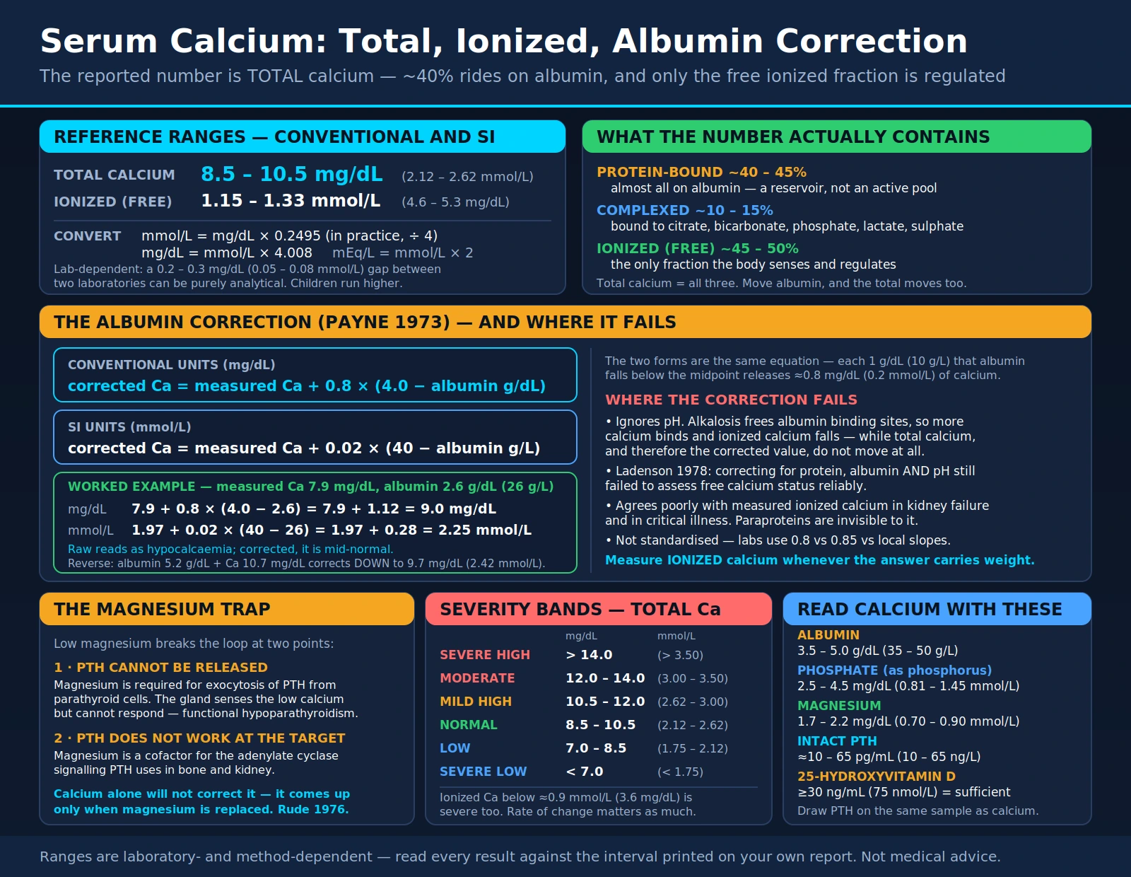
Serum calcium is one of the most frequently measured analytes in medicine — it appears on every basic and comprehensive metabolic panel — and it is also one of the most frequently misread. The number on the report is total calcium, which sums three physically different pools: calcium bound to albumin, calcium complexed to small anions, and the free ionized calcium that is the only fraction the body actually senses and regulates. Because roughly 40 percent of total calcium is stuck to albumin, anything that moves albumin moves total calcium without changing the biologically active fraction at all. That single fact explains most of the false alarms and most of the missed diagnoses. This page covers what the two calcium measurements mean, when each is used, how the albumin-correction formula works and where it fails, what raises and lowers calcium, and why calcium is close to uninterpretable without parathyroid hormone, phosphate, vitamin D, magnesium, and albumin measured alongside it.
Table of Contents
- What Serum Calcium Measures
- Total Calcium vs Ionized Calcium
- Reference Ranges and Units
- The Albumin Correction and Its Limits
- When the Test Is Ordered
- Causes of Hypercalcaemia
- Symptoms and Severity of Hypercalcaemia
- Causes of Hypocalcaemia
- Symptoms and Signs of Hypocalcaemia
- Magnesium Depletion: The Calcium That Will Not Correct
- Reading Calcium With PTH, Phosphate, Vitamin D and Albumin
- Pre-Analytical and Sample-Handling Pitfalls
- References
- Connections
- Featured Videos
What Serum Calcium Measures
An adult body contains roughly 1–1.2 kg of calcium, but about 99 percent of it is locked into the mineral phase of bone as hydroxyapatite. Everything a blood test can see lives in the remaining one percent — the calcium dissolved in extracellular fluid. That circulating pool is tiny, but it is defended more tightly than almost any other analyte in the body, because free calcium ions set the threshold at which nerves fire, muscles contract, hormones are secreted, and the clotting cascade proceeds.
Circulating calcium exists in three forms, and a standard "serum calcium" result is the sum of all three:
- Protein-bound calcium (~40–45%). Almost all of this is bound to albumin, with a small contribution from globulins. It is a reservoir, not an active pool — bound calcium cannot cross membranes or bind receptors.
- Complexed calcium (~10–15%). Calcium paired with small diffusible anions: bicarbonate, citrate, phosphate, lactate and sulphate. It is filterable at the glomerulus but not biologically active.
- Ionized (free) calcium (~45–50%). The physiologically active fraction. This is what the calcium-sensing receptor on the parathyroid gland monitors, and it is the fraction the entire calcium-regulating system exists to hold constant.
The control loop is short and fast. Parathyroid chief cells carry a calcium-sensing receptor (CaSR) that reads ionized calcium continuously. A fall in ionized calcium releases parathyroid hormone (PTH) within seconds; PTH then raises calcium by three routes — it mobilises calcium from bone, increases calcium reabsorption in the distal renal tubule, and stimulates renal 1α-hydroxylase to convert 25-hydroxyvitamin D into calcitriol (1,25-dihydroxyvitamin D), which in turn drives intestinal calcium absorption. PTH simultaneously dumps phosphate into the urine, which is why phosphate so often moves in the opposite direction to calcium and is such a useful interpretive clue. A rise in ionized calcium suppresses PTH by the same receptor.
Because this loop is fast and multi-layered, a persistently abnormal calcium almost always means something has broken the loop itself — a gland, a kidney, a receptor, a tumour, or a drug — rather than a dietary quirk. Dietary calcium intake, on its own, essentially never moves serum calcium in a person with intact parathyroid and kidney function. This is the single most common misconception patients bring to a calcium result.
Total Calcium vs Ionized Calcium
These are two different tests measuring two different things, and they are not interchangeable.
Total calcium is measured colorimetrically on a general chemistry analyser, most often with an o-cresolphthalein complexone or Arsenazo III dye method. It is cheap, fast, requires no special handling, and is bundled into the basic metabolic panel (BMP) and comprehensive metabolic panel (CMP). It is the right first test for essentially everyone in an outpatient setting.
Ionized calcium (also written iCa or free calcium) is measured directly with an ion-selective electrode, usually on a blood-gas analyser. It reports the active fraction and is completely independent of albumin. It is more demanding pre-analytically — the sample must be handled anaerobically and analysed promptly, because pH drives the result — and it is not part of routine panels.
Ionized calcium is preferred, and total calcium is unreliable, in the following situations:
- Marked hypoalbuminaemia. Cirrhosis, nephrotic syndrome, protein-losing enteropathy, malnutrition, prolonged critical illness. Total calcium is low; ionized calcium is frequently normal.
- Critical illness and the intensive care unit. Sepsis, shock, and acid–base derangement all distort protein binding. Correction formulas perform poorly in exactly this population.
- Massive transfusion or apheresis. Citrate anticoagulant chelates calcium; total calcium can look normal while ionized calcium falls sharply, because the chelated calcium is still counted in the total.
- Cardiopulmonary bypass, liver transplantation, and large-volume plasma exchange. Same citrate mechanism, minute-to-minute changes.
- Chronic kidney disease and dialysis. Albumin, pH, and complexing anions are all abnormal at once. Guideline bodies including KDIGO note the limitations of albumin-adjusted calcium in this group.
- Acid–base disorders. Alkalosis increases calcium binding to albumin and lowers ionized calcium without changing total calcium — the mechanism behind tetany during hyperventilation.
- Multiple myeloma and other paraprotein disorders. Abnormal immunoglobulins can bind calcium and produce a raised total calcium with a normal ionized calcium (pseudohypercalcaemia).
- Borderline results that will drive a major decision. When a total calcium sits just outside the reference interval and the answer determines whether someone gets a parathyroid scan or a cancer work-up, measuring the free fraction directly removes the guesswork.
Conversely, total calcium is entirely adequate for routine screening, for tracking a known and stable hyperparathyroid patient, and for any ambulatory patient with a normal albumin. The practical rule is that the albumin result decides which calcium number you can trust.
Reference Ranges and Units
Reference ranges are laboratory-dependent. They vary with the analytical method, the calibrator, the instrument platform, and the reference population the laboratory used to establish them. Always read a result against the interval printed on that laboratory's own report, and be cautious about comparing values drawn at different laboratories — a difference of 0.2–0.3 mg/dL (0.05–0.08 mmol/L) between two labs can be purely analytical. The figures below are typical adult intervals, given for orientation only.
Total Serum Calcium — Adults (mg/dL)
Total Serum Calcium — Adults (mmol/L, SI)
Ionized (Free) Calcium — Adults (mmol/L)
Ionized (Free) Calcium — Adults (mg/dL)
Unit conversion. Calcium has an atomic mass of 40.08, so:
- mmol/L = mg/dL × 0.2495 (in practice, divide mg/dL by 4)
- mg/dL = mmol/L × 4.008
- Some laboratories, particularly in the United States, also report mEq/L: mEq/L = mmol/L × 2, because calcium is divalent.
Severity bands for total calcium (conventional and SI, laboratory-dependent):
- Severe hypocalcaemia: below about 7.0 mg/dL (1.75 mmol/L), or ionized calcium below about 0.9 mmol/L (3.6 mg/dL). Symptomatic in most people; clinicians treat this as urgent.
- Hypocalcaemia: 7.0–8.5 mg/dL (1.75–2.12 mmol/L).
- Normal: approximately 8.5–10.5 mg/dL (2.12–2.62 mmol/L).
- Mild hypercalcaemia: 10.5–12.0 mg/dL (2.62–3.00 mmol/L). Frequently asymptomatic and found incidentally.
- Moderate hypercalcaemia: 12.0–14.0 mg/dL (3.00–3.50 mmol/L).
- Severe hypercalcaemia / hypercalcaemic crisis: above 14.0 mg/dL (3.50 mmol/L). A medical emergency regardless of symptoms.
Age matters. Reference intervals in children and adolescents run higher than adult intervals, because growing bone is actively mineralising; a calcium of 10.6 mg/dL (2.65 mmol/L) that would prompt investigation in a 50-year-old can be entirely normal in a 12-year-old. Neonates have their own intervals and their own physiology. Age-specific reference data, not adult data, must be used in paediatrics.
Companion analytes and their typical adult intervals, given here because calcium is never read alone:
- Albumin: 3.5–5.0 g/dL (35–50 g/L)
- Phosphate (as phosphorus): 2.5–4.5 mg/dL (0.81–1.45 mmol/L)
- Magnesium: 1.7–2.2 mg/dL (0.70–0.90 mmol/L)
- Intact PTH: approximately 10–65 pg/mL (10–65 ng/L) — assay-dependent and highly variable between platforms
- 25-hydroxyvitamin D: sufficiency generally defined at or above 30 ng/mL (75 nmol/L); deficiency below 20 ng/mL (50 nmol/L)
The Albumin Correction and Its Limits
Because roughly 40 percent of total calcium rides on albumin, a low albumin drags total calcium down without touching the ionized fraction. A patient with cirrhosis and an albumin of 2.5 g/dL (25 g/L) can show a total calcium of 8.0 mg/dL (2.00 mmol/L) and be perfectly calcium-replete. The albumin-correction formula — usually called the Payne formula after the 1973 British Medical Journal paper that introduced it — is an attempt to estimate what the total calcium would have been if albumin were normal.
Conventional units (mg/dL):
Corrected calcium = measured calcium + 0.8 × (4.0 − albumin in g/dL)
SI units (mmol/L):
Corrected calcium = measured calcium + 0.02 × (40 − albumin in g/L)
The two forms are the same equation. Each 1 g/dL (10 g/L) that albumin falls below the reference midpoint releases approximately 0.8 mg/dL (0.2 mmol/L) of calcium from protein binding, so that much is added back.
A worked example. Measured total calcium 7.9 mg/dL (1.97 mmol/L); albumin 2.6 g/dL (26 g/L).
- Conventional: 7.9 + 0.8 × (4.0 − 2.6) = 7.9 + 0.8 × 1.4 = 7.9 + 1.12 = 9.0 mg/dL
- SI: 1.97 + 0.02 × (40 − 26) = 1.97 + 0.28 = 2.25 mmol/L
The raw result reads as hypocalcaemia; the corrected result is mid-normal. In the opposite direction, a dehydrated patient with an albumin of 5.2 g/dL (52 g/L) and a calcium of 10.7 mg/dL (2.67 mmol/L) corrects down to 9.7 mg/dL (2.42 mmol/L) — the apparent hypercalcaemia was albumin, not calcium.
Where the formula fails
The correction is a population-derived regression, not a measurement, and it has been repeatedly shown to misclassify individual patients. Its assumptions break down in the very populations where clinicians most want an answer:
- It ignores pH. Hydrogen ions compete with calcium for albumin binding sites. Alkalosis strips hydrogen off albumin, freeing binding sites, so more calcium binds and ionized calcium falls — with no change in total calcium and therefore no change in the corrected value. Acidosis does the reverse. Ladenson and colleagues demonstrated in 1978 that correcting for protein, albumin and pH still failed to assess free calcium status reliably, and that finding has held up.
- It ignores globulins and paraproteins. In myeloma, in chronic inflammation, and in liver disease, the non-albumin protein fraction changes independently. Calcium bound to a paraprotein is invisible to an albumin-based correction.
- It performs poorly in chronic kidney disease. Comparisons in renal failure have found albumin-adjusted calcium to agree poorly with directly measured ionized calcium, which is why nephrology guidance leans toward measuring the free fraction when the answer will change management.
- It performs poorly in critical illness. Sepsis simultaneously drops albumin, shifts pH, raises lactate and citrate, and alters capillary permeability — four independent violations of the formula's assumptions at once.
- It is not standardised. Different laboratories use different constants (0.8 versus 0.85 versus locally derived slopes) and different albumin assays. Bromocresol green and bromocresol purple albumin methods give systematically different numbers, which propagates straight into the corrected calcium.
- It misclassifies in both directions. A large tertiary-hospital study published in Clinical Chemistry in 2018 found that albumin-adjusted calcium misclassified calcium status in a clinically meaningful fraction of patients when compared against measured ionized calcium — labelling normocalcaemic patients as abnormal and, more dangerously, normalising genuinely abnormal ones.
The practical position most laboratories and guidelines have converged on: use corrected calcium as a screening adjustment in ambulatory patients with mild albumin abnormalities, and measure ionized calcium directly whenever the correction is being asked to carry real clinical weight — critical illness, renal failure, paraproteinaemia, acid–base derangement, or a borderline result that will trigger imaging, surgery, or a cancer work-up.
One further caution: a "corrected calcium" is a calculated field. If a laboratory reports both, the corrected number can differ from what a clinician would calculate by hand if the lab uses a different constant. When tracking a patient over time, compare like with like — raw calcium against raw calcium, corrected against corrected, and ideally always from the same laboratory.
When the Test Is Ordered
Serum calcium is ordered so routinely that most abnormal results are found by accident rather than by suspicion. Mild hypercalcaemia in particular is usually discovered on a metabolic panel drawn for something else entirely — a fact that changed the presenting picture of primary hyperparathyroidism from a bone-and-stone disease to an asymptomatic biochemical finding once automated chemistry panels became routine in the 1970s.
Calcium is deliberately ordered when a clinician is evaluating:
- Any routine metabolic panel — calcium is a standard component of both the BMP and the CMP.
- Kidney disease of any stage — calcium, phosphate, PTH and vitamin D together define the CKD–mineral and bone disorder picture and are monitored serially.
- Kidney stones or nephrocalcinosis — recurrent calcium stones warrant a calcium and PTH check to exclude primary hyperparathyroidism.
- Osteoporosis, unexplained fractures, or bone pain — part of the standard secondary-cause screen before osteoporosis is called primary.
- Known or suspected malignancy — particularly breast, lung, renal cell, myeloma and lymphoma; hypercalcaemia in a cancer patient is prognostically significant.
- Neck surgery — checked in the hours and days after thyroidectomy or parathyroidectomy, because the parathyroid glands can be bruised, devascularised or removed.
- Perioral or finger tingling, cramps, or tetany — classic hypocalcaemic symptoms.
- Confusion, fatigue, constipation, or excessive thirst and urination — classic hypercalcaemic symptoms, often dismissed as ageing or depression.
- An unexplained short or long QT interval on the ECG, or new-onset seizures.
- Pancreatitis — both as a cause (hypercalcaemia can trigger it) and as a consequence (saponification consumes calcium).
- Malabsorption — coeliac disease, Crohn's disease, chronic pancreatitis, post-bariatric anatomy.
- Drug monitoring — lithium, thiazides, high-dose vitamin D, teriparatide, denosumab, bisphosphonates, cinacalcet.
- Critical illness — ionized calcium specifically, often several times daily during resuscitation or massive transfusion.
- Sarcoidosis and other granulomatous disease — granulomas make calcitriol outside the kidney's control loop.
The test requires an ordinary venous blood draw. Fasting is not strictly required for total calcium, but many laboratories prefer a fasting morning sample for consistency, and it is usual to avoid calcium supplements and calcium-containing antacids on the morning of the draw. Ionized calcium requires the specific handling described in the pre-analytical section below.
Causes of Hypercalcaemia
Two diagnoses account for the large majority of all hypercalcaemia — commonly cited as roughly 90 percent of cases in most clinical series: primary hyperparathyroidism and malignancy. They separate cleanly on a single test. Primary hyperparathyroidism raises calcium with a PTH that is high or inappropriately within the reference range; malignancy raises calcium with a PTH that is suppressed. The clinical settings differ too: primary hyperparathyroidism is typically an incidental, mild, chronic finding in an outpatient, while malignant hypercalcaemia is usually higher, rises faster, and occurs in someone who is visibly unwell.
PTH-dependent hypercalcaemia (PTH high or inappropriately normal)
- Primary hyperparathyroidism — the most common cause in the outpatient population. About 80–85 percent of cases are a single benign adenoma; most of the rest is four-gland hyperplasia, and parathyroid carcinoma accounts for well under one percent. Incidence rises with age and is markedly higher in women after menopause. Signature: high calcium, high or inappropriately normal PTH, low or low-normal phosphate, normal-to-raised 24-hour urine calcium.
- Tertiary hyperparathyroidism — long-standing secondary hyperparathyroidism in advanced kidney disease becomes autonomous, so the glands over-secrete even after the stimulus is corrected, classically after a kidney transplant. Calcium rises where it used to be low.
- Familial hypocalciuric hypercalcaemia (FHH) — an inactivating calcium-sensing receptor mutation resets the thermostat upward. Calcium mildly high, PTH normal or slightly high, and the distinguishing feature is low urinary calcium excretion (calcium-to-creatinine clearance ratio typically below 0.01). This matters: FHH is benign and lifelong, and mistaking it for primary hyperparathyroidism leads to an unnecessary neck operation. Hypercalcaemia since youth, or a family history of it, should raise the question.
- Lithium therapy — shifts the calcium-sensing receptor set-point, producing a hyperparathyroid picture that sometimes persists after the drug is stopped.
- MEN1, MEN2A, and familial isolated hyperparathyroidism — consider in younger patients, in multi-gland disease, and with a family history of pituitary, pancreatic or thyroid tumours.
PTH-independent hypercalcaemia (PTH suppressed)
- Humoral hypercalcaemia of malignancy — the commonest malignant mechanism, driven by tumour secretion of parathyroid hormone-related peptide (PTHrP) acting on the PTH receptor. Classic in squamous cell carcinomas (lung, head and neck, oesophagus, cervix), renal cell carcinoma, and some breast cancers. PTH suppressed, PTHrP measurable.
- Osteolytic metastases and marrow malignancy — direct bone destruction with cytokine-driven osteoclast activation; typical of myeloma, breast cancer with bone metastases, and lymphoma.
- Calcitriol-secreting lymphoma — some Hodgkin and non-Hodgkin lymphomas express 1α-hydroxylase, raising 1,25-dihydroxyvitamin D outside renal control.
- Granulomatous disease — sarcoidosis is the archetype; tuberculosis, histoplasmosis, berylliosis and granulomatosis with polyangiitis behave alike. Macrophages in granulomas make calcitriol without the kidney's negative feedback, so calcium rises and often worsens with sun exposure or vitamin D supplementation.
- Vitamin D toxicity — sustained very high-dose supplementation, compounding errors, or mislabelled products; 25-hydroxyvitamin D markedly elevated, PTH suppressed. A rising cause as high-dose over-the-counter products spread.
- Vitamin A toxicity — excess retinol, including high-dose retinoid therapy, stimulates bone resorption.
- Calcium-alkali syndrome (historically milk-alkali) — large calcium carbonate intake plus absorbable alkali produces hypercalcaemia, metabolic alkalosis and renal impairment, each reinforcing the others.
- Thyrotoxicosis — thyroid hormone accelerates bone turnover; mild hypercalcaemia occurs in a minority.
- Adrenal insufficiency and phaeochromocytoma (directly, and as part of MEN2A).
- Immobilisation — prolonged bed rest, spinal cord injury or casting uncouples resorption from formation; risk is highest where turnover is already high (adolescents, Paget disease, post-fracture).
- Thiazide diuretics — reduce urinary calcium excretion. Rarely causative alone, but frequently unmask underlying primary hyperparathyroidism, which is why the drug is often paused and calcium rechecked.
- Recovery phase of rhabdomyolysis or acute kidney injury — calcium deposited in damaged muscle is remobilised.
Pseudohypercalcaemia is a separate category and must be excluded before any work-up. It refers to a raised total calcium with a normal ionized calcium, caused by high albumin (dehydration, prolonged tourniquet), high globulins or a calcium-binding paraprotein in myeloma, or thrombocytosis. Directly measuring ionized calcium settles the question in one test.
Symptoms and Severity of Hypercalcaemia
The classical teaching mnemonic is "stones, bones, abdominal groans, and psychiatric moans," to which "thrones" is often added for the polyuria. The mnemonic is memorable but misleading about frequency: most mild hypercalcaemia today is asymptomatic and picked up incidentally. Symptoms broadly track both the height of the calcium and, importantly, how fast it rose — a calcium of 13 mg/dL (3.25 mmol/L) that developed over three days in a cancer patient produces far more illness than the same number reached over three years in hyperparathyroidism.
- Renal — "stones and thrones": polyuria and polydipsia (high calcium causes a nephrogenic diabetes insipidus by blocking the kidney's concentrating mechanism); dehydration and volume depletion, which raise calcium further in a self-reinforcing loop that is the central problem in severe hypercalcaemia; calcium oxalate and calcium phosphate stones; nephrocalcinosis; reduced kidney function, acutely from volume depletion and chronically from nephrocalcinosis.
- Skeletal — "bones": bone pain; reduced bone density in long-standing hyperparathyroidism, particularly at cortical sites such as the distal third of the radius; fragility fractures. Osteitis fibrosa cystica — brown tumours, subperiosteal resorption, the "salt and pepper" skull — is now rare where routine biochemistry screening catches the disease early.
- Gastrointestinal — "groans": constipation, often the earliest and most persistent complaint; anorexia, nausea and vomiting; vague abdominal pain, dyspepsia and peptic ulcer disease (calcium stimulates gastrin); acute pancreatitis, uncommon but well recognised.
- Neuropsychiatric — "moans": fatigue, weakness and lethargy, the most common symptom overall and the one most often attributed to something else; poor concentration and short-term memory; depression, anxiety and irritability, a genuinely common feature of mild primary hyperparathyroidism that frequently improves after treatment; confusion, delirium, stupor and, at severe levels, coma; proximal muscle weakness and hyporeflexia.
- Cardiovascular: a shortened QT interval — the electrocardiographic signature of hypercalcaemia; hypertension; bradyarrhythmias and, at very high levels, bundle branch block and arrest; increased digoxin sensitivity; vascular and valvular calcification with long-standing disease.
What clinicians monitor. In mild, stable, asymptomatic hypercalcaemia the usual approach is serial measurement of calcium, creatinine and eGFR, periodic bone density testing, and imaging for kidney stones, with attention to whether the calcium is trending upward. In severe hypercalcaemia, calcium is monitored frequently alongside renal function, volume status, electrolytes and the ECG. Nothing on this page is treatment advice — management decisions belong with the treating clinician.
Causes of Hypocalcaemia
The first question in any low calcium is whether it is real. A low total calcium with a normal albumin-corrected or ionized calcium is a protein artefact, not hypocalcaemia, and low albumin is by a wide margin the most common reason a calcium result comes back low. Once genuine hypocalcaemia is established, the second question is what PTH is doing, because that splits the causes into two groups.
Low or inappropriately normal PTH (the parathyroid axis has failed)
- Post-surgical hypoparathyroidism — the single most common cause. Follows total thyroidectomy, extensive neck dissection, or parathyroid surgery when the glands are removed, devascularised or bruised. May be transient (hours to weeks) or permanent; calcium is checked routinely after neck surgery for exactly this reason.
- Autoimmune hypoparathyroidism — isolated, or part of autoimmune polyglandular syndrome type 1 alongside adrenal insufficiency and mucocutaneous candidiasis.
- Genetic and developmental causes — DiGeorge (22q11.2 deletion) syndrome, and activating calcium-sensing receptor mutations causing autosomal dominant hypocalcaemia: the mirror image of FHH, with the thermostat reset downward and inappropriately high urinary calcium.
- Infiltrative and destructive causes — haemochromatosis (iron), Wilson disease (copper), metastatic infiltration, neck radiation.
- Magnesium depletion — causes both impaired PTH secretion and PTH resistance, and is the cause most often missed. Covered in its own section below.
- Severe hypermagnesaemia — typically iatrogenic, as during magnesium infusion for pre-eclampsia; very high magnesium activates the calcium-sensing receptor and suppresses PTH.
- Hungry bone syndrome — after parathyroidectomy for severe hyperparathyroidism, previously resorbing bone takes up calcium, phosphate and magnesium at high rates, driving all three down for days to weeks despite a functioning parathyroid remnant.
High PTH (the parathyroid axis is working but losing)
- Vitamin D deficiency or insufficiency — the most common cause worldwide. Without calcitriol, intestinal absorption falls, calcium drifts down, and PTH rises to compensate (secondary hyperparathyroidism). Calcium is often still normal at this stage, held there by the rising PTH at the expense of bone. Drivers: low sun exposure, dark skin at high latitude, malabsorption (coeliac, Crohn's, chronic pancreatitis, bariatric surgery), liver disease impairing 25-hydroxylation, and anticonvulsants (phenytoin, phenobarbital, carbamazepine) that accelerate vitamin D catabolism.
- Chronic kidney disease — a double hit: the failing kidney makes less calcitriol (less calcium absorbed) and excretes less phosphate (phosphate rises and precipitates with calcium). Both push calcium down and PTH up. This is the CKD–mineral and bone disorder complex, and the reason calcium, phosphate, PTH and vitamin D are monitored together at intervals set by CKD stage.
- Pseudohypoparathyroidism — genuine end-organ resistance to PTH: calcium low, phosphate high, PTH markedly elevated. The hormone is present but the target tissues cannot hear it. Albright hereditary osteodystrophy is the classic phenotype.
- Acute pancreatitis — free fatty acids released by lipase saponify calcium around the inflamed pancreas; the degree of hypocalcaemia is a recognised severity marker.
- Tumour lysis syndrome — massive cell breakdown after chemotherapy releases intracellular phosphate; calcium phosphate precipitates and calcium falls, alongside hyperkalaemia, hyperuricaemia and acute kidney injury.
- Rhabdomyolysis — calcium deposits into damaged muscle acutely, and is released again during recovery, sometimes causing rebound hypercalcaemia.
- Massive transfusion, apheresis and citrate anticoagulation — citrate chelates ionized calcium. Total calcium may look normal because the chelated fraction is still counted; only ionized calcium reveals it.
- Osteoblastic metastases — prostate and some breast cancers consume calcium into rapidly forming bone.
- Drugs — bisphosphonates and denosumab (especially with unrecognised vitamin D deficiency or renal impairment), cinacalcet, foscarnet, phosphate preparations, loop diuretics, proton pump inhibitors (via magnesium depletion), aminoglycosides, cisplatin, calcineurin inhibitors.
- Sepsis and critical illness — ionized hypocalcaemia is very common in intensive care and multifactorial: cytokine effects, magnesium depletion, citrate load and altered protein binding all contribute.
- Acute respiratory alkalosis — hyperventilation raises pH, increases calcium binding to albumin, and lowers ionized calcium without lowering total calcium. This is why a panic attack can produce genuine tetany while the routine chemistry panel looks entirely normal — a case where only ionized calcium tells the truth.
Symptoms and Signs of Hypocalcaemia
Hypocalcaemia is fundamentally a disease of nerve and muscle excitability. Free calcium ions stabilise the resting membrane potential of excitable cells; when calcium falls, sodium channels open more readily, and nerves and muscles begin to fire without being asked to. As with hypercalcaemia, the rate of change matters as much as the absolute number — a slowly developing chronic hypocalcaemia can be strikingly well tolerated, while a rapid fall of the same magnitude produces florid tetany.
- Neuromuscular — the dominant picture: perioral numbness and tingling around the mouth and lips, characteristically the first symptom; paraesthesiae of the fingertips and toes; muscle cramps in the calves, hands and feet; carpopedal spasm (involuntary flexion of the wrist and metacarpophalangeal joints with extended fingers), the hallmark of tetany; laryngospasm and bronchospasm with stridor and a sensation of throat closure, one of the dangerous manifestations; generalised or focal seizures in people with no epilepsy history; hyperreflexia.
- Chvostek sign — tapping over the facial nerve just anterior to the ear twitches the ipsilateral facial muscles. Easy to elicit but not specific; a proportion of people with entirely normal calcium show it.
- Trousseau sign — inflating a blood pressure cuff above systolic pressure for about three minutes provokes carpal spasm. Less comfortable but considerably more specific than Chvostek.
- Cardiac: a prolonged QT interval — the mirror image of hypercalcaemia's short QT — with the associated risk of torsades de pointes; reduced myocardial contractility and, in severe or prolonged cases, a reversible cardiomyopathy and heart failure; hypotension refractory to fluids and pressors in critical illness, where correcting ionized calcium is part of resuscitation.
- Psychiatric and cognitive: anxiety, irritability, emotional lability, depression, confusion, and in severe chronic cases psychosis. Papilloedema and raised intracranial pressure are described.
Chronic hypocalcaemia — the slow damage:
- Cataracts, a well-recognised long-term consequence of untreated hypoparathyroidism, and basal ganglia calcification on CT, sometimes with parkinsonian features or movement disorder.
- Dry, scaly skin; coarse, brittle hair; brittle, ridged nails.
- Dental abnormalities when hypocalcaemia occurs during tooth development in childhood — enamel hypoplasia, delayed eruption, root defects.
- Rickets in children and osteomalacia in adults when the driver is vitamin D deficiency: bone pain, proximal muscle weakness, waddling gait, and in children bowed legs and growth failure.
What clinicians monitor. In established hypoparathyroidism, the monitored quantities typically include calcium (often albumin-corrected or ionized), phosphate, magnesium, creatinine and eGFR, and 24-hour urinary calcium — the last because the goal is a calcium in the low-normal range that controls symptoms without driving excessive urinary calcium excretion and kidney damage. Frequency of monitoring is set by the treating clinician and by guideline recommendations from bodies such as the European Society of Endocrinology.
Magnesium Depletion: The Calcium That Will Not Correct
This deserves a section of its own because it is the most commonly missed cause of persistent hypocalcaemia, and because missing it leads directly to a specific, avoidable clinical failure: a calcium that will not come up no matter how much calcium is given.
Magnesium sits at two separate points in the calcium control loop, and low magnesium breaks both at once:
- Impaired PTH secretion. Magnesium is required for the exocytosis of PTH from parathyroid chief cells. In magnesium depletion, the gland senses the low calcium perfectly well but cannot release hormone in response. PTH is therefore low or inappropriately normal — a picture indistinguishable on paper from hypoparathyroidism, which is why it is called functional hypoparathyroidism.
- End-organ PTH resistance. Even the PTH that does get released works poorly, because magnesium is a cofactor for the adenylate cyclase signalling that PTH uses at the bone and kidney. Bone and kidney simply do not respond normally.
Rude, Oldham and Singer characterised this dual mechanism in human magnesium deficiency in 1976, and the finding has been reproduced consistently since. The practical consequence is unambiguous: hypocalcaemia caused by magnesium depletion is refractory to calcium replacement and resolves only when magnesium is replaced. A patient can receive repeated calcium without the calcium moving; the moment magnesium is corrected, PTH rises and calcium normalises, often within a day.
The corollary is a rule of practice: any hypocalcaemia that does not respond as expected should prompt a magnesium measurement, and in many settings magnesium is checked at the outset alongside calcium, phosphate and PTH rather than only after failure.
Who is magnesium-depleted
- Proton pump inhibitor use, particularly long-term. PPI-induced hypomagnesaemia is a recognised drug effect and often develops after months to years of therapy.
- Loop and thiazide diuretics — renal magnesium wasting.
- Chronic alcohol use — poor intake, renal wasting, and gastrointestinal losses together. Alcohol-related hypomagnesaemia is common and often severe.
- Chronic diarrhoea, malabsorption, and short bowel — coeliac disease, Crohn's disease, and post-bariatric anatomy.
- Poorly controlled diabetes — osmotic diuresis drags magnesium out.
- Nephrotoxic and magnesium-wasting drugs — cisplatin, aminoglycosides, amphotericin B, calcineurin inhibitors (ciclosporin, tacrolimus), and cetuximab.
- Refeeding syndrome — magnesium, phosphate and potassium all shift intracellularly as nutrition is restored.
- Genetic renal magnesium-wasting disorders such as Gitelman syndrome.
A caveat about the magnesium test itself
Serum magnesium is an imperfect marker of magnesium status. Only about 1 percent of total body magnesium is extracellular, so serum magnesium can sit within the reference interval while intracellular stores are meaningfully depleted. A normal serum magnesium therefore does not fully exclude magnesium deficiency as a contributor to refractory hypocalcaemia, and clinicians sometimes treat empirically on the basis of the clinical picture. Red-cell magnesium and magnesium loading tests exist but are not routinely available. See the site's Magnesium Test page for the detail.
The mirror situation: very high magnesium — almost always from intravenous magnesium therapy — also suppresses PTH, by activating the calcium-sensing receptor. So both ends of the magnesium range can lower calcium, for opposite reasons.
Reading Calcium With PTH, Phosphate, Vitamin D and Albumin
A calcium result in isolation answers almost nothing. The same 10.8 mg/dL (2.70 mmol/L) can mean a parathyroid adenoma, an occult malignancy, sarcoidosis, a benign inherited receptor variant, or a tight tourniquet, and the tests that distinguish them are inexpensive and widely available. The reverse is equally true and is why this page exists: PTH cannot be interpreted without a simultaneous calcium. A PTH of 70 pg/mL is normal-ish in isolation, appropriate and expected if calcium is low, and diagnostic of primary hyperparathyroidism if calcium is high. Ideally both are drawn from the same blood sample, because both move.
The core panel
- Albumin — decides whether the total calcium can be trusted at all, and drives the correction formula. The CMP includes it automatically.
- Intact PTH — the single most informative companion test; splits every calcium abnormality into PTH-dependent and PTH-independent.
- Phosphate — PTH is phosphaturic, so phosphate usually moves opposite to PTH activity. Low or low-normal phosphate with high calcium supports hyperparathyroidism; high phosphate with low calcium points toward hypoparathyroidism, PTH resistance, or kidney disease.
- Magnesium — mandatory in any unexplained or refractory hypocalcaemia, for the reasons above.
- 25-hydroxyvitamin D — the storage form and the correct measure of vitamin D status. Low values explain hypocalcaemia with high PTH; deficiency also blunts the calcium rise in hyperparathyroidism, sometimes masking it.
- 1,25-dihydroxyvitamin D (calcitriol) — not a general status test. Ordered when granulomatous disease or a calcitriol-producing lymphoma is suspected, where it is high while PTH is suppressed.
- Creatinine and eGFR — kidney function shapes the whole calcium–phosphate–PTH axis, and is also a target organ for hypercalcaemic damage.
- 24-hour urine calcium, or calcium-to-creatinine clearance ratio — separates FHH (low urinary calcium, ratio typically below 0.01) from primary hyperparathyroidism, and tracks stone risk in treated hypoparathyroidism.
- PTHrP — when hypercalcaemia has a suppressed PTH and malignancy is suspected.
- Alkaline phosphatase — a rough index of bone turnover; raised in osteitis fibrosa cystica, osteomalacia, bone metastases and Paget disease.
- Serum and urine protein electrophoresis — when myeloma is possible, both as a cause of true hypercalcaemia and of paraprotein pseudohypercalcaemia.
The interpretive patterns
High calcium:
- High calcium + high or inappropriately normal PTH + low/low-normal phosphate + normal-to-high urine calcium → primary hyperparathyroidism.
- High calcium + high or normal PTH + low urine calcium (ratio <0.01) → consider familial hypocalciuric hypercalcaemia; lithium produces a similar biochemistry.
- High calcium + suppressed PTH + raised PTHrP → humoral hypercalcaemia of malignancy.
- High calcium + suppressed PTH + raised 1,25-dihydroxyvitamin D with normal 25-hydroxyvitamin D → granulomatous disease or lymphoma.
- High calcium + suppressed PTH + markedly raised 25-hydroxyvitamin D → vitamin D toxicity.
- High calcium + suppressed PTH + normal vitamin D metabolites → consider thyrotoxicosis, immobilisation, vitamin A excess, adrenal insufficiency, calcium-alkali syndrome.
- High total calcium + normal ionized calcium → pseudohypercalcaemia from high albumin, paraprotein, or a pre-analytical artefact.
Low calcium:
- Low calcium + low or inappropriately normal PTH + high phosphate + normal kidney function → hypoparathyroidism (most often post-surgical).
- Low calcium + low or inappropriately normal PTH + low magnesium → magnesium-dependent functional hypoparathyroidism. Will not correct until magnesium is replaced.
- Low calcium + high PTH + low phosphate + low 25-hydroxyvitamin D → vitamin D deficiency with secondary hyperparathyroidism.
- Low calcium + high PTH + high phosphate + reduced eGFR → chronic kidney disease–mineral and bone disorder.
- Low calcium + markedly high PTH + high phosphate + normal kidney function and normal vitamin D → pseudohypoparathyroidism (PTH resistance).
- Low total calcium + normal ionized calcium + low albumin → not hypocalcaemia; a protein artefact.
- Normal total calcium + low ionized calcium + alkalosis → binding shift, as in hyperventilation or citrate load.
A last note on normal-calcaemic states. Normocalcaemic primary hyperparathyroidism — a persistently raised PTH with a consistently normal calcium, once vitamin D deficiency, kidney disease, malabsorption and calcium-losing drugs have been excluded — is a recognised entity discussed in the International Workshop guidelines. It is a diagnosis of exclusion, and it depends entirely on having the whole panel rather than a single number.
Pre-Analytical and Sample-Handling Pitfalls
A surprising share of abnormal calcium results are created between the needle and the analyser. Because the physiological range is so narrow, small artefacts produce results that cross decision thresholds, and each one can trigger an expensive and anxious work-up. Before investigating an unexpected calcium, it is standard practice to repeat the test with careful technique.
At the draw
- Prolonged tourniquet time — the most common and most preventable artefact. Venous stasis beyond about a minute drives fluid out of the vessel while proteins stay behind; albumin rises, and because 40 percent of calcium is albumin-bound, total calcium rises with it. Short-term venous stasis has been shown to significantly alter routine chemistry results. Release the tourniquet as soon as blood flow is established.
- Fist clenching and pumping — repeated contraction releases potassium and lactate locally and lowers local pH, shifting calcium off albumin and spuriously raising ionized calcium.
- Posture — standing shifts fluid out of the vascular space and concentrates albumin, typically raising total calcium by roughly 0.2–0.5 mg/dL (0.05–0.12 mmol/L). A patient sampled seated in clinic and later supine on a ward has changed two things at once.
- Hydration status — dehydration concentrates albumin and raises total calcium; intravenous fluids dilute it. This is behind much of the transient hypercalcaemia seen in unwell patients.
- Drawing above a running intravenous line — dilution, or contamination with calcium- or citrate-containing fluid. Sample from the opposite arm.
- Tube contamination and order of draw — EDTA carry-over from a lavender-top tube chelates calcium and produces a spuriously very low (sometimes near-zero) calcium, classically alongside a spuriously high potassium. An implausibly low calcium in a well patient is an EDTA artefact until proven otherwise; citrate from a blue-top tube does the same less dramatically.
Handling, analysis, and patient-side factors
- Ionized calcium must be handled anaerobically. Air exposure lets carbon dioxide escape, pH rises, more calcium binds to albumin, and measured ionized calcium falls falsely. Samples are drawn into an appropriate syringe or tube, bubbles expelled, capped, and analysed promptly.
- Heparin choice matters for ionized calcium — liquid heparin both dilutes the sample and binds calcium; electrolyte-balanced (calcium-titrated) heparin syringes exist specifically to avoid this, and the difference is clinically meaningful.
- Delay and temperature — prolonged storage at room temperature allows glycolysis, lowering pH and shifting ionized calcium upward. Prompt analysis, or transport on ice where the laboratory specifies it, is required.
- Gadolinium-based MRI contrast interferes with certain colorimetric calcium methods and can produce a strikingly falsely low total calcium for a day or so after a contrast-enhanced scan — worth asking about when a calcium is inexplicably low.
- Haemolysis, icterus, lipaemia and paraproteins all interfere with colorimetric assays to varying degrees; paraproteins can push the total either way depending on method, on top of binding calcium.
- Method differences between laboratories. Total calcium is not perfectly harmonised across platforms; serial monitoring is most meaningful on the same analyser.
- Calcium supplements and calcium-containing antacids shortly before the draw can transiently raise calcium; it is usual to hold them on the morning of the test. Thiazides and lithium raise calcium and are often paused before repeat testing, when clinically safe.
- High-dose biotin does not affect the calcium measurement itself (a chemistry method) but does interfere with many immunoassays, including some PTH and 25-hydroxyvitamin D assays — exactly the results calcium is read against. Recent neck surgery, transfusion, contrast imaging and current intravenous fluids are all worth stating on the request form.
The general principle: an isolated abnormal calcium in an otherwise well patient should be repeated before it is investigated, ideally fasting, without a prolonged tourniquet, and with albumin measured on the same sample.
References
Key Research Papers and Guidelines
- Payne RB, Little AJ, Williams RB, Milner JR. Interpretation of serum calcium in patients with abnormal serum proteins. British Medical Journal. 1973;4(5893):643–646. PMID 4758544
- Payne RB, Carver ME, Morgan DB. Interpretation of serum total calcium: effects of adjustment for albumin concentration on frequency of abnormal values and on detection of change in the individual. Journal of Clinical Pathology. 1979;32(1):56–60. PMID 429580
- Ladenson JH, Lewis JW, Boyd JC. Failure of total calcium corrected for protein, albumin, and pH to correctly assess free calcium status. Journal of Clinical Endocrinology and Metabolism. 1978;46(6):986–993. PMID 45478
- Smith JD, Wilson S, Schneider HG. Misclassification of calcium status based on albumin-adjusted calcium: studies in a tertiary hospital setting. Clinical Chemistry. 2018;64(12):1713–1722. PMID 30352866
- Baird GS. Ionized calcium. Clinica Chimica Acta. 2011;412(9–10):696–701. PMID 21238441
- Gøransson LG, Skadberg Ø, Bergrem H. Albumin-corrected or ionized calcium in renal failure? What to measure? Nephrology Dialysis Transplantation. 2005;20(10):2126–2129. PMID 16030044
- Bushinsky DA, Monk RD. Electrolyte quintet: calcium. The Lancet. 1998;352(9124):306–311. PMID 9690425
- Minisola S, Pepe J, Piemonte S, Cipriani C. The diagnosis and management of hypercalcaemia. BMJ. 2015;350:h2723. PMID 26037642
- Turner JJO. Hypercalcaemia — presentation and management. Clinical Medicine (London). 2017;17(3):270–273. PMID 28572230
- Stewart AF. Hypercalcemia associated with cancer. New England Journal of Medicine. 2005;352(4):373–379. PMID 15673803
- Goldner W. Cancer-related hypercalcemia. Journal of Oncology Practice. 2016;12(5):426–432. PMID 27170690
- Bilezikian JP, Bandeira L, Khan A, Cusano NE. Hyperparathyroidism. The Lancet. 2018;391(10116):168–178. PMID 28923463
- Walker MD, Silverberg SJ. Primary hyperparathyroidism. Nature Reviews Endocrinology. 2018;14(2):115–125. PMID 28885621
- Bilezikian JP, Khan AA, Silverberg SJ, El-Hajj Fuleihan G, et al. Evaluation and management of primary hyperparathyroidism: summary statement and guidelines from the Fifth International Workshop. Journal of Bone and Mineral Research. 2022;37(11):2293–2314. PMID 36245251
- Christensen SE, Nissen PH, Vestergaard P, Heickendorff L, et al. Discriminative power of three indices of renal calcium excretion for the distinction between familial hypocalciuric hypercalcaemia and primary hyperparathyroidism. Clinical Endocrinology (Oxford). 2008;69(5):713–720. PMID 18410554
- Hannan FM, Kallay E, Chang W, Brandi ML, Thakker RV. The calcium-sensing receptor in physiology and in calcitropic and noncalcitropic diseases. Nature Reviews Endocrinology. 2018;15(1):33–51. PMID 30443043
- Cooper MS, Gittoes NJL. Diagnosis and management of hypocalcaemia. BMJ. 2008;336(7656):1298–1302. PMID 18535072
- Shoback D. Hypoparathyroidism. New England Journal of Medicine. 2008;359(4):391–403. PMID 18650515
- Brandi ML, Bilezikian JP, Shoback D, Bouillon R, et al. Management of hypoparathyroidism: summary statement and guidelines. Journal of Clinical Endocrinology and Metabolism. 2016;101(6):2273–2283. PMID 26943719
- Bollerslev J, Rejnmark L, Marcocci C, Shoback DM, et al. European Society of Endocrinology clinical guideline: treatment of chronic hypoparathyroidism in adults. European Journal of Endocrinology. 2015;173(2):G1–G20. PMID 26160136
- Rude RK, Oldham SB, Singer FR. Functional hypoparathyroidism and parathyroid hormone end-organ resistance in human magnesium deficiency. Clinical Endocrinology (Oxford). 1976;5(3):209–224. PMID 182417
- Ayuk J, Gittoes NJL. Contemporary view of the clinical relevance of magnesium homeostasis. Annals of Clinical Biochemistry. 2014;51(Pt 2):179–188. PMID 24402002
- Ketteler M, Block GA, Evenepoel P, Fukagawa M, et al. Executive summary of the 2017 KDIGO Chronic Kidney Disease–Mineral and Bone Disorder (CKD-MBD) guideline update: what's changed and why it matters. Kidney International. 2017;92(1):26–36. PMID 28646995
- Holick MF, Binkley NC, Bischoff-Ferrari HA, Gordon CM, et al. Evaluation, treatment, and prevention of vitamin D deficiency: an Endocrine Society clinical practice guideline. Journal of Clinical Endocrinology and Metabolism. 2011;96(7):1911–1930. PMID 21646368
- Lippi G, Salvagno GL, Montagnana M, Brocco G, et al. Influence of short-term venous stasis on clinical chemistry testing. Clinical Chemistry and Laboratory Medicine. 2005;43(8):869–875. PMID 16201899
- Coiffier B, Altman A, Pui CH, Younes A, Cairo MS. Guidelines for the management of pediatric and adult tumor lysis syndrome: an evidence-based review. Journal of Clinical Oncology. 2008;26(16):2767–2778. PMID 18509186
Live PubMed Searches
- Ionized calcium versus albumin-adjusted calcium — PubMed literature search
- Albumin-corrected calcium formula accuracy — PubMed literature search
- Hypercalcaemia differential diagnosis and PTH — PubMed literature search
- Humoral hypercalcaemia of malignancy and PTHrP — PubMed literature search
- Refractory hypocalcaemia and magnesium depletion — PubMed literature search
- Post-thyroidectomy hypoparathyroidism and calcium — PubMed literature search
- Familial hypocalciuric hypercalcaemia and calcium-creatinine clearance ratio — PubMed literature search
- Sarcoidosis, hypercalcaemia and calcitriol — PubMed literature search
- CKD–mineral and bone disorder: calcium, phosphate and PTH — PubMed literature search
- Pre-analytical variables in serum calcium: tourniquet and posture — PubMed literature search
- EDTA contamination and spurious hypocalcaemia — PubMed literature search
- Gadolinium contrast interference with calcium assays — PubMed literature search
- Normocalcaemic primary hyperparathyroidism — PubMed literature search
- Citrate anticoagulation and ionized calcium in massive transfusion — PubMed literature search
External Authoritative Resources
- MedlinePlus — Calcium Blood Test
- Testing.com (formerly Lab Tests Online, ADLM/AACC) — Calcium
- Endotext (NCBI Bookshelf) — Hypocalcemia: Diagnosis and Treatment
- KDIGO — Chronic Kidney Disease–Mineral and Bone Disorder Guideline
- NIH Office of Dietary Supplements — Calcium Fact Sheet for Health Professionals
Connections
- All Lab Tests
- Parathyroid Hormone (PTH) Test
- Albumin Test
- Vitamin D Test (25-Hydroxyvitamin D)
- Magnesium Test
- Comprehensive Metabolic Panel
- Basic Metabolic Panel
- Kidney Function Tests
- Calcium — Essential Mineral
- Hypocalcemia (Low Calcium)
- Hypercalcemia (High Calcium)
- Magnesium
- Magnesium Deficiency
- Phosphorus
- Vitamin D3 (Cholecalciferol)
- Hyperparathyroidism
- Primary Hyperparathyroidism
- Secondary Hyperparathyroidism
- Hypoparathyroidism
- Chronic Kidney Disease
- Kidney Stones
- Osteoporosis
- Sarcoidosis
- Bone Loss and Osteoporosis
- Calcium and Kidney Stones
- Calcium & PTH Homeostasis — interactive animation
- Bone Remodeling & Calcium Balance — interactive animation