Orthostatic Intolerance in Myalgic Encephalomyelitis/Chronic Fatigue Syndrome (ME/CFS)
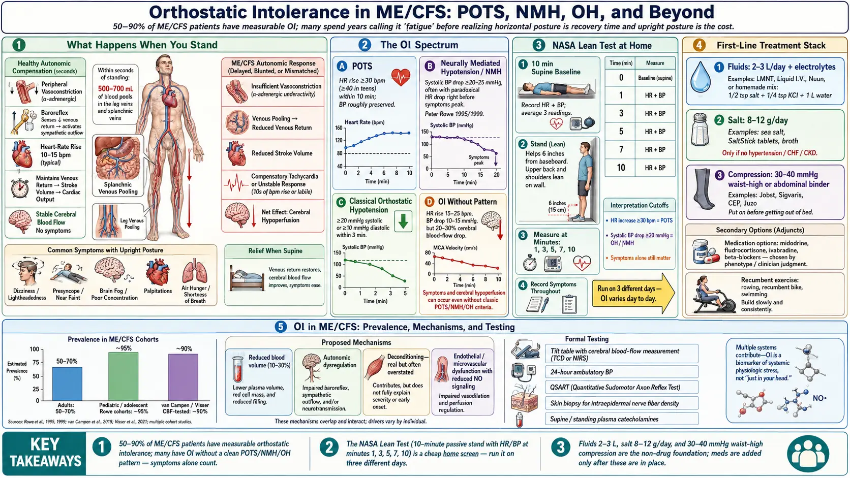
Table of Contents
- What Orthostatic Intolerance Is
- How Common It Is in Myalgic Encephalomyelitis/Chronic Fatigue Syndrome
- The Spectrum: POTS, NMH, OH, and OI-Without-Pattern
- Why Upright Posture Worsens Myalgic Encephalomyelitis/Chronic Fatigue Syndrome
- Mechanisms — What Is Actually Going Wrong
- The NASA Lean Test at Home
- Tilt Table, 24-Hour Blood Pressure, and Other Formal Testing
- First-Line Treatment: Fluids, Salt, Compression
- Medications — What, When, and How Much
- Recumbent Exercise and Why Upright GET Fails
- Practical Tips for Daily Life
- Key Research Papers
- Connections
- Featured Videos
What Orthostatic Intolerance Is
Orthostatic intolerance (OI) is the umbrella term for symptoms that appear or worsen when you stand up and improve when you lie back down. In healthy physiology, standing shifts about 500–700 mL of blood into the legs and splanchnic veins within the first few seconds. The autonomic nervous system answers by tightening blood vessels and nudging heart rate up by 10–15 beats per minute, keeping brain perfusion steady. In orthostatic intolerance, that response is broken, overshoots, or simply never comes — and the brain ends up underperfused.
The symptoms are what you would expect from a brain that is not getting enough blood: lightheadedness, brain fog, blurred vision, fatigue that gets dramatically worse the longer you stand, racing or pounding heart, nausea, shakiness, and sometimes fainting. In myalgic encephalomyelitis/chronic fatigue syndrome (ME/CFS) the picture is even messier, because the fatigue, cognitive fog, and post-exertional malaise of the underlying illness blur the edges of the OI syndrome. Many patients spend years describing “fatigue” without realizing that a huge fraction of it is driven by the act of staying upright.
How Common It Is in Myalgic Encephalomyelitis/Chronic Fatigue Syndrome
Depending on the criteria used and whether testing is passive (lean test, tilt table) or symptom-based, 50–90% of myalgic encephalomyelitis/chronic fatigue syndrome patients have measurable orthostatic intolerance. Pediatric and adolescent cohorts skew toward the higher end — studies from Peter Rowe at Johns Hopkins have consistently found that nearly all teenagers with myalgic encephalomyelitis/chronic fatigue syndrome develop OI on tilt testing. Adult cohorts trend lower, but in the large cohort of 429 patients studied by van Campen and Visser in the Netherlands, 90% of myalgic encephalomyelitis/chronic fatigue syndrome patients overall showed an abnormal fall in brain blood flow on tilt testing, including 82% of those whose heart rate and blood pressure stayed normal — that is, without meeting criteria for postural orthostatic tachycardia syndrome (POTS) or orthostatic hypotension.
Given these figures, most people with myalgic encephalomyelitis/chronic fatigue syndrome who are tested show some form of OI.
The Spectrum: POTS, NMH, OH, and OI-Without-Pattern
Orthostatic intolerance is not one thing. It is a family of patterns on the same axis, and myalgic encephalomyelitis/chronic fatigue syndrome patients can sit anywhere on it — or can show more than one pattern across different days. The four main categories:
Postural Orthostatic Tachycardia Syndrome (POTS). Heart rate rises by at least 30 beats per minute (40 bpm in adolescents) within 10 minutes of standing, without a meaningful drop in blood pressure. Standing HR typically hits 120 bpm or higher. Patients feel racing heart, lightheadedness, air hunger, shaking, and overwhelming fatigue. See the dedicated postural orthostatic tachycardia syndrome page for the deeper clinical picture.
Neurally Mediated Hypotension (NMH). Also called neurocardiogenic syncope or vasovagal syndrome in its faintier form. Blood pressure drops by at least 20–25 mmHg systolic (or below 90 mmHg absolute) during prolonged upright posture, often with a paradoxical drop in heart rate right before symptoms peak. NMH tends to produce delayed symptoms — you feel fine for 10, 20, 30 minutes, then suddenly sweaty, nauseous, foggy, and about to fall over. Peter Rowe’s landmark 1995 and 1999 studies established NMH as a major OI pattern in myalgic encephalomyelitis/chronic fatigue syndrome, sometimes more common than postural orthostatic tachycardia syndrome in adults.
Classical Orthostatic Hypotension (OH). Sustained drop of 20 mmHg systolic or 10 mmHg diastolic within 3 minutes of standing. Less common in myalgic encephalomyelitis/chronic fatigue syndrome than postural orthostatic tachycardia syndrome or NMH. When present, it tends to point toward autonomic neuropathy, medication effects, or severe dehydration rather than primary myalgic encephalomyelitis/chronic fatigue syndrome physiology.
Orthostatic intolerance without a defined pattern. The most common and most underdiagnosed category. Heart rate rises only 15–25 bpm. Blood pressure drops 10–15 mmHg. Neither number hits the diagnostic cutoff. But the patient’s symptoms — cognitive fog, leg heaviness, fatigue, breathlessness — appear only upright and vanish supine. Cerebral blood flow measurements during tilt show 20–30% reductions in these patients even when HR and blood pressure (BP) look normal. If you have been told your tilt test was “negative” but you still can’t tolerate standing, this is very likely what you have.
Why Upright Posture Worsens Myalgic Encephalomyelitis/Chronic Fatigue Syndrome
Every myalgic encephalomyelitis/chronic fatigue syndrome patient eventually notices the same thing: the day gets worse as it goes on, and horizontal time is recovery time. This is not an accident of mood or effort. It is biology.
When you stand, blood pools in dependent veins. In a healthy person the nervous system compensates in seconds. In myalgic encephalomyelitis/chronic fatigue syndrome the compensation is sluggish, incomplete, or wrong, and cerebral blood flow drops. Reduced cerebral blood flow is not a theory — it has been measured directly with transcranial Doppler and extracranial Doppler ultrasound by the van Campen/Visser group and by Medow and Stewart’s lab at New York Medical College. The brain starves, and the symptoms we call “brain fog,” “cognitive fatigue,” and even “post-exertional malaise” in part reflect that ongoing hypoperfusion.
There is also a feedback loop. Upright posture in an OI patient provokes sympathetic overactivation — adrenaline surges to try to compensate — which itself feels awful (racing heart, tremor, sweating, anxiety) and consumes adenosine triphosphate (ATP) at a cellular level. In myalgic encephalomyelitis/chronic fatigue syndrome, where cellular energy production is already impaired, every hour upright is an energy debt you cannot easily repay. That is why many moderately affected patients can function for two or three hours upright and then collapse for the rest of the day.
Mechanisms — What Is Actually Going Wrong
There is no single cause of OI in myalgic encephalomyelitis/chronic fatigue syndrome. Four mechanisms overlap in most patients.
1. Low blood volume (hypovolemia). Some studies have found that a portion of myalgic encephalomyelitis/chronic fatigue syndrome patients carry less circulating blood volume than healthy controls, with red cell mass and plasma volume both reduced. Less blood in the tank means less to spare when gravity pulls it south. This is the reasoning behind fluid and salt loading as a first-line treatment — refilling the tank.
Low blood volume is not the whole story, though. A 2026 study measured total blood volume directly in 49 adults with myalgic encephalomyelitis/chronic fatigue syndrome: blood volume was low in 35%, normal in 43% and high in 22%, and red blood cell volume was low in 49%. About 55% had at least one abnormal result on a 10-minute lean test, but these abnormal results were no more common in the low-volume group than in the others. The authors concluded that blood-volume category was not linked to the lean-test findings, which points to the nerves, blood vessels, breathing and brain circulation as further contributors.
2. Autonomic dysfunction. The autonomic nervous system — the part that runs blood vessels, heart rate, digestion, and sweat without your conscious input — is dysregulated in myalgic encephalomyelitis/chronic fatigue syndrome. Some vessels fail to constrict on demand. Others constrict inappropriately. Sympathetic tone is often high at rest and paradoxically low when needed. Small-fiber neuropathy is found in a meaningful subset and may contribute.
3. Deconditioning (real, but overstated). Yes, months or years of being horizontal weakens the muscle pump in the legs and reduces cardiac stroke volume. But deconditioning alone does not explain why an acute viral illness can flip a previously athletic person into severe OI overnight. Deconditioning is a contributor, not the driver. Treating it as the driver — the error at the heart of graded exercise therapy — is what makes patients worse, not better. Measured data point the same way: in a 2021 study of 199 myalgic encephalomyelitis/chronic fatigue syndrome patients, brain blood flow fell during tilt testing by the same amount whether an exercise test showed no, mild or severe deconditioning, and the authors concluded that orthostatic intolerance in these patients “is not caused by deconditioning”.
4. Endothelial and microvascular dysfunction. Newer work suggests that the small blood vessels themselves behave abnormally in myalgic encephalomyelitis/chronic fatigue syndrome, with reduced endothelial nitric oxide signaling and impaired flow-mediated dilation. This may overlap with the post-viral endothelial injury seen after other acute infections. A 2024 study of 534 myalgic encephalomyelitis/chronic fatigue syndrome patients whose heart rate and blood pressure stayed normal on a tilt test found that 91% had an abnormal fall in both the heart’s output (cardiac output) and brain blood flow, falling in an almost one-to-one ratio. In healthy people the brain’s blood vessels widen to protect its blood flow when cardiac output drops; the authors suggest this widening is missing in most of these patients, which might point to a problem with the lining of the blood vessels (endothelial dysfunction).
The NASA Lean Test at Home
You do not need a tilt table to get the first useful answer. The NASA Lean Test (also called the 10-minute stand test) is a protocol you can run at home with a basic blood-pressure cuff and a watch. It was originally developed to assess orthostatic tolerance in astronauts returning from spaceflight and is now the workhorse screening tool for OI in myalgic encephalomyelitis/chronic fatigue syndrome and postural orthostatic tachycardia syndrome clinics.
Protocol.
- Lie flat on your back for 10 minutes. Do nothing. At the end, record HR and blood pressure three times, one minute apart, and average them. This is your supine baseline.
- Stand up. Lean your upper back against a wall with your heels about 6 inches (15 cm) from the baseboard. Do not move your legs. Do not shift weight. The lean minimizes the muscle-pump effect that masks OI during active standing.
- At minutes 1, 3, 5, 7, and 10, record HR and blood pressure. Note any symptoms — lightheadedness, visual changes, cognitive slowing, chest tightness, nausea.
- Stop early and lie down at any point if you feel faint, have tunnel vision, or start to lose awareness. Symptoms count even if numbers do not.
Interpretation. Calculate maximum HR rise over baseline and maximum blood pressure drop over baseline. A HR rise of 30+ bpm (40+ in teens) suggests postural orthostatic tachycardia syndrome. A systolic blood pressure drop of 20+ mmHg (or 25+ for NMH criteria) or a diastolic drop of 10+ mmHg suggests orthostatic hypotension or NMH. Symptoms alone, with smaller numeric changes, still count as orthostatic intolerance. The written record of numbers and symptoms is what a clinician reviews.
Repeating the test on three different days gives a fuller picture. OI is notoriously day-to-day variable — salt intake, sleep, menstrual cycle phase, and hydration all move the numbers. A single negative test does not rule out OI when the symptoms are clear.
Tilt Table, 24-Hour Blood Pressure, and Other Formal Testing
Tilt table testing is the formal version. You lie strapped to a motorized table that tilts to 60–70 degrees upright for 30–45 minutes while HR and blood pressure are recorded continuously. Some centers add cerebral blood flow measurement via transcranial Doppler or extracranial Doppler — this is the single biggest diagnostic advance in OI in the last decade and the only way to catch the OI-without-pattern group. Not every center measures cerebral blood flow; most community cardiology practices do not.
Tilt testing is typically used when the home lean test is equivocal, when medications are being considered, or when an insurance carrier or disability application demands an objective result. The test is uncomfortable — some myalgic encephalomyelitis/chronic fatigue syndrome patients describe being knocked out for weeks by the provocation — so its cost is weighed against what the answer will change.
24-hour ambulatory blood pressure monitoring catches patterns the clinic misses: non-dipping at night, morning surges, paradoxical hypertension, and “white coat” normal readings that hide everyday OI. Useful when symptoms do not match the in-office numbers.
Other adjuncts: QSART (quantitative sudomotor axon reflex testing) for small-fiber neuropathy, skin biopsy for intraepidermal nerve fiber density, and plasma catecholamines (supine and standing) when hyperadrenergic postural orthostatic tachycardia syndrome is suspected.
First-Line Treatment: Fluids, Salt, Compression
Specialist clinics describe three non-drug measures as the foundation, used before any prescription. They are cheap and broadly safe, and patients and clinics describe them as helpful for many people.
Fluids: 2–3 liters per day. Plain water is not enough — without sodium to hold it in the vasculature, extra water is simply urinated out. The usual target described is 2–3 L of fluid, with most of it paired with salt. Patients use commercial electrolyte drink mixes or a homemade recipe of ½ tsp salt + ¼ tsp potassium chloride + 1 L water. Intake is spread through the day; drinking 2 L at once triggers a diuresis (a rush of urine) and much of it is lost.
Salt: 8–12 grams per day. That is 3–5 times the average Western intake and can feel absurd the first week. Methods patients describe include sea salt in cooking, generously salted meals, 1 g salt tablets when taste tolerance is limited, and broth or bouillon between meals. High salt intake raises blood pressure, and it is a hazard for people with high blood pressure (hypertension), heart failure, or kidney disease; clinics monitor blood pressure during the ramp-up. Most OI patients run chronically low-normal blood pressure and handle the salt load without trouble.
Compression: 30–40 mmHg, waist-high. Knee-high compression is not enough. The blood that pools during standing pools mostly in the splanchnic veins of the abdomen and the large veins of the thighs, not the calves. Effective compression therefore needs to reach at least thigh-high, and ideally includes abdominal binders or waist-high pantyhose at 30–40 mmHg. Patients describe putting graduated compression garments on before getting out of bed — once upright and pooling, pulling them on is a wrestling match and much of the benefit is lost.
Medications — What, When, and How Much
Specialists describe the non-drug measures as coming first; when symptoms remain disabling after 4–8 weeks of consistent salt, fluid, and compression, medications are the next step they describe. No single drug works for everyone; the choice depends on which physiological pattern is dominant. These drugs are usually prescribed by a specialist — a dysautonomia cardiologist, autonomic neurologist, or myalgic encephalomyelitis/chronic fatigue syndrome-experienced internist.
Fludrocortisone. A mineralocorticoid that tells the kidneys to retain sodium and water, expanding blood volume. Typical dose 0.05–0.2 mg once daily in the morning. It is used for NMH, POTS with low blood volume, and classical OH. In myalgic encephalomyelitis/chronic fatigue syndrome, however, the randomized trial by Rowe and colleagues (100 adults with ME/CFS and NMH, 0.1 mg a day or placebo for 9 weeks) found it no better than placebo: 14% on fludrocortisone and 10% on placebo improved by at least 15 points on a 100-point wellness scale (P = .76). Common side effects: headache, leg swelling, low potassium (often offset with potassium-rich foods such as bananas or with prescription potassium chloride), and a small increase in blood pressure that can be beneficial. Weeks are required for full effect.
Midodrine. An alpha-1 agonist that directly constricts blood vessels. Typical dose 2.5–10 mg three times daily, timed to active hours — first dose on rising, then every 3–4 hours, with the last dose at least 4 hours before bed because of the risk of high blood pressure when lying down (supine hypertension). The effect is fast (30–60 minutes) and short (3–4 hours). Side effects: scalp tingling (a signature), piloerection, and urinary retention. Used for postural orthostatic tachycardia syndrome and NMH where vasoconstriction is the missing piece.
Low-dose propranolol. A non-selective beta blocker. Typical dose 10–20 mg two to four times daily. Counterintuitive — you would think slowing the heart in a patient whose HR already runs high would worsen the situation — but in postural orthostatic tachycardia syndrome and hyperadrenergic patterns, low-dose propranolol reduces the sympathetic surge that drives symptoms and often improves both HR and subjective tolerance. Clinicians describe starting very low (2.5–5 mg) in myalgic encephalomyelitis/chronic fatigue syndrome; some patients are exquisitely sensitive.
Ivabradine. Selectively slows the sinus node without affecting blood pressure or contractility. Typical dose 2.5–7.5 mg twice daily. Increasingly used for pure postural orthostatic tachycardia syndrome — it lowers HR without the fatigue that beta blockers cause in some patients. Expensive and often requires prior authorization; generic availability is expanding. A 2021 randomized, placebo-controlled crossover trial enrolled 22 people with hyperadrenergic postural orthostatic tachycardia syndrome for one month on each treatment: heart rate fell and physical and social functioning scores improved on ivabradine, with no significant slow heart rate (bradycardia) or low blood pressure. That trial was not done in myalgic encephalomyelitis/chronic fatigue syndrome.
Pyridostigmine. An acetylcholinesterase inhibitor originally used in myasthenia gravis. Typical dose 30–60 mg two to three times daily. Boosts parasympathetic tone, modestly lowers standing HR, and many myalgic encephalomyelitis/chronic fatigue syndrome patients report that it also improves fatigue and muscle endurance. In a 2022 randomized, placebo-controlled trial in 45 ME/CFS patients, a single 60 mg dose given between two exercise tests raised peak oxygen uptake by 13.3 mL/min, while it fell by 40.2 mL/min after placebo, and cardiac output held up better than after placebo — a one-dose exercise-test result, not a long-term trial of fatigue. Side effects: cramping, loose stools, excess saliva; often dose-limiting.
Droxidopa. A synthetic norepinephrine precursor used mainly for neurogenic orthostatic hypotension. Typical dose 100–600 mg three times daily. Expensive and usually insurance-restricted; usually reserved for patients in whom midodrine has not worked and who have documented OH on testing.
Desmopressin. For selected patients with pronounced diuresis-driven symptoms. Used intermittently — not daily — because of the risk of low blood sodium (hyponatremia). It is prescribed by specialists.
A note on timing. Most OI medications have narrow therapeutic windows and interact. It is entirely normal to need a combination — e.g., fludrocortisone daily to expand volume, midodrine three times a day to constrict vessels, and low-dose propranolol to rein in sympathetic surges. Patients and clinicians describe having one prescriber coordinate the regimen to keep it coherent.
Recumbent Exercise and Why Upright GET Fails
Patients with myalgic encephalomyelitis/chronic fatigue syndrome have been harmed for decades by well-meaning physical therapists prescribing graded exercise therapy (GET) — the steady upward push of upright aerobic exercise, on the theory that deconditioning is the driver. In myalgic encephalomyelitis/chronic fatigue syndrome with OI, upright GET attacks exactly the posture the patient cannot tolerate. It provokes post-exertional malaise, worsens cerebral hypoperfusion, and pushes many patients from moderate into severe disease. The 2021 NICE guidelines formally removed GET as a recommended treatment for this reason, and the U.S. Centers for Disease Control and Prevention (CDC) has done the same.
What can help is recumbent reconditioning done within a tight energy envelope (see pacing and the energy envelope). The protocol most commonly cited is CHOP’s (Children’s Hospital of Philadelphia) modified Dallas Protocol: it starts with a recumbent bike or rowing machine, or floor-based exercises like leg lifts and bridges, for 2–5 minutes at a time, two or three times a week. Heart rate is kept well below the anaerobic threshold identified on 2-day CPET (typically HR < 110 bpm in deconditioned adults). The protocol progresses only when the current level is tolerated without post-exertional malaise for two full weeks. Many patients never progress past recumbent work — recumbent fitness is still fitness.
The key reframe: exercise in myalgic encephalomyelitis/chronic fatigue syndrome is not about deconditioning reversal. It is about protecting what autonomic function remains without triggering a crash.
Practical Tips for Daily Life
Measures that patients and specialist clinics describe; most of them cost nothing.
- Raising the head of the bed 4–6 inches. Wooden blocks under the head-end bedposts, not just extra pillows. Sleeping head-up trains the kidneys to retain more sodium overnight and reduces the morning blood pressure drop. Many patients describe it as buying an extra hour of functional morning time.
- Cross-leg standing. In line at the store or on a platform, crossing one leg tightly in front of the other and squeezing. The muscle contraction pushes venous blood upward. Widely used by trained vocalists and flight attendants for the same reason.
- Squatting. When presyncope (near-fainting) hits, patients describe dropping into a full squat rather than sitting. Squatting mobilizes far more venous blood back into circulation than sitting and aborts many near-faint episodes in seconds.
- Counter-maneuvers. Clenching buttocks and thigh muscles for 30 seconds, gripping an object tightly, or tensing abdominal muscles. These isometric contractions raise blood pressure acutely — useful when you feel symptoms coming and cannot sit down yet.
- Small, frequent meals. Large meals divert blood to the gut and worsen postprandial OI. Many patients describe six small meals as easier than three large ones.
- Heat worsens OI. Hot showers, saunas, hot yoga, and midday summer heat all dilate peripheral vessels and drop blood pressure. Patients describe warm rather than hot showers, and mornings rather than afternoons, as easier.
- Alcohol and cannabis both worsen OI. Both vasodilate. The “one glass of wine” that used to be harmless may now knock you horizontal for the evening.
- Tracking with a cuff and a strap. A cheap home blood pressure cuff plus a wrist heart-rate strap used a few times a week shows whether a regimen is working more reliably than a symptom diary alone.
Key Research Papers
- Rowe PC, Calkins H, DeBusk K, et al. Fludrocortisone acetate to treat neurally mediated hypotension in chronic fatigue syndrome: a randomized controlled trial. JAMA. 2001;285(1):52–59. — doi:10.1001/jama.285.1.52
- Stewart JM. Orthostatic intolerance in pediatrics. J Pediatr. 2002;140(4):404–411. — doi:10.1067/mpd.2002.122727
- van Campen CLMC, Verheugt FWA, Rowe PC, Visser FC. Cerebral blood flow is reduced in ME/CFS during head-up tilt testing even in the absence of hypotension or tachycardia: a quantitative, controlled study using Doppler echography. Clin Neurophysiol Pract. 2020;5:50–58. — doi:10.1016/j.cnp.2020.01.003
- Rowe PC, Underhill RA, Friedman KJ, et al. Myalgic encephalomyelitis/chronic fatigue syndrome diagnosis and management in young people: a primer. Front Pediatr. 2017;5:121. — doi:10.3389/fped.2017.00121
- van Campen CLMC, Rowe PC, Visser FC. Deconditioning does not explain orthostatic intolerance in ME/CFS (myalgic encephalomyelitis/chronic fatigue syndrome). J Transl Med. 2021;19(1):193. — doi:10.1186/s12967-021-02819-0
- van Campen CLMC, Verheugt FWA, Rowe PC, Visser FC. The cardiac output-cerebral blood flow relationship is abnormal in most myalgic encephalomyelitis/chronic fatigue syndrome patients with a normal heart rate and blood pressure response during a tilt test. Healthcare (Basel). 2024;12(24):2566. — doi:10.3390/healthcare12242566
- Yamazaki T, Mancini DM, Blate M, et al. Total blood volume and red blood cell volume are not associated with orthostatic intolerance in adults with myalgic encephalomyelitis/chronic fatigue syndrome. Physiol Rep. 2026;14(17):e71087. — doi:10.14814/phy2.71087
- Joseph P, Pari R, Miller S, et al. Neurovascular dysregulation and acute exercise intolerance in myalgic encephalomyelitis/chronic fatigue syndrome: a randomized, placebo-controlled trial of pyridostigmine. Chest. 2022;162(5):1116–1126. — doi:10.1016/j.chest.2022.04.146
- Taub PR, Zadourian A, Lo HC, et al. Randomized trial of ivabradine in patients with hyperadrenergic postural orthostatic tachycardia syndrome. J Am Coll Cardiol. 2021;77(7):861–871. — doi:10.1016/j.jacc.2020.12.029
PubMed Topic Searches
The following PubMed topic searches return current peer-reviewed work on orthostatic intolerance in myalgic encephalomyelitis/chronic fatigue syndrome, postural orthostatic tachycardia syndrome, and autonomic dysfunction:
- PubMed: Orthostatic intolerance and chronic fatigue syndrome
- PubMed: POTS and ME/CFS overlap
- PubMed: Neurally mediated hypotension in ME/CFS
- PubMed: Cerebral blood flow during tilt testing in ME/CFS
- PubMed: Blood volume and hypovolemia in ME/CFS
- PubMed: Fludrocortisone for orthostatic intolerance
- PubMed: Midodrine for POTS and orthostatic intolerance
- PubMed: Ivabradine in postural tachycardia syndrome
- PubMed: Pyridostigmine for autonomic orthostatic symptoms
- PubMed: NASA lean test for orthostatic intolerance
- PubMed: Compression garments in orthostatic intolerance
Connections
- Neurology
- Myalgic Encephalomyelitis/Chronic Fatigue Syndrome Overview
- Postural Orthostatic Tachycardia Syndrome
- Postural Orthostatic Tachycardia Syndrome Subtypes
- Tilt-Table and NASA Lean Test
- Salt and Hydration Protocol
- Postural Orthostatic Tachycardia Syndrome Medications Guide
- 2-Day CPET and Objective Testing
- Pacing and the Energy Envelope
- Post-Exertional Malaise
- Severe and Very Severe Myalgic Encephalomyelitis
- Post-Viral Triggers: Epstein-Barr Virus and Others
- Myalgic Encephalomyelitis/Chronic Fatigue Syndrome Diagnostic Criteria
- Low Dose Naltrexone, Abilify and Experimental Therapies
- Mitochondrial Dysfunction and Energy Metabolism
- Mast Cell Activation Syndrome
- Fibromyalgia
- Lightheadedness
- Fatigue
- Mast Cell Activation Syndrome (MCAS)
- Fibromyalgia
- Post-Exertional Malaise Explained
- 2-Day CPET and Objective Testing
- Pacing and the Energy Envelope
- Severe and Very Severe Myalgic Encephalomyelitis