Lauterbur & Mansfield: MRI, and What the Scan Actually Shows
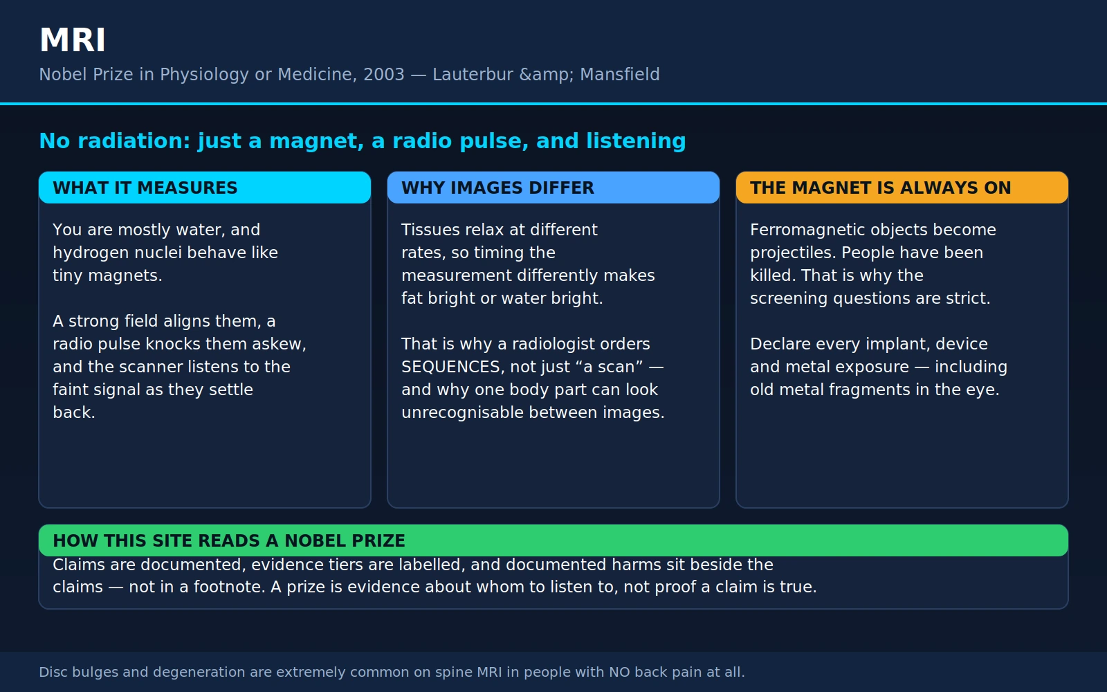
In 2003 the Nobel Assembly gave the Prize in Physiology or Medicine to an American chemist and a British physicist — neither of them a physician — "for their discoveries concerning magnetic resonance imaging." Between them, Paul Lauterbur and Peter Mansfield had worked out how to take a measurement that until then produced a single number about a test tube and turn it into a picture of the inside of a living person, without a single X-ray.
This page is written for the person who has been sent for an MRI, or who has just had one and is holding a report full of words like "disc bulge," "T2 hyperintensity" and "incidental finding." It covers the physics in plain language, because the plain-language explanations you find elsewhere are usually too vague to be useful; then it covers the four things that actually change how a scan goes for you — safety, contrast, incidental findings, and what to ask beforehand.
Table of Contents
- The Prize and the Two Men
- What MRI Actually Measures
- Lauterbur's Idea: The Gradient
- Mansfield's Contribution: Making It Fast
- T1, T2, and Why Two Images Look Nothing Alike
- What MRI Is Best For — and When CT Wins
- Safety: The Magnet Is Always On
- Gadolinium Contrast, Honestly
- Incidental Findings and Over-Imaging
- fMRI: What It Shows and What It Doesn't
- What to Ask Before an MRI
- Where Medicine Agrees, and What's Debated
- Key Research Papers
- Connections
- Featured Videos
1. The Prize and the Two Men
The 2003 Nobel Prize in Physiology or Medicine was shared by Paul Christian Lauterbur (1929–2007) and Sir Peter Mansfield (1933–2017). The citation was short: for their discoveries concerning magnetic resonance imaging. It is one of the few medicine prizes given for a machine rather than a mechanism, a molecule or a microbe — and it is one of the few whose product almost everyone reading this has either had, or will have.
Paul Lauterbur, and the napkin
Lauterbur was born in Sidney, Ohio, and trained as a chemist. Through the 1950s and 1960s he worked with nuclear magnetic resonance — NMR — which by then was a standard laboratory tool for chemists. You put a sample in a strong magnet, hit it with radio waves, listen to what comes back, and deduce something about the molecules inside. It was analytical chemistry, not imaging. The whole sample gave one answer.
In September 1971, by his own account, Lauterbur was in a restaurant in Pittsburgh — he described it as a fast-food place, over a hamburger — when the idea arrived: if the magnetic field were deliberately made uneven, different parts of the sample would resonate at different frequencies, and the frequency would tell you where the signal came from. He wrote it down on a paper napkin. That evening he bought a notebook, wrote the idea up properly, and had it signed and dated by a witness.
The napkin story is worth flagging as exactly what it is: Lauterbur's own telling, repeated by him over the years. It is not independently documented, and origin stories of this kind tend to get smoother with each retelling. What is documented is the notebook entry and, eighteen months later, the paper.
Peter Mansfield, who left school at fifteen
Mansfield's path is worth stating plainly, because it is unusual for a Nobel laureate and it is routinely glossed over. He was born in Lambeth, south London, and left school at fifteen. His first job was as a printer's assistant. He worked afterwards as a scientific assistant at a rocket propulsion establishment, did his National Service in the Army, and only then went back to education — taking the qualifications he had never sat, earning a physics degree at Queen Mary College, London, in 1959, and a PhD in 1962. He spent essentially his whole academic career at the University of Nottingham. He was knighted in 1993 and shared the Nobel Prize ten years after that.
There is no moral to extract from this beyond the obvious one, which is that the pipeline that selects fifteen-year-olds out of science is not a good predictor of anything.
The omission: Raymond Damadian
This prize carries one of the most publicly contested omissions in the history of the Nobel. Raymond Damadian (1936–2022), an American physician and scientist, was not included, and he objected loudly and at length.
The facts, as far as they are documented:
- 1971. Damadian published a single-author paper in Science showing that NMR relaxation times — T1 and T2, the two numbers that describe how quickly nuclei return to equilibrium — differed measurably between malignant tumours and normal tissue. Working in rats, he measured six normal tissues (muscle, kidney, stomach, intestine, brain and liver) and two malignant tumours (Walker sarcoma and Novikoff hepatoma) and found the tumour values fell distinctly outside the normal range. He also measured two benign fibroadenomas, whose T1 values were like muscle rather than like the cancers. That paper is real, it is his, and it predates Lauterbur's by two years.
- 1972. He filed a patent application covering apparatus and a method for detecting cancer in tissue by NMR, granted in 1974.
- 1977. His group at the State University of New York, Downstate, built a whole-body superconducting scanner they nicknamed Indomitable and produced a cross-sectional scan of a human chest. He went on to found FONAR Corporation, a commercial MRI manufacturer.
- 1988. Damadian and Lauterbur jointly received the United States National Medal of Technology. Damadian was inducted into the National Inventors Hall of Fame the following year.
- 2003. After the Nobel announcement, Damadian bought full-page advertisements in major American newspapers, including The New York Times and The Washington Post, under the headline "The Shameful Wrong That Must Be Righted," arguing that the prize could not honestly be given for MRI without him.
The case for including him, as his supporters made it: he was first to publish that NMR could distinguish diseased from healthy tissue, which is the medical premise of the entire field; he was first to patent a medical NMR scanning apparatus; and his group produced an early whole-body human scan. Without a medical reason to image the body, the imaging methods are a physics curiosity.
The case against, as it was argued at the time: the 1971 finding was a measurement, not an image, and it contained no method for locating a signal in space — which is precisely the problem Lauterbur solved and Mansfield made fast. The T1/T2 difference between tumour and normal tissue also turned out to be too non-specific to serve as a cancer test in the way Damadian originally proposed; clinical MRI does not diagnose cancer by relaxation-time thresholds. And the field-focusing technique Damadian's own scanner used was not the technique that became clinical MRI; gradient encoding and Fourier reconstruction were.
Two structural facts sit underneath the argument and are not in dispute: a Nobel Prize may be shared by at most three people, and the Nobel Committee never explains an omission and seals its deliberations for fifty years. Some of Damadian's supporters alleged that his public young-earth creationism counted against him. That claim cannot be tested against any evidence, precisely because of the secrecy rule, and no member of the committee has confirmed it — it should be recorded as an allegation, not as a finding.
This site keeps an honest record of contested Nobel decisions rather than a tidy one. We are not going to adjudicate this one. Both accounts above are accurate; which of them you weigh more heavily depends on whether you think a prize for MRI is a prize for the medical idea or for the imaging method, and reasonable people have never agreed on that.
2. What MRI Actually Measures
Almost every popular explanation of MRI stops at "it uses magnets and radio waves," which is true and tells you nothing. Here is the mechanism in full, in ordinary words.
Step one: you are mostly water, and water has hydrogen in it
An adult body is roughly 60% water by mass, and water is two hydrogen atoms bonded to an oxygen. Fat contains a great deal of hydrogen too. The nucleus of ordinary hydrogen is a single proton, and there are something on the order of 1027 of them in an adult. This matters because a proton has a property called spin, and a spinning charge behaves like an extraordinarily small bar magnet — it has a north and a south end.
Normally those tiny magnets point in every direction at random and cancel out. You are not magnetic.
Step two: a very strong magnet lines a few of them up
Put the body inside a powerful, uniform magnetic field and a very slight majority of those proton magnets settle into alignment with it. The excess is tiny — parts per million — but there are so many protons that the leftover adds up to a small net magnetisation pointing along the field.
Clinical scanners run at 1.5 tesla or 3 tesla. The Earth's magnetic field is about 50 microtesla, so a 1.5 T scanner is on the order of thirty thousand times stronger than the field that moves a compass needle. That number is the reason for everything in the safety section below.
Aligned protons do not sit still; they wobble around the field direction like a spun top that is starting to lean, at a rate called the Larmor frequency that is strictly proportional to the field strength. For hydrogen the constant is about 42.6 megahertz per tesla. At 1.5 T that is roughly 64 MHz; at 3 T, roughly 128 MHz. Those are radio frequencies — the FM broadcast band sits at 88–108 MHz, right between them. An MRI scanner is, quite literally, a very large and very precisely tuned radio.
Step three: a radio pulse knocks them over
Transmit a pulse of radio waves at exactly the Larmor frequency and the protons absorb energy and tip away from alignment — they resonate. That is the "resonance" in magnetic resonance. Nothing else in the body responds, because nothing else is tuned to that frequency at that field strength.
Step four: they relax, and the scanner listens
Switch the pulse off and the protons drift back into alignment, releasing the energy they absorbed as a faint radio signal that receiver coils pick up. That returning whisper is the entire raw data of an MRI. Everything you see on the images is reconstructed from how strong that signal is and how quickly it fades, point by point through the body.
What is not involved
No X-rays. No gamma rays. No radioactive tracer. No ionising radiation of any kind, which means no radiation dose and no radiation-related cancer risk, and no cumulative lifetime budget to worry about. A patient can have twenty MRIs without a dose ledger, which is not true of CT or nuclear medicine.
The energies involved are radio-frequency — the same part of the spectrum as a mobile phone or a television transmitter — and they are far too low to break a chemical bond or damage DNA. This is the single most important practical thing about MRI, and it is why the risks that do exist are entirely different in kind from radiation risks: they are mechanical, thermal and chemical, and they are covered in sections 7 and 8.
3. Lauterbur's Idea: The Gradient
Here is the problem Lauterbur solved, and it is worth understanding because it is the whole trick.
In a perfectly uniform magnetic field, every proton in the body resonates at the same frequency. You transmit your pulse, you listen, and you get back one signal that is the sum of everything in the coil. It tells you how much hydrogen is present overall and something about the average environment it sits in. It cannot tell you whether the signal came from the liver or the kidney, because there is nothing in the measurement that encodes position. Damadian's 1971 measurement had exactly this limitation: it could tell tumour tissue from normal tissue in a sample, but it had no way of saying where in a body a tumour was.
The insight
Make the magnetic field deliberately non-uniform — stronger at one end of the body than the other, varying smoothly in between. Because resonance frequency is strictly proportional to field strength, position now maps onto frequency. A proton at the head end of the gradient sings at a slightly higher pitch than one at the foot end. Listen to the returning signal, break it into its constituent frequencies, and you have broken the body into slices along that direction. Do the same with gradients in other directions and you can locate a signal in three dimensions.
That is it. That is what turned a bulk chemical measurement into a picture. Every MRI scanner ever built contains three sets of gradient coils — for the head-foot, left-right and front-back directions — that switch on and off many times a second during a scan. The banging, clicking and buzzing that patients complain about is the sound of those coils being kicked mechanically by the forces on them as their currents switch. The noise is Lauterbur's idea, audibly at work.
Zeugmatography
Lauterbur named the technique zeugmatography, from the Greek zeugma, a yoking or joining — a reference to the joining of the two magnetic fields, the strong uniform one and the weak gradient. The name did not survive, and it is easy to see why. The field settled on "NMR imaging," then dropped the word "nuclear" in the early 1980s when hospitals concluded that patients would hear "nuclear" and think of radioactivity. Hence magnetic resonance imaging: a name chosen partly for marketing, describing a technology whose defining virtue is that it involves no radiation at all.
The paper Nature turned down
Lauterbur wrote the work up and submitted it to Nature, which rejected it. By his account the objection was that the work was not of sufficiently broad significance. He appealed, revised the manuscript, and it appeared on 16 March 1973 as a two-page communication: "Image Formation by Induced Local Interactions: Examples Employing Nuclear Magnetic Resonance."
The image in that paper is not a picture of a person or an organ. It is a reconstruction of small tubes of ordinary water sitting inside a container of heavy water — a deliberately simple object whose true shape was known in advance, so that the reconstructed picture could be checked against reality. That was the point. The claim being made was not "look at this anatomy," it was "the spatial structure came out right."
Two pages, one author, one rejection, and the beginning of a technology that now runs somewhere in the region of a hundred million examinations a year worldwide.
4. Mansfield's Contribution: Making It Fast
Lauterbur's method worked, and it was agonisingly slow. Early images took many minutes to hours to acquire, during which the subject had to stay perfectly still. That is survivable for a test tube. It is not survivable for a beating heart, a breathing chest, a swallowing throat, a restless child, or a person in pain. Without a solution to speed, MRI would have stayed where NMR already was: a laboratory instrument.
The mathematics: signal and space
Mansfield, working at Nottingham, came at the problem as a physicist rather than a chemist. With P. K. Grannell he published in 1973 — in a solid-state physics journal, months after Lauterbur — an analysis showing that the NMR signal collected under gradients and the spatial structure of the object stand in a Fourier relationship to one another. In plain terms: the raw data an MRI collects is not a picture and does not look remotely like one. It is a set of measurements in a mathematical space (now universally called k-space), and the image is obtained by applying a Fourier transform to it.
This sounds abstract and is intensely practical. Once you know that scanning means "filling in k-space," you can start asking how to fill it in efficiently — which paths through it are fastest, which parts matter most, which parts can be skipped or guessed. Essentially every acceleration technique developed since, up to and including the compressed-sensing and machine-learning reconstructions in scanners today, is an answer to that question.
Echo-planar imaging
Mansfield's own answer, published in 1977, was echo-planar imaging (EPI): rather than exciting the tissue once per line of data, excite it once and then sweep the gradients back and forth extremely rapidly to collect an entire image plane from a single excitation.
EPI reduced the acquisition of one image from minutes to a fraction of a second. It demanded gradient hardware far beyond what existed in 1977, and it took roughly fifteen years for commercial scanners to catch up with the idea. When they did, it changed what MRI was for:
- Diffusion-weighted imaging — the sequence that shows an acute stroke — is built on EPI. It has to be, because it must freeze out any bulk motion of the head to measure the microscopic motion of water.
- Functional MRI is built on EPI. Tracking a signal that changes over seconds requires whole-brain images every couple of seconds.
- Cardiac and abdominal imaging in a single breath-hold became possible, which is what made MRI usable in the torso at all.
Mansfield's group also published early demonstrations of NMR imaging applied to human anatomy, including a 1977 paper with A. A. Maudsley in the British Journal of Radiology — one of the earliest appearances of this physics in a clinical journal, which is a marker of the moment the field stopped being physics and started being medicine.
If Lauterbur made the image possible, Mansfield made it practical. The prize is correctly a shared one.
5. T1, T2, and Why Two Images of the Same Knee Look Nothing Alike
This is the section that answers the question patients most often have when they look at their own images: why does the same body part look completely different on two pictures taken half an hour apart?
Two clocks, not one
When the radio pulse stops, the protons return to equilibrium by two independent processes, running on two different clocks:
- T1 — longitudinal relaxation. How fast the magnetisation re-aligns with the main magnetic field, giving its absorbed energy back to its surroundings. Think of it as how quickly the tissue "recharges" and is ready to be pulsed again.
- T2 — transverse relaxation. How fast the protons, which started out wobbling in step with each other, fall out of step and cancel each other out. Think of it as how quickly the signal "loses coherence" and fades.
Every tissue has its own characteristic T1 and T2, set by how tightly its water molecules are held. Water in free fluid — cerebrospinal fluid, urine, a joint effusion, the fluid in inflamed tissue — tumbles freely and has a long T1 and a long T2. Fat has a short T1. Water bound up in dense structures like tendon, ligament and cortical bone has a very short T2, which is why those structures are almost black on nearly every sequence.
The radiographer chooses which clock to look at
By changing the timing of the pulses — how long between pulses (TR) and how long you wait before listening (TE) — you can make the image depend mostly on T1, mostly on T2, or mostly on how much hydrogen is present. Nothing about the patient changes. Only the question being asked changes.
- T1-weighted: fat is bright, free fluid is dark. Good anatomy; this is the "normal-looking" picture where the brain looks like a brain. Also the sequence on which gadolinium contrast shows up bright.
- T2-weighted: free fluid is bright. Since most disease processes — inflammation, oedema, infection, many tumours, acute injury — involve extra water, T2 is where pathology tends to glow. The old radiology aphorism is that T2 shows you where the trouble is and T1 shows you what it looks like.
- FLAIR (fluid-attenuated inversion recovery): a T2-weighted image with the signal from plain cerebrospinal fluid deliberately nulled to black. Without it, a bright lesion sitting next to a bright ventricle is invisible. FLAIR is why multiple sclerosis plaques around the ventricles are so conspicuous on brain MRI, and it is standard on essentially every brain protocol.
- Diffusion-weighted imaging (DWI): sensitive to the microscopic random motion of water molecules. In an acute ischaemic stroke the affected cells swell and water can no longer move freely, so the infarct becomes bright on DWI within minutes — long before it appears on conventional imaging, and long before CT shows anything. The 1992 study by Warach and colleagues showed this directly in patients: diffusion imaging revealed infarcts sooner than conventional T2 spin-echo imaging, and four hyperacute infarcts were visible only on diffusion. DWI is also heavily used in cancer imaging, because densely packed tumour tissue restricts diffusion too.
- Fat suppression (STIR and similar): switch fat off so that bright fat cannot hide bright fluid. Essential in musculoskeletal imaging, where bone marrow oedema from an occult fracture is bright fluid sitting inside bright fatty marrow.
- Gradient-echo and susceptibility-weighted imaging: exquisitely sensitive to blood products, calcium and iron. This is how tiny old microbleeds are found.
- MR angiography: images blood vessels, often with no injected contrast at all, by exploiting the fact that blood is moving and stationary tissue is not.
Why this matters to you as a patient
Three practical consequences follow.
First, your doctor does not order "an MRI." They order a protocol — a named list of sequences chosen for a specific question. "MRI lumbar spine" and "MRI lumbar spine for suspected infection" are different examinations of the same anatomy. If the clinical question given on the request is wrong or vague, the wrong sequences get run, and the answer you needed may simply not be in the images. When you are told "the MRI was normal," it is legitimate to ask what question the scan was set up to answer.
Second, the same body part genuinely does look unrecognisably different from one image to the next in the same session. This is not an error and it is not two different scans. It is the same tissue interrogated with different timing.
Third, this flexibility is MRI's real advantage over every other imaging method. A CT scanner measures one physical property — how much X-ray a tissue absorbs — and everything on the image is a shade of that one variable. MRI can be tuned to water content, fat content, molecular motion, blood flow, blood oxygenation, iron content, chemical composition and more, from the same machine in the same sitting. That is why an MRI takes 30 to 60 minutes while a CT takes seconds: you are not taking one picture, you are running a battery of physically different experiments.
6. What MRI Is Genuinely Best For — and When CT Wins
MRI is not "the better scan." It is a different scan, and there are common, urgent situations in which asking for an MRI would be a mistake.
MRI is the right tool for
- Brain and spinal cord. Nothing else comes close for soft-tissue contrast inside the skull and spinal canal. Multiple sclerosis, brain tumours, epilepsy, dementia workup, spinal cord compression, cranial nerve problems.
- Soft tissue generally — muscle, tendon, ligament, cartilage, nerve, bone marrow. A torn meniscus, a ruptured cruciate ligament, a rotator cuff tear, a soft-tissue mass.
- Bone marrow, as distinct from bone. This is a subtle but important one: CT shows the mineralised skeleton better, but MRI is far better at what is going on inside the bone. Marrow oedema from a stress fracture, early osteomyelitis, and marrow infiltration by tumour are often invisible on CT and obvious on MRI.
- Tumour characterisation and local staging — how far a rectal or cervical or prostate cancer extends, whether a liver lesion is a haemangioma or something worse.
- Liver and pelvis. Multiparametric prostate MRI, endometriosis, uterine and adnexal masses, fistulas, perianal disease.
- Cardiac function and tissue characterisation. Cardiac MRI measures ventricular volumes and ejection fraction with a precision echocardiography cannot match, and it can distinguish scar from viable muscle, and infiltration from inflammation, in a way no other test does non-invasively. See Cardiology.
- Anyone who should not accumulate radiation dose — children, pregnant patients (without contrast), and people with conditions requiring repeated lifelong imaging such as Crohn's disease or MS.
CT is the right tool for
- Speed. A CT of the head takes seconds. An MRI takes half an hour or more, during which the patient is inside a tube, out of easy reach, and cannot have ferromagnetic monitoring or resuscitation equipment nearby. For an unstable patient this is decisive.
- Acute bleeding. Fresh blood in the brain, abdomen or chest is what CT does best, and it is why every suspected stroke gets a CT first — not to find the stroke, but to rule out a haemorrhage before clot-busting treatment can be given.
- Cortical bone and fractures. Complex fracture patterns, facial and skull-base bones, spinal fracture geometry before surgery.
- Lung. Air-filled lung has almost no signal on MRI. CT is the lung imaging method.
- Major trauma, where a whole-body CT in a couple of minutes answers a dozen questions at once.
- Kidney stones and many acute abdomens, where CT is faster, cheaper and more available.
- Anyone who cannot have an MRI for the reasons in the next section.
The honest summary: CT is fast, cheap, available, excellent for bone, blood and lung, and costs you radiation. MRI is slow, expensive, less available, unrivalled for soft tissue, and costs you radiation not at all. Ultrasound, which is fast, cheap, radiation-free and portable but limited by bone and gas, remains the right first answer more often than either.
7. Safety: The Magnet Is Always On
This is the section that matters most on the day of your scan.
The headline advantage, stated once more
MRI uses no ionising radiation. There is no dose, no cumulative risk, no reason to ration scans across a lifetime on radiation grounds. If you have been advised to have an MRI rather than a CT specifically to avoid radiation, that advice is sound.
The headline hazard: the magnet never switches off
People assume the magnet turns on when the scan starts, like an X-ray tube. It does not. A clinical MRI magnet is superconducting — its coils are cooled with liquid helium to a few degrees above absolute zero, where they carry current with no resistance. The current, once established, circulates indefinitely. The field is at full strength overnight, at weekends, during maintenance, and during a power cut. Removing it requires a "quench," a deliberate emergency procedure that boils off the helium, takes the scanner out of service for days and costs a great deal of money. It is not something anyone does to retrieve a dropped object.
The consequence is that any ferromagnetic object brought into the room is accelerated toward the bore and becomes a projectile. Documented cases involve oxygen and nitrous oxide cylinders, wheelchairs, floor polishers, mop buckets, stretchers, chairs, tool boxes, scissors, hair clips, keys and oxygen regulators. A 2001 report in the American Journal of Roentgenology described projectile accidents caused by ferromagnetic gas cylinders taken into MR suites — the specific hazard that killed a six-year-old boy in a New York hospital that same year, when an oxygen cylinder was pulled into the bore while he was inside the scanner.
This is why the screening questionnaire is repetitive and why staff will ask you the same questions three times and make you change into a gown and empty your pockets even though you have "nothing metal." The American College of Radiology's MR safe practice guidance defines a four-zone access system around the magnet room, with screening required before anyone crosses into the controlled zones. Take it seriously; it exists because of deaths.
Implants and devices
Every implant falls into one of three labels: MR Safe (poses no known hazard in any MR environment), MR Conditional (safe under specified conditions — a maximum field strength, a maximum rate of gradient switching, a maximum radiofrequency power, sometimes a specific body position or a waiting period), or MR Unsafe. The great majority of modern implants are conditional, which means the scan can go ahead if the department knows exactly what you have and follows the manufacturer's conditions.
- Cardiac pacemakers and defibrillators. "A pacemaker means no MRI" is outdated. Most devices implanted in recent years are explicitly MR-conditional and are scanned routinely with the device reprogrammed for the duration. Beyond that, a 2017 study in the New England Journal of Medicine scanned 1,509 patients who had legacy pacemakers or defibrillators — older devices never labelled MR-conditional — across 2,103 examinations at 1.5 tesla under a prespecified safety protocol. No long-term clinically significant adverse events occurred. In nine examinations (0.4%) the device reset to a backup mode, and in eight of those nine the reset was transient. This is a strong result, but note what it does and does not license: it was performed under a strict protocol with cardiology support, not as an ordinary walk-in scan. If you have a cardiac device, MRI is very often possible — ask, do not assume.
- Cerebral aneurysm clips. Older ferromagnetic clips are a genuine contraindication. Modern clips are typically titanium and MR conditional. If you have one, the department needs documentation of the exact model.
- Cochlear implants. Many current models are MR conditional, some requiring the internal magnet to be removed or the head tightly bandaged, and some are restricted to 1.5 T. Some older implants are unsafe. Again: model-specific.
- Neurostimulators, deep-brain stimulators, spinal cord stimulators, insulin and drug pumps — almost all conditional, all requiring specific programming or removal.
- Stents, coils, filters, heart valves, joint replacements, plates, screws, and surgical clips — the overwhelming majority are non-ferromagnetic and safe. They may still cause a local distortion of the image around themselves.
- Metal fragments in the eye. This is the one occupational history that must never be skipped. If you have ever ground, drilled or welded metal without eye protection, say so. A fragment in the orbit can move and blind you. Departments will often obtain plain orbital radiographs first.
- Shrapnel, bullets and retained fragments anywhere else — declare them.
Heating: tattoos, patches and cosmetics
The transmitted radio pulses deposit a small amount of energy in tissue as heat, which is monitored and limited by the scanner (the specific absorption rate, SAR). It is not a problem in ordinary tissue. It can be a problem where a conductive loop concentrates it.
Tattoo pigments can contain iron oxide, and there are documented cases of tattoo sites heating, tingling, swelling or burning during a scan — including a published case report of a burn over lower-limb tattoos in a professional footballer during a pelvic MRI. This is uncommon, not a reason to cancel a scan, and mostly affects large, dark, older tattoos. Tell the radiographer where your tattoos are and report any heating immediately; they can stop the sequence. The same applies to permanent cosmetics and cosmetic tattooing around the eyes and lips.
Transdermal patches — nicotine, hormone, opioid and others — sometimes have a metallic backing foil that can heat and burn. They should be removed before the scan and replaced afterwards. Tell them what you are wearing.
Also remove or declare: hearing aids, removable dentures, wigs and hairpieces with metal clips, magnetic eyelashes, body piercings, and anything containing a battery.
Noise
The gradient coils are loud — commonly over 100 decibels, comparable to a power tool at close range. Hearing protection is mandatory, not optional, and you should insist on it if it is not offered. Ear plugs, ear defenders, or headphones playing music are all standard.
Claustrophobia and the bore
A conventional scanner bore is a tube roughly 60 cm across, and you are inside it, sometimes with a coil close over the body part being scanned. A substantial minority of people find this difficult, and it is nothing to be embarrassed about.
Options genuinely exist: wide-bore scanners (typically 70 cm), short-bore scanners where much of the body sits outside the tube, open scanners with a C-shaped or vertical-field design, feet-first or prone positioning for many examinations, prism glasses or mirrors that let you see out of the bore, music, a companion in the room, and oral or intravenous sedation.
Be aware that the evidence for the newer designs is more modest than the marketing. A randomised trial of 174 patients with elevated claustrophobia scores compared a short-bore scanner against an open scanner: claustrophobic events occurred in 39% of the short-bore group and 26% of the open group — a difference that did not reach statistical significance (P = 0.08). The authors' own conclusion was blunt: even modern scanner designs do not prevent claustrophobia. What the trial did show is that events happened sooner in the short-bore group, and that people who had an event were more likely to report worse claustrophobia seven months later. The practical lesson is to raise the problem before the appointment, when planning and premedication are still possible, rather than discovering it on the table.
Pregnancy
MRI without contrast is generally regarded as acceptable in pregnancy when there is a clinical indication, and is often preferred precisely because it avoids radiation. Gadolinium contrast crosses the placenta and is generally avoided in pregnancy unless the benefit is considered to outweigh an uncertain risk. Tell the department if you are or might be pregnant.
8. Gadolinium Contrast, Honestly
Start with the fact that gets lost: most MRI examinations do not need contrast at all. Routine knee, shoulder, spine and brain-screening scans are usually done without it. Contrast is added when the question is about blood supply, breakdown of the blood-brain barrier, inflammation, infection or tumour behaviour.
What it is
Gadolinium is a lanthanide metal that is strongly paramagnetic — it shortens the T1 of nearby water dramatically, so tissue that takes it up turns bright on T1-weighted images. The free gadolinium ion is toxic, so it is never given free: it is wrapped inside a chelating molecule that holds it and is excreted by the kidneys. Gadolinium-based contrast agents differ in how tightly they hold the metal. Linear agents wrap it in an open-chain molecule; macrocyclic agents wrap it in a closed ring and hold it far more securely. That distinction turns out to matter for both concerns below.
Nephrogenic systemic fibrosis
In 2006 two independent reports linked a rare and severe fibrosing condition — skin thickening and hardening that could progress to contractures and internal organ involvement, sometimes fatal — to gadolinium contrast given to patients with severe kidney impairment. Grobner's paper in Nephrology Dialysis Transplantation proposed gadolinium as a specific trigger; Marckmann and colleagues in the Journal of the American Society of Nephrology implicated gadodiamide, a linear agent, in a series of cases. The mechanism is thought to be retention of the agent in a patient who cannot clear it, allowing gadolinium to dissociate from a loosely binding chelate.
The response was rapid and, by the standards of drug safety, effective. Renal function is now checked before contrast in at-risk patients, the highest-risk linear agents were restricted or withdrawn in many countries, and macrocyclic agents became standard. The result is that NSF has become vanishingly rare. A 2020 systematic review and meta-analysis in JAMA Internal Medicine pooled 16 studies covering 4,931 patients with stage 4 or 5 chronic kidney disease who received a modern group II agent and found zero cases of unconfounded NSF — a pooled incidence of 0%, with the upper bound of the 95% confidence interval at 0.07%. The authors' conclusion is worth quoting in substance: the diagnostic harm of withholding a needed contrast-enhanced scan in this population may well outweigh the residual NSF risk.
If you have significantly reduced kidney function, expect the department to check your eGFR and to discuss it. That is the system working, not a red flag.
Gadolinium retention in the brain
This is the live concern, and it deserves to be stated exactly, because both the reassurance and the alarm circulating online overstate what is known.
What is documented. In 2014, Kanda and colleagues reported in Radiology that patients who had received more gadolinium doses showed progressively higher signal in two specific brain structures — the dentate nucleus of the cerebellum and the globus pallidus — on unenhanced T1-weighted images, meaning images taken with no contrast in the body at that moment. The correlation with cumulative number of administrations was strong and highly significant. Subsequent work, including tissue analysis at autopsy, confirmed that gadolinium is genuinely deposited in brain tissue, that it happens even in people with normal kidney function, and that it happens more with linear than with macrocyclic agents.
What is not established. Whether any of this causes harm. No consistent neurological syndrome, cognitive deficit or disease has been shown to result from retained gadolinium in patients with normal renal function. That is not the same as "it is proven safe" — it is a genuine open question that has been studied and has not produced a signal. A 2017 summary of the evidence with recommendations in The Lancet Neurology, and a 2018 research roadmap in Radiology arising from a joint NIH, ACR and RSNA workshop, both reach the same position: retention is real, the clinical significance is unknown, and the sensible response is to use the lowest effective dose, prefer macrocyclic agents, and not give contrast that is not needed.
Regulators took the same view. The US Food and Drug Administration required a class warning and a patient Medication Guide about retention in 2017 while stating that it had not found evidence of harm outside NSF; the European Medicines Agency suspended several linear agents.
The honest summary for a patient: a small amount of gadolinium is retained in the body, including in brain tissue, after contrast-enhanced MRI. It is visible on imaging and measurable in tissue. Nobody has demonstrated that it makes anybody ill. If you need a contrast-enhanced scan for a real clinical question, have it. If you are being offered contrast routinely and nobody can tell you what question it answers, ask.
Other reactions
Acute allergic-type reactions to gadolinium agents occur but are considerably less common than with the iodinated contrast used for CT. Mild reactions — hives, nausea — are the usual form. Severe reactions are rare. Some patients report transient discomfort or a cold sensation at the injection site, which is expected.
9. Incidental Findings and Over-Imaging
This section contains what may be the single most useful fact on this page.
MRI's sensitivity is a genuine double-edged property. A test that can see almost everything will see a great deal that does not matter, and there is no marker on the image distinguishing a finding that explains your symptoms from one that has been sitting there quietly for twenty years.
The spine, in people with no pain at all
This has been studied repeatedly, in different countries, decades apart, with consistent results.
- Boden and colleagues, 1990. Sixty-seven people who had never had low-back pain, sciatica or neurogenic claudication were scanned; three neuroradiologists read the images blinded to symptoms. About a third had a substantial abnormality. Among those under sixty, 20% had a herniated nucleus pulposus — a herniated disc — and one had spinal stenosis. Among those sixty and over, around 57% of scans were abnormal: 36% with a herniated disc and 21% with spinal stenosis. Disc degeneration or bulging was present at at least one level in 35% of the twenty-to-thirty-nine-year-olds and in all but one of the sixty-to-eighty-year-olds.
- Jensen and colleagues, 1994, New England Journal of Medicine. Ninety-eight asymptomatic people. Only 36% had normal discs at every level. Fifty-two per cent had a bulge at at least one level, 27% a protrusion, 1% an extrusion. Thirty-eight per cent had more than one abnormal disc. The authors' conclusion: given how common both these findings and back pain are, discovering a bulge or protrusion on the MRI of somebody with back pain "may frequently be coincidental."
- Brinjikji and colleagues, 2015. A systematic review pooling 33 studies and 3,110 asymptomatic individuals, producing age-specific prevalence curves. Disc degeneration rose from 37% at age 20 to 96% at age 80. Disc bulge rose from 30% to 84%. Disc protrusion from 29% to 43%. Annular fissures from 19% to 29%. Their conclusion: many of these imaging features are simply part of normal ageing and are unassociated with pain.
Read those numbers again. A degenerated disc on a spine MRI in a fifty-year-old is a finding roughly as diagnostic as grey hair. It is present in most people that age who feel completely well. It may be the cause of your pain. It may equally have nothing to do with it. The image alone cannot tell you which, and a report that lists it is not making a claim that it can.
The knee tells the same story
A 2012 population-based study in the BMJ from the Framingham Osteoarthritis Study imaged 710 people over fifty who had no radiographic evidence of knee osteoarthritis at all. Any abnormality was found in 89% of them. Osteophytes in 74%, cartilage damage in 69%, bone marrow lesions in 52%. And critically: "any abnormality" was present in 86–88% of the painless knees, against 90–97% of the painful ones. A meniscal tear or a cartilage defect on a knee MRI in a person over fifty is close to a normal finding.
The brain, too
The Rotterdam Study scanned 2,000 people from the general population, mean age 63. Asymptomatic brain infarcts — strokes nobody knew had happened — were present in 7.2%. Cerebral aneurysms in 1.8%. Benign primary tumours, mostly meningiomas, in 1.6%. If you scan a hundred ordinary older adults' brains for any reason, you will find something unexpected in a meaningful number of them, and then you have to decide what to do about it.
What happens when you image back pain early
This is where the sensitivity stops being a curiosity and starts affecting outcomes.
A 2003 randomised controlled trial in JAMA assigned 380 primary-care patients whose doctors had ordered lumbar X-rays to receive either the X-rays or rapid MRI instead. At twelve months, back-related disability was essentially identical in the two groups. Every secondary outcome — pain bothersomeness, pain frequency, physical functioning, quality of life — was statistically indistinguishable. Ten patients in the MRI group had lumbar spine operations against four in the X-ray group; that difference had a confidence interval crossing zero, so it is suggestive rather than proven. Costs were higher in the MRI arm. The authors' conclusion was that substituting rapid MRI for radiographs "may offer little additional benefit to patients, and it may increase the costs of care because of the increased number of spine operations."
A 2013 study in Spine looked at 555 workers with acute, disabling, work-related low back pain in whom, after review of the clinical records, early MRI was not indicated by guidelines. Those who received an MRI within thirty days had markedly lower rates of coming off disability and, on average, $12,948 to $13,816 higher medical costs than those who did not, after adjustment for demographics and severity. Even among people with minimal ongoing disability, costs ran $7,643 to $8,584 higher. The authors describe an "iatrogenic effect" — harm caused by the medical process itself.
The important caveat: that second study is observational, not randomised. Doctors do not order scans at random, and it is always possible that something unmeasured about the patients who got early scans also predicted worse outcomes. The authors excluded cases where imaging might have been indicated and adjusted for severity, which helps, but confounding by indication cannot be ruled out by design. The randomised trial and the cohort study point the same direction, which is what makes the finding credible; neither alone would be enough.
What this means for you
Clinical guidelines across countries now advise against routine imaging for uncomplicated low back pain in the first four to six weeks, in the absence of specific warning signs. Those warning signs — the reasons an early scan is right — include significant trauma, a history of cancer, unexplained weight loss, fever or suspected infection, intravenous drug use, immunosuppression, progressive neurological weakness, and any symptom of cauda equina compression: numbness in the saddle region, new bladder or bowel dysfunction, or bilateral leg symptoms. Cauda equina symptoms are an emergency and should be assessed the same day.
If none of those apply, and you are being told to wait rather than scan, that is not your doctor being cheap. It is the evidence. And if you have already been scanned and the report lists disc degeneration, bulges, osteophytes and facet arthropathy, the right question is not "how bad is that?" but "how many people my age without pain have exactly that?" — and the numbers above are the answer.
See also Low Back Pain and Herniated Disc.
10. fMRI: What It Shows and What It Doesn't
Functional MRI is the source of most of the brain images you have seen in newspapers, and most of what those articles claimed about them was overstated. It is worth separating what fMRI genuinely does from what it is sold as doing.
What is actually measured
fMRI does not measure neurons firing. It measures the blood-oxygen-level-dependent (BOLD) signal, and the chain from neural activity to that signal has several links in it.
Haemoglobin's magnetic properties depend on whether it is carrying oxygen. Deoxygenated haemoglobin is paramagnetic and disturbs the local magnetic field; oxygenated haemoglobin is not. Ogawa and colleagues demonstrated in 1990 that this difference produces measurable MRI contrast in the brain. When a region of the brain becomes more active, local blood flow increases — and it increases more than oxygen consumption does, so the proportion of deoxygenated haemoglobin actually falls, and the signal goes slightly up.
Three properties of that chain constrain everything fMRI can claim:
- It is indirect. It is a haemodynamic measure standing in for an electrical one.
- It is slow. The blood-flow response peaks several seconds after the neural event, while the neural event itself lasts milliseconds.
- It is small. The signal change is typically a few per cent at most, which is why fMRI requires repeated trials and heavy statistics to see anything at all.
None of this makes fMRI worthless. It makes it a measurement with a known and quite narrow resolution, whose conclusions live or die on the statistics applied to it.
Where fMRI has genuinely delivered
🟢 Presurgical mapping. Before removing a brain tumour or an epileptic focus, fMRI helps localise language and motor areas so the surgeon knows what to avoid. This is a real clinical use with real benefit, and it is individual-patient mapping rather than group inference.
🟢 Basic neuroscience. Large-scale functional organisation, resting-state networks, the discovery that the brain is highly active during "rest," and a great deal of systems neuroscience that could not have been done any other way in living humans.
🟢 Disorders of consciousness. Careful studies have detected consistent, task-driven brain responses in a small number of behaviourally unresponsive patients, which has changed how some of these patients are assessed. This is a research-grade finding with major implications, not a routine test.
Where the claims outrun the evidence
🟡 "This region lights up when you feel X." Reverse inference — concluding from activity in a region that a person is having a particular experience — is far weaker than it sounds, because most brain regions participate in many tasks. The amygdala does not mean fear; the insula does not mean disgust.
🔴 Commercial brain scans for psychiatric diagnosis. Clinics selling functional brain imaging to diagnose ADHD, depression, anxiety or "brain type," and to guide treatment, are ahead of the evidence. There is no validated fMRI or functional imaging test that diagnoses a psychiatric disorder in an individual, and no professional body endorses one.
🔴 Lie detection. Commercial fMRI-based lie detection has been marketed for two decades. Its validation rests largely on laboratory tasks in cooperative volunteers instructed to lie about trivial matters; real-world accuracy in motivated subjects is unestablished, countermeasures are plausible, and courts in the United States have generally excluded it.
The dead salmon
The standard cautionary demonstration in this field is the dead salmon, and it deserves telling accurately.
Craig Bennett and colleagues placed a dead Atlantic salmon, purchased from a market, in an MRI scanner and ran a standard fMRI experiment on it — showing it photographs of humans in social situations and "asking" it to judge the emotion each person was experiencing. They then analysed the data exactly as a great many published fMRI studies did at the time, using an uncorrected statistical threshold. A cluster of apparently active voxels duly appeared in the salmon's brain cavity.
The salmon was not thinking about anything. The point was arithmetic: an fMRI scan divides the brain into tens of thousands of voxels and runs a statistical test in every one of them. At a conventional threshold, a few per cent of those tests will be positive by chance alone. Without a correction for multiple comparisons, you will find "activity" in a dead fish.
On the status of the work, honestly: the salmon was presented as a poster at the 2009 Organization for Human Brain Mapping meeting and later written up in a small satirical venue, the Journal of Serendipitous and Unexpected Results. It has no PubMed record and is not a peer-reviewed journal article, and anyone citing it as one is mistaken. The same authors made the serious version of the argument in a peer-reviewed commentary in Social Cognitive and Affective Neuroscience in 2009, which laid out the principled methods — familywise error rate and false discovery rate control — and noted that papers using improperly corrected methods were still being published every month.
That concern has not gone away. In 2016 Eklund, Nichols and Knutsson used real resting-state data to run three million random "task" group analyses through the standard fMRI software packages. For a nominal 5% familywise error rate, they found the common parametric methods conservative for voxel-level inference but invalid for cluster-level inference — the form of inference most task fMRI papers rely on — while a non-parametric permutation test performed correctly. The likely culprit was an assumption about the spatial smoothness of noise that does not hold in real data. That paper carries a published correction in the same journal.
The upshot is not that fMRI is fake. It is that an fMRI result is a statistical claim before it is a biological one, and the correct question about any brain-imaging headline is what correction was applied and how many subjects were scanned.
11. What to Ask Before an MRI
Before the appointment is booked
- "Will the result change what we do?" This is the single best question to ask about any test. If the answer is a clear yes — it determines whether you have surgery, whether a drug is started, whether a cancer is staged one way or another — have the scan. If the answer is vague, ask what the plan is under each possible result. A scan whose every outcome leads to the same next step is a scan that will mostly generate incidental findings.
- "What specific question is the scan meant to answer?" This determines the protocol. It should be on the request form, and it should be more specific than "back pain."
- "Do I need contrast, and why?" Many scans do not. If contrast is planned, there should be a reason expressible in one sentence. If you have reduced kidney function, ask whether it has been checked and which agent will be used.
- "Is there existing imaging that can be compared?" A prior scan turns an ambiguous finding into a stable one or a changing one, which is often the whole answer. Bring discs or arrange transfer of prior studies from other hospitals.
- Cost and authorisation. Ask what the scan will cost, whether prior authorisation is needed, and whether a free-standing imaging centre is an option — in some health systems the price difference between a hospital and an outpatient centre for an identical scan is large.
Before you go in
- Declare every implant, device and metal exposure — without exception. Pacemaker or defibrillator; neurostimulator or deep-brain stimulator; cochlear implant; aneurysm clip; insulin or drug pump; stents, coils or filters; heart valve; joint replacement; plates, screws or wires; IUD; surgical staples or clips; dental implants and removable dentures; hearing aids; shrapnel or bullets; and any history of grinding, drilling or welding metal without eye protection, which raises the question of a retained fragment in the eye. Bring your implant identification card if you have one; the exact model number is what the department needs.
- Mention tattoos, permanent makeup and transdermal patches. Patches usually come off; tattoos usually stay, but the staff should know where they are.
- "How long will it take, and how will I be positioned?" Knowing it is twenty minutes rather than an hour changes how the time feels. Ask whether you can go in feet-first, whether the scan can be done prone, and whether the machine is a wide-bore.
- If enclosed spaces are difficult, say so now, not on the table. Ask about an open or wide-bore scanner, a mirror or prism glasses, music, having someone in the room, and whether an oral sedative can be prescribed in advance. All of these need arranging beforehand.
- If you are in pain, ask about timing your analgesia so that you are comfortable during the half hour you must lie still. Movement is the commonest reason a scan has to be repeated.
- Say if you might be pregnant.
Afterwards
- Ask for a copy of the report and the images. You are entitled to them in most health systems, and they matter for any future comparison.
- Ask which findings are relevant and which are incidental. A report may list eight things, of which one answers the question and seven are ordinary age-related changes. Getting the radiologist's or clinician's ranking is far more useful than reading the list alone.
- Ask what happens next for anything unexpected. An incidental finding usually needs either nothing, one follow-up scan at a stated interval, or a referral. Which of the three it is should be stated explicitly rather than left open.
- Report any burning, heating or pain that occurred during the scan. It is uncommon, it should be documented, and it changes how the next scan is set up.
12. Where Medicine Agrees, and What's Debated
🟢 Where mainstream medicine agrees
- MRI involves no ionising radiation and carries no radiation risk. Not contested by anyone.
- The static magnetic field is permanently on, and ferromagnetic objects are lethal in the magnet room. Universal, and the basis of formal safety zoning.
- MRI is the imaging method of choice for brain, spinal cord and soft tissue; CT is the method of choice for acute haemorrhage, cortical bone, lung, and any situation where speed matters.
- Degenerative findings on spine and knee MRI are extremely common in people without symptoms. Replicated across four decades and multiple populations.
- Routine early imaging for uncomplicated low back pain without red flags is not recommended, and is now a standard item on "do not do" lists across specialty societies.
- Nephrogenic systemic fibrosis is real, is linked to gadolinium in severe renal impairment, and has become extremely rare since linear agents were restricted and renal screening became standard.
- Modern MR-conditional cardiac devices can be scanned safely under manufacturer conditions; blanket refusal on the basis of "you have a pacemaker" is out of date.
- fMRI requires correction for multiple comparisons. No longer arguable.
🟡 What remains genuinely debated
- The clinical significance of gadolinium retention in the brain. Deposition is established. Harm is not. Some patient groups report symptoms attributed to retained gadolinium; no controlled study has established a syndrome, and the absence of a demonstrated effect is not the same as a demonstration of no effect. Practice has moved toward macrocyclic agents and lowest-effective-dose regardless.
- Where exactly to set the imaging threshold in back pain. Everyone agrees red flags warrant imaging and that day-one scans for ordinary mechanical back pain do not help. The boundary in between — persistent pain at six weeks, radicular pain that is improving slowly, the patient who cannot work — is a real clinical judgement, and reasonable guidelines differ.
- How much MRI should drive decisions in knee osteoarthritis. Given that most people over fifty have meniscal and cartilage findings whether or not their knee hurts, how much weight an MRI should carry in deciding on arthroscopy remains contested — and the trial evidence for arthroscopic partial meniscectomy in degenerative tears has been repeatedly unfavourable.
- Whether legacy (non-conditional) cardiac devices should be scanned routinely. The 2017 NEJM data are reassuring under protocol; how widely that generalises outside centres with cardiology support is a live question.
- Clinical claims for fMRI. Presurgical mapping is accepted. Beyond that — psychiatric diagnosis, treatment selection, prognosis, lie detection — the claims are contested and the commercial ones are, in the view of most academic neuroimagers, unsupported.
- Screening whole-body MRI for healthy people. Marketed directly to consumers and expanding rapidly. It generates incidental findings at high rates, and no randomised evidence shows it reduces mortality. Its advocates argue early detection must help; its critics point to the Rotterdam numbers above and ask what happens to the 7% with a silent infarct and the 1.8% with an aneurysm.
13. Key Research Papers
Every citation below was checked against PubMed or Crossref: journal, year, volume and pages confirmed against the record, and the abstract read for every finding stated on this page. Where a paper carries a published erratum or correction, that is disclosed. Where a foundational item has no PubMed record, that is stated rather than papered over with a plausible-looking link.
The founding papers
- Lauterbur PC. Image formation by induced local interactions: examples employing nuclear magnetic resonance. Nature, 1973;242(5394):190–191. The gradient paper — two pages, single author, initially rejected by the journal that published it. Verified via Crossref; this paper has no PubMed record, as PubMed's coverage of 1973 Nature is incomplete. Do not trust any PMID offered for it.
- Mansfield P, Grannell PK. NMR "diffraction" in solids? Journal of Physics C: Solid State Physics, 1973;6(22):L422–L426. The Fourier / reciprocal-space analysis underlying k-space. Verified via Crossref; a solid-state physics journal, not indexed in PubMed.
- Mansfield P. Multi-planar image formation using NMR spin echoes. Journal of Physics C: Solid State Physics, 1977;10(3):L55–L58. Echo-planar imaging — a whole image plane from one excitation, the basis of modern DWI and fMRI. Verified via Crossref; not in PubMed.
- Mansfield P, Maudsley AA. Medical imaging by NMR. British Journal of Radiology, 1977;50(591):188–194. PMID 849520. One of the earliest appearances of NMR imaging in a clinical journal.
- Damadian R. Tumor detection by nuclear magnetic resonance. Science, 1971;171(3976):1151–1153. PMID 5544870. The disputed priority paper: T1 and T2 relaxation times in six normal rat tissues versus Walker sarcoma and Novikoff hepatoma, with tumour values falling outside the normal range. A measurement, not an image.
- Ogawa S, Lee TM, Kay AR, Tank DW. Brain magnetic resonance imaging with contrast dependent on blood oxygenation. Proceedings of the National Academy of Sciences USA, 1990;87(24):9868–9872. PMID 2124706. The BOLD effect — the physical basis of every fMRI study since.
- Warach S, Chien D, Li W, Ronthal M, Edelman RR. Fast magnetic resonance diffusion-weighted imaging of acute human stroke. Neurology, 1992;42(9):1717–1723. PMID 1513459. Thirty-two patients; diffusion imaging showed infarcts earlier than conventional T2 imaging, and four hyperacute infarcts were visible only on diffusion. An erratum was published in Neurology 1992;42(11):2192.
Incidental findings and over-imaging
- Boden SD, Davis DO, Dina TS, Patronas NJ, Wiesel SW. Abnormal magnetic-resonance scans of the lumbar spine in asymptomatic subjects. A prospective investigation. Journal of Bone and Joint Surgery (American), 1990;72(3):403–408. PMID 2312537. Sixty-seven people who had never had back pain; 20% of those under sixty had a herniated disc, and about 57% of scans in those sixty and over were abnormal.
- Jensen MC, Brant-Zawadzki MN, Obuchowski N, Modic MT, Malkasian D, Ross JS. Magnetic resonance imaging of the lumbar spine in people without back pain. New England Journal of Medicine, 1994;331(2):69–73. PMID 8208267. Ninety-eight asymptomatic people; only 36% had normal discs at all levels; 52% had a bulge, 27% a protrusion.
- Brinjikji W, Luetmer PH, Comstock B, et al. Systematic literature review of imaging features of spinal degeneration in asymptomatic populations. American Journal of Neuroradiology, 2015;36(4):811–816. PMID 25430861. Thirty-three studies, 3,110 asymptomatic people; disc degeneration in 37% at age 20 rising to 96% at age 80.
- Guermazi A, Niu J, Hayashi D, et al. Prevalence of abnormalities in knees detected by MRI in adults without knee osteoarthritis: population based observational study (Framingham Osteoarthritis Study). BMJ, 2012;345:e5339. PMID 22932918. Any abnormality in 89% of 710 people over fifty with no radiographic knee osteoarthritis — and in 86–88% of the painless knees.
- Vernooij MW, Ikram MA, Tanghe HL, et al. Incidental findings on brain MRI in the general population. New England Journal of Medicine, 2007;357(18):1821–1828. PMID 17978290. Two thousand people, mean age 63: asymptomatic infarcts in 7.2%, aneurysms in 1.8%, benign tumours in 1.6%.
- Jarvik JG, Hollingworth W, Martin B, et al. Rapid magnetic resonance imaging vs radiographs for patients with low back pain: a randomized controlled trial. JAMA, 2003;289(21):2810–2818. PMID 12783911. Three hundred and eighty patients; near-identical twelve-month disability. Ten spine operations in the MRI arm versus four — a difference whose confidence interval crossed zero, so suggestive rather than established — and higher costs.
- Webster BS, Bauer AZ, Choi Y, Cifuentes M, Pransky GS. Iatrogenic consequences of early magnetic resonance imaging in acute, work-related, disabling low back pain. Spine, 2013;38(22):1939–1946. PMID 23883826. Retrospective cohort of 555 workers in whom early MRI was not indicated; early-MRI groups had much lower rates of leaving disability and $12,948–$13,816 higher costs. Observational (level of evidence 3) — confounding by indication cannot be excluded by design.
Gadolinium contrast
- Grobner T. Gadolinium — a specific trigger for the development of nephrogenic fibrosing dermopathy and nephrogenic systemic fibrosis? Nephrology Dialysis Transplantation, 2006;21(4):1104–1108. PMID 16431890. The report that first identified gadolinium as the trigger for NSF. An erratum was published in Nephrol Dial Transplant 2006;21(6):1745; cite the primary record above.
- Marckmann P, Skov L, Rossen K, et al. Nephrogenic systemic fibrosis: suspected causative role of gadodiamide used for contrast-enhanced magnetic resonance imaging. Journal of the American Society of Nephrology, 2006;17(9):2359–2362. PMID 16885403. The independent case series implicating a specific linear agent.
- Kanda T, Ishii K, Kawaguchi H, Kitajima K, Takenaka D. High signal intensity in the dentate nucleus and globus pallidus on unenhanced T1-weighted MR images: relationship with increasing cumulative dose of a gadolinium-based contrast material. Radiology, 2014;270(3):834–841. PMID 24475844. The paper that opened the retention question: signal intensity correlated with number of prior administrations.
- Gulani V, Calamante F, Shellock FG, Kanal E, Reeder SB. Gadolinium deposition in the brain: summary of evidence and recommendations. The Lancet Neurology, 2017;16(7):564–570. PMID 28653648. Note: an authors' reply letter by the same group exists at Lancet Neurol 2017;16(12):955–956 — that letter is not this paper.
- Woolen SA, Shankar PR, Gagnier JJ, MacEachern MP, Singer L, Davenport MS. Risk of nephrogenic systemic fibrosis in patients with stage 4 or 5 chronic kidney disease receiving a group II gadolinium-based contrast agent: a systematic review and meta-analysis. JAMA Internal Medicine, 2020;180(2):223–230. PMID 31816007. Sixteen studies, 4,931 patients, zero cases of NSF; upper bound of the 95% confidence interval 0.07%.
Safety
- ACR Committee on MR Safety; Greenberg TD, Hoff MN, et al. ACR guidance document on MR safe practices: updates and critical information 2019. Journal of Magnetic Resonance Imaging, 2020;51(2):331–338. PMID 31355502. The safety-zone framework, screening requirements and implant handling.
- Chaljub G, Kramer LA, Johnson RF 3rd, Johnson RF Jr, Singh H, Crow WN. Projectile cylinder accidents resulting from the presence of ferromagnetic nitrous oxide or oxygen tanks in the MR suite. American Journal of Roentgenology, 2001;177(1):27–30. PMID 11418392. The projectile hazard, documented.
- Nazarian S, Hansford R, Rahsepar AA, et al. Safety of magnetic resonance imaging in patients with cardiac devices. New England Journal of Medicine, 2017;377(26):2555–2564. PMID 29281579. Note carefully: this studied 1,509 patients with legacy, non-MR-conditional devices — not MR-conditional ones — across 2,103 scans at 1.5 T under a prespecified protocol. No long-term clinically significant adverse events; device reset in 0.4% of examinations, transient in eight of nine.
- Ross JR, Matava MJ. Tattoo-induced skin "burn" during magnetic resonance imaging in a professional football player: a case report. Sports Health, 2011;3(5):431–434. PMID 23016039. A single documented case — the hazard is real and uncommon.
- Enders J, Zimmermann E, Rief M, et al. Reduction of claustrophobia with short-bore versus open magnetic resonance imaging: a randomized controlled trial. PLoS ONE, 2011;6(8):e23494. PMID 21887259. One hundred and seventy-four patients with elevated claustrophobia scores: events in 39% (short-bore) versus 26% (open), P = 0.08 — not significant. The authors conclude that even modern scanner designs do not prevent claustrophobia.
fMRI and statistics
- Bennett CM, Wolford GL, Miller MB. The principled control of false positives in neuroimaging. Social Cognitive and Affective Neuroscience, 2009;4(4):417–422. PMID 20042432. A peer-reviewed commentary by the authors of the dead-salmon demonstration, arguing for familywise-error or false-discovery-rate control. The salmon itself is not a peer-reviewed paper — it was a 2009 Organization for Human Brain Mapping poster, later written up in the Journal of Serendipitous and Unexpected Results, and it has no PubMed record. This commentary is the citable version of the argument.
- Eklund A, Nichols TE, Knutsson H. Cluster failure: why fMRI inferences for spatial extent have inflated false-positive rates. Proceedings of the National Academy of Sciences USA, 2016;113(28):7900–7905. PMID 27357684. Three million random group analyses through SPM, FSL and AFNI: parametric methods conservative for voxelwise inference and invalid for clusterwise inference; non-parametric permutation testing correct. A correction was published at PNAS 2016;113(33):E4929.
Live PubMed Searches
- Magnetic resonance imaging — history and development
- Gadolinium retention in the brain
- Lumbar MRI findings in asymptomatic people
- Early imaging for low back pain — outcomes
- fMRI multiple-comparisons correction
14. Connections
- Notable Doctors — the full index of physicians and scientists profiled on this site.
- Nobel Prize in Physiology or Medicine — the complete roll, and the pattern of who was left out.
- Cormack & Hounsfield — the 1979 prize for computed tomography, the other imaging Nobel and MRI's constant point of comparison.
- Prizes That Aged Badly — the site's honest record of Nobel decisions that did not survive scrutiny.
- Willem Einthoven — the electrocardiogram, the first great instrument prize in medicine.
- Rosalyn Yalow — radioimmunoassay, another physics technique that rebuilt clinical medicine from the laboratory bench.
- Hubel, Wiesel & Sperry — how visual cortex was mapped before anyone could image a living brain.
- Cajal & Golgi — the original argument about what brain images actually show.
- Neurology — the specialty MRI changed most completely.
- Stroke — why CT comes first and diffusion-weighted MRI comes next.
- Multiple Sclerosis — a diagnosis whose modern criteria are written in the language of MRI lesions.
- Orthopedics — where incidental findings do the most damage.
- Low Back Pain — read section 9 above before agreeing to an early scan.
- Herniated Disc — common on imaging in people who feel entirely well.
- Osteoarthritis — and the Framingham knee data on what MRI finds in painless joints.
- Oncology — tumour characterisation and staging, MRI's other stronghold.
- Cardiology — cardiac MRI, and the pacemaker question.