Brenner, Horvitz and Sulston: The Worm, and the Cells That Are Meant to Die
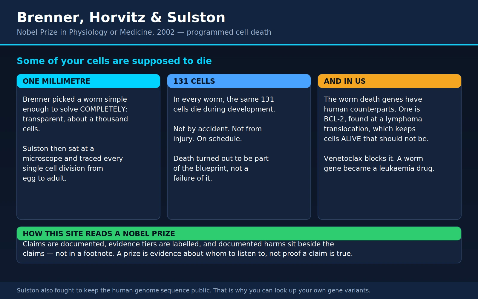
Table of Contents
- Overview
- Choosing the Animal
- Sulston's Lineage: Every Cell, From Egg to Adult
- The Cells That Always Die
- Horvitz and the Death Genes
- Apoptosis Versus Necrosis, Practically
- Too Little Cell Death: Cancer, BCL-2 and Venetoclax
- Too Much Cell Death: The Failures Nobody Advertises
- How the Immune System Uses It
- Sulston, the Genome, and the Fight Over Free Data
- What Model Organisms Are Actually For
- Where Mainstream Medicine Agrees
- What Remains Debated
- Key Research Papers
- Connections
- Featured Videos
1. Overview
In 2002 the Nobel Prize in Physiology or Medicine went to three men — Sydney Brenner, H. Robert Horvitz and John E. Sulston — "for their discoveries concerning genetic regulation of organ development and programmed cell death." That sentence is doing a lot of quiet work. What the three of them actually established is that dying is something your cells are built to do. Not as an accident. Not as a failure of maintenance. As a scheduled, gene-controlled, precisely timed part of being alive.
They found it in a worm about a millimetre long. And the reason a worm could answer that question is the second story on this page, and the more useful one, because it is the story of how scientists choose what to study — and of the honest limits of what an animal experiment can tell you about a person.
The three came at it from different angles. Brenner (1927–2019), a South African-born molecular biologist working at the Medical Research Council Laboratory of Molecular Biology in Cambridge, made the founding decision: he picked the animal. Sulston (1942–2018), an English chemist-turned-biologist in the same laboratory, spent years at a microscope building the map. Horvitz (born 1947), an American who arrived in Brenner's lab as a postdoctoral researcher and later spent his career at MIT, found the genes that switch the dying on and off — and then found that we have the same ones.
The last part is why this page belongs on a health site rather than only in a history of biology. A gene named ced-9 in a nematode turned out to be the same gene as BCL-2, which was first noticed because it sits at a chromosome break in human lymphoma. A gene named ced-3 in the same worm turned out to belong to the family we now call the caspases, the enzymes that dismantle a dying human cell. Twenty-some years after the worm work, a drug built to block BCL-2 — venetoclax — became a standard treatment for chronic lymphocytic leukaemia and for acute myeloid leukaemia in older patients. That is a real, traceable line from a nematode gene to a pill, and this page follows it end to end.
2. Choosing the Animal
By the early 1960s Sydney Brenner had already helped establish some of the central facts of molecular biology — the triplet genetic code, the existence of messenger RNA. And he came to a conclusion that sounds obvious once stated and was radical at the time: the interesting unsolved problems were no longer about molecules. They were about development and about the nervous system. How does a single fertilised egg turn into an organised animal with the right parts in the right places? How does a nervous system get wired?
Nobody had a method for that. So Brenner did something unusual. Instead of designing a better experiment, he designed a better subject. He went looking for an animal simple enough that the question could be answered completely rather than partially.
His requirements were brutal and specific. The animal had to be small, so it would fit under a microscope whole. It had to be transparent, so you could watch cells inside a living, undissected body. It had to breed fast, so genetics would be practical. It had to be cheap to keep. It had to have few enough cells that a person could conceivably count all of them. And, ideally, it should reproduce by self-fertilisation, so that a mutation would breed true without any mating being arranged.
What met all of that was a soil nematode: Caenorhabditis elegans. About one millimetre long. Transparent from end to end. It eats bacteria, so you grow it on a petri dish smeared with E. coli. It goes from egg to reproducing adult in about three days at room temperature, and lives two to three weeks. Most individuals are self-fertilising hermaphrodites, with males appearing rarely — which means one worm can found an entire genetically identical population, and it also means you have a way to do crosses when you want them.
Brenner's founding paper, The genetics of Caenorhabditis elegans, appeared in Genetics in 1974. Its abstract is almost aggressively unglamorous: methods for isolating, complementing and mapping mutants; about 300 chemically induced mutants affecting behaviour and morphology; about a hundred genes defined; 77 of those genes altering how the animal moves. There is no grand claim in it. What it actually did was hand the world a working genetic toolkit for an animal that nobody had studied.
Here is the methodological point, and it is worth holding onto. Brenner treated the choice of organism as the experiment. He bet that a complete answer in a simple animal would be worth more than a partial answer in a complicated one — that if you could describe one animal exhaustively, the principles you extracted would generalise, whereas a fragment of a mouse would stay a fragment. That bet is not obviously correct. It could have failed. Nematodes could have turned out to run development on machinery that vertebrates abandoned half a billion years ago. Section 11 deals honestly with where the bet paid and where it did not. But the bet paid enough that it changed how biology is done, and C. elegans went on to earn a share of three Nobel Prizes in Physiology or Medicine within a decade — 2002 for this work, 2006 for RNA interference, and 2024 for microRNA.
3. Sulston's Lineage: Every Cell, From Egg to Adult
Now the part that is hard to convey without sounding like exaggeration.
John Sulston sat down at a microscope and traced the ancestry of every single cell in the animal. Not a sample. Not a representative region. Every cell, from the fertilised egg to the adult.
Understand what that involved. There were no fluorescent markers for this, no automated imaging, no software that tracked objects between frames. There was a microscope with Nomarski differential-interference optics, a living transparent worm on a slide, and a person watching. Cells divide; the daughters move; they are small, crowded, and look much like one another. To know that the cell you are watching now is the same cell you were watching four hours ago, you have to not look away. Sulston watched in shifts, hours at a time, day after day, drawing what he saw by hand and building the family tree cell by cell.
It came out in two enormous instalments, and the two are easy to confuse, so here they are separately:
- Sulston & Horvitz, 1977, in Developmental Biology, covered the post-embryonic lineages — everything that happens after the larva hatches, through four larval stages, to the adult. Robert Horvitz was Sulston's collaborator on this, and it is where their partnership begins.
- Sulston, Schierenberg, White & Thomson, 1983, also in Developmental Biology, covered the embryonic lineage — zygote to newly hatched larva. That paper's first sentence records what it completed: with this, "the entire cell lineage of this organism is now known."
The 1983 paper reports that 671 cells are generated during embryogenesis. The adult hermaphrodite ends up with 959 somatic cells — a figure still stated as a plain fact in the current literature, for instance in a 2017 Genetics review of the worm's cell biology, which describes development "from a one cell-stage embryo to a fertile hermaphrodite with 959 somatic nuclei." The rest of the adult is germline.
The adult male is built differently — it has a specialised mating tail the hermaphrodite does not — and its somatic count is higher. The number usually quoted is 1,031, and it traces to Sulston, Albertson & Thomson's 1980 paper on male post-embryonic development. That paper is indexed in PubMed with no abstract, and no indexed abstract anywhere states the figure, so this page reports it as the number commonly cited rather than as one it has confirmed.
But the count is not the achievement. The invariance is.
The lineage is the same in every animal. Not statistically similar — the same. Cell number 419 in this worm came from the same ancestor by the same sequence of divisions as cell number 419 in that worm, and it ends up in the same place doing the same job. You can point at any cell in an adult C. elegans and say precisely where it came from, which divisions produced it, when each of those divisions happened, and which cells are its sisters and cousins. There is no other animal for which that sentence is true.
Sulston himself was careful about what the map meant. The 1983 paper notes that although the relationship between a cell's ancestry and its fate is fixed, the correlation between them "lacks much obvious pattern" — most neurons come from embryonic ectoderm, but some come from mesoderm, and a few are sisters to muscle cells. Lineage boundaries do not tidily match functional ones. Cell-ablation experiments — killing a specific cell with a laser and watching what fails to happen — showed substantial autonomy: many cells follow their programme largely on their own account. Their conclusion was that the lineage itself is a real determinant of what a cell becomes, not merely a record of what happened.
Two other products of the same effort deserve a mention because they show what a complete map makes possible. Horvitz and Sulston used it in 1980 to isolate the first cell-lineage mutants — animals whose cells divided in the wrong pattern, which you can only recognise if you already know the right pattern. And in 1986, White, Southgate, Thomson and Brenner published the complete structure of the worm's nervous system: every neuron, every connection, reconstructed from serial electron micrographs and running to 340 printed pages. It was the first complete wiring diagram of any nervous system, and it remains the only one for an animal that behaves.
4. The Cells That Always Die
Buried in the lineage was the finding that made this a medical story.
Some of the cells in the map are born and then die. Not damaged cells. Not sick cells. Cells that divide normally, exist for a while, and then, at a particular moment, shrink, condense, break up and disappear — eaten by a neighbour.
The 1983 embryonic paper states it flatly: of the 671 cells generated during embryogenesis, 113 in the hermaphrodite undergo programmed death (111 in the male). Add the deaths that occur after hatching, and the total in the hermaphrodite is 131 — the figure the field still uses, and the one you find stated in current primary literature, for instance a 2018 Genetics paper describing "the 131 cells that are 'programmed' to die during C. elegans development."
Now the part that reframes everything. It is the same 131 cells every time. The same identities, the same lineage positions, the same approximate moment in development. Worm after worm after worm.
Think about what that rules out. If cell death were injury, it would happen to different cells in different animals, depending on what each animal bumped into. If it were wear and tear, it would be variable. If it were a defect, it would be rare and idiosyncratic. Instead it is stereotyped to the level of the individual cell — which is only possible if the information about which cells die, and when, is written into the animal's genes.
So death, at the level of a single cell, is not the failure state. It is a function. It is on the blueprint.
That is a genuinely different way to think about biology, and it changes what questions are worth asking about human disease. If a cell dying is an accident, then a disease of too much cell death is a disease of accidents, and there is nothing to study but the accident. If a cell dying is a programme, then it has genes, and the genes have switches, and the switches can be broken in both directions — stuck on, or stuck off. Everything in sections 7 through 9 follows from that one shift.
The idea did not come out of nowhere. Pathologists had been noticing for years that dying cells sometimes look orderly rather than ruptured. In 1972, John Kerr, Andrew Wyllie and Alastair Currie published a paper in the British Journal of Cancer proposing a name for it: apoptosis, from a Greek word for the dropping of leaves from a tree or petals from a flower. Their abstract argued it was "an active, inherently programmed phenomenon" playing "a complementary but opposite role to mitosis in the regulation of animal cell populations" — and that it occurs in normal adult tissue turnover, in embryonic development, spontaneously inside untreated tumours, and during tumour regression after treatment.
It was, by any reasonable standard, right. It also went comparatively unnoticed for years, because a description is not a mechanism. What the worm supplied was the mechanism.
5. Horvitz and the Death Genes
Horvitz's contribution was to ask the question that only made sense once Sulston's map existed: if 131 specific cells die on schedule, is there a gene whose job is to kill them — and what happens if you break it?
He went hunting for mutant worms in which cells that should have died were still there. Because the lineage was known cell by cell, a surviving cell was visible as an anomaly rather than lost in noise. He found them, and the genes were named ced, for cell death abnormal.
The 1986 paper by Hilary Ellis and Robert Horvitz in Cell laid out the first two. The normal function of ced-3 and ced-4 is required to initiate programmed cell death. Knock either one out and the cells that should have died do not die — and, crucially, they do not linger as debris either. They survive and differentiate, taking on the identity of cells they are related to. The death programme had been removed, and the cells simply went on being cells.
Then the finding that gets skipped in most retellings, and which the abstract states plainly: ced-3 and ced-4 mutant worms "appear grossly normal in morphology and behavior." A nematode carrying 131 cells it was supposed to have deleted crawls around, eats, and reproduces. Programmed cell death, in this animal, is not required for development to work.
A third gene worked the other way. ced-9 is a survival gene: its job is to protect cells that are supposed to live. Break ced-9 and cells die that should not have. So the system is an ordered pathway with an accelerator and a brake — a killing arm (ced-3, ced-4) held in check by a protective arm (ced-9), with an upstream trigger gene, egl-1, that releases the brake in the cells selected to die.
Then came the payoff, and it arrived from two directions at once.
In 1992, David Vaux, Irving Weissman and Stuart Kim reported in Science that expressing the human bcl-2 gene inside C. elegans reduced the number of programmed cell deaths. A human gene, dropped into a nematode, worked the nematode's brake pedal.
In 1993, Junying Yuan, Shai Shaham, Stephane Ledoux, Hilary Ellis and Horvitz cloned ced-3 and found that the protein it encodes resembles a mammalian enzyme called interleukin-1β-converting enzyme — a cysteine protease. They proposed that CED-3 acts as a protease to initiate cell death in the worm, and that cysteine proteases do the same job in mammals. They were right; that protein family is now called the caspases, and they are the demolition crew of the dying cell.
In 1994, Michael Hengartner and Horvitz cloned ced-9 itself and found it encodes a 280-amino-acid protein with sequence and structural similarity to the mammalian proto-oncogene bcl-2. Overexpressing bcl-2 mimicked ced-9's protective effect and rescued worms whose own ced-9 was broken. Their conclusion: the two are homologues, and "the molecular mechanism of programmed cell death has been conserved from nematodes to mammals."
Sit with what that means. BCL-2 was not discovered by anyone studying worms or development. It was discovered by cancer researchers, in human lymphoma cells, as a gene sitting at a chromosome breakpoint. It had a cancer name and a cancer history. And it turned out to be the same gene as a nematode's cell-survival switch — interchangeable enough that each version works inside the other species' cells.
Two research communities that had no reason to talk to each other had been describing the same molecule from opposite ends. That is the moment the worm stopped being a curiosity and became a machine for understanding human disease.
The correspondence runs all the way down the pathway. ced-9 corresponds to the BCL-2 family. ced-3 corresponds to the caspases. ced-4 corresponds to a human protein called APAF-1. egl-1 corresponds to the human "BH3-only" proteins. The worm's four-gene sketch is the human system with the labels changed.
6. Apoptosis Versus Necrosis, Practically
Cells can die two broadly different ways, and the difference is not academic. It determines whether the tissue around them gets inflamed, and inflammation is what you feel, what scars, and what a great deal of medicine is spent managing.
Apoptosis is a controlled demolition. The cell shrinks rather than swells. Its chromatin condenses and its DNA is cut into fragments. Critically, the outer membrane stays intact throughout. The cell packages itself into sealed, membrane-bound parcels — apoptotic bodies — which neighbouring cells and macrophages recognise and swallow whole. Nothing spills. The 1972 Kerr paper described exactly this two-stage sequence, decades before the genes were known: first condensation and fragmentation into well-preserved membrane-bound pieces, then uptake by other cells and digestion inside them.
It is more than merely non-inflammatory. In 1998 Valerie Fadok and colleagues showed in the Journal of Clinical Investigation that macrophages which have eaten apoptotic cells actively suppress inflammatory signalling, through mediators including TGF-β, prostaglandin E2 and platelet-activating factor. Clearing an apoptotic cell does not just avoid raising an alarm; it damps one down.
Necrosis is a burst pipe. The cell swells, the membrane fails, and the contents — enzymes, proteins, mitochondrial fragments, DNA — spill into the surrounding tissue. The immune system reads all of that as evidence of injury, because normally it only ever sees those molecules when something has gone badly wrong. Inflammation follows: swelling, heat, pain, recruitment of immune cells, and, eventually, scar.
Why the distinction matters to a patient:
- It explains why routine cell turnover is silent. Your body replaces an enormous number of cells every day — gut lining, skin, blood cells, immune cells. Estimates of the daily total run into the tens of billions, though that figure is a calculation rather than a measurement and should be treated as an order-of-magnitude claim. Because almost all of it happens by apoptosis, none of it inflames anything. You do not feel your intestinal lining being replaced.
- It explains why a heart attack leaves a scar and a bruise mostly does not. Necrotic death in the core of an infarct spills contents, triggers inflammation, and heals with fibrous tissue that does not contract. Scarred heart muscle is the reason a heart attack has permanent consequences even after the artery is reopened.
- It explains part of how cancer treatment works. Chemotherapy and radiation do not mostly dissolve tumour cells. They inflict damage that pushes cells to trigger their own apoptotic programme. A tumour whose apoptotic machinery is disabled — which is common — can absorb the same damage and not die. This is one of the mechanisms of treatment resistance, and it is why a drug that restores the ability to die (section 7) can work where more cell-killing damage does not.
- It explains why "cell death" on a lab report is not one thing. Tests that detect tissue damage — troponin after heart injury, liver enzymes, lactate dehydrogenase — are largely detecting leakage. They report necrosis well and apoptosis poorly, because an apoptotic cell does not leak.
One correction to a common oversimplification: this is not a clean binary. Since the worm work, biologists have identified several other regulated ways for a cell to die — necroptosis, pyroptosis, ferroptosis — some of which are programmed like apoptosis but inflammatory like necrosis. The apoptosis-versus-necrosis contrast is the right first model. It is not the last word.
7. Too Little Cell Death: Cancer, BCL-2 and Venetoclax
This is the cleanest bench-to-bedside line in the whole story, so it is worth telling in order.
1984. Yoshihide Tsujimoto, working with Carlo Croce and colleagues at the Wistar Institute, cloned the DNA at a chromosome breakpoint found in B-cell cancers. Follicular lymphoma very often carries a translocation, written t(14;18), that joins a gene on chromosome 18 to the immunoglobulin heavy-chain locus on chromosome 14. Their Science paper reported that the probe identified a gene locus at chromosome 18q21 which was "unrelated to known oncogenes" and might matter in the origin of these tumours. That gene was named bcl-2 — B-cell lymphoma 2.
Why the translocation matters. The immunoglobulin locus is one of the most furiously active regions of the genome in a B cell — it has to be, since antibody production is the cell's job. Moving bcl-2 next to it does not change the gene. It changes how loudly it is switched on. The cell ends up permanently flooded with a protein it should only be making in measured amounts.
1988. David Vaux, Suzanne Cory and Jerry Adams, in Melbourne, established what BCL-2 actually does — and it was not what anyone expected of a cancer gene. Writing in Nature, they put human bcl-2 into mouse blood-cell precursors. In cells deprived of the growth factor interleukin-3, which normally causes them to die, bcl-2 kept them alive — but the surviving cells "persisted in a G0 state, rather than proliferating." They stayed alive without dividing. Combined with a deregulated c-myc gene, bcl-2 did drive proliferation and tumour formation, but on its own its contribution was survival. Their conclusion was that bcl-2 "provided a distinct survival signal to the cell and may contribute to neoplasia by allowing a clone to persist until other oncogenes... become activated."
That was a new kind of cancer gene. Until then the mental model of an oncogene was an accelerator: a gene that makes cells divide too fast, which is what Bishop and Varmus's work on cellular oncogenes had opened up. BCL-2 does not push the accelerator. It disables the brakes on survival — it stops cells from dying when they should. A population of cells that will not die accumulates, and every extra day it survives is another day to acquire a second mutation.
1992–1994. The worm work identified ced-9 as the same gene, in an animal with no blood, no immune system and no capacity for cancer. That did two things: it proved BCL-2 is not a cancer protein that happens to affect survival, but a survival protein that happens to cause cancer when overexpressed — and it gave researchers a whole pathway to target instead of a single gene.
2016 onwards. If a cancer survives because BCL-2 is holding the death programme shut, then a drug that unsticks BCL-2 should let the cell finish a job it has already started. Venetoclax is that drug: a small molecule that binds BCL-2 and displaces the pro-death proteins it was sequestering.
The first substantial human results, published in the New England Journal of Medicine in 2016, came from a phase 1 dose-escalation study in 116 patients with relapsed or refractory chronic lymphocytic leukaemia or small lymphocytic lymphoma. Most had already had multiple treatments and 89% had poor-prognosis features. Ninety-two of the 116 (79%) responded; complete remissions occurred in 20%. The response rate held up in the hardest subgroups, including patients with deletion of chromosome 17p. It also produced a warning that has shaped how the drug is given ever since: clinical tumour lysis syndrome occurred in 3 of the first 56 patients, with one death, because killing a large mass of leukaemia cells very quickly floods the bloodstream with their contents. After the dosing schedule was changed to a slow weekly ramp-up, it did not occur in any of the next 60 patients.
The randomised confirmation came in 2018 with the MURANO trial, also in the New England Journal of Medicine. Three hundred and eighty-nine patients with relapsed or refractory CLL were randomly assigned to venetoclax for up to two years plus rituximab for the first six months, or to bendamustine plus rituximab for six months. After a median follow-up of 23.8 months, two-year progression-free survival was 84.9% with venetoclax-rituximab versus 36.3% with bendamustine-rituximab (hazard ratio 0.17; 95% CI 0.11 to 0.25). Among patients with 17p deletion — historically the worst genetic subgroup in CLL — it was 81.5% versus 27.8%. Grade 3 or 4 neutropenia was more common with venetoclax; febrile neutropenia and infections were less common than with bendamustine; grade 3 or 4 tumour lysis syndrome occurred in 3.1%.
Then acute myeloid leukaemia. The VIALE-A trial, published in 2020, enrolled 431 previously untreated AML patients who could not have standard intensive induction chemotherapy — because they were 75 or older, or had other conditions making it unsafe. Median age was 76. They received azacitidine plus either venetoclax or placebo. At a median follow-up of 20.5 months, median overall survival was 14.7 months with venetoclax versus 9.6 months with placebo (hazard ratio for death 0.66; 95% CI 0.52 to 0.85). Complete remission occurred in 36.7% versus 17.9%. The cost was real: febrile neutropenia in 42% versus 19%, and infections of any grade in 85% versus 67%.
Read those numbers honestly. Venetoclax is not a cure for either disease. VIALE-A bought a median of about five additional months of life in a group of frail, elderly patients who previously had almost nothing, at the price of substantial infection risk. MURANO is a much larger effect but measures time without progression, in a relapsed population, over roughly two years. Both are genuine, both are practice-changing, and neither is the word "cured."
What makes this section worth its length is not the size of the effect. It is that the chain holds at every link: a chromosome break in human lymphoma, a gene that keeps cells alive rather than making them divide, the same gene found independently in a nematode, a conserved pathway mapped in an animal that cannot get cancer, and then a molecule designed against the human protein that works in randomised trials. Very little in medicine runs that cleanly from mechanism to medicine.
8. Too Much Cell Death: The Failures Nobody Advertises
If too little apoptosis produces cancer, the symmetry is irresistible: too much apoptosis should produce degeneration, and blocking it should treat degeneration. That symmetry is where a great deal of money and hope has gone, and the results are sobering. This section is deliberately less encouraging than the last one.
What is well supported. Excessive or inappropriate apoptosis is implicated in a long list of conditions: Alzheimer's disease, Parkinson's disease, amyotrophic lateral sclerosis, Huntington's disease, the zone of injured-but-not-yet-dead tissue around a stroke or a heart attack, some inherited anaemias, and the loss of CD4 T cells in untreated HIV infection. Dying cells in these conditions frequently show apoptotic features, caspases are activated, and in laboratory animals blocking the pathway often reduces the damage.
Label the tier plainly: that is mechanistic and animal-model evidence. It is strong evidence that apoptosis is happening. It is much weaker evidence that apoptosis is the cause rather than the final common consequence of some upstream problem — and it is not evidence at all that blocking it helps a person.
What has repeatedly failed. Two trials tell the story better than any summary.
NXY-059 in stroke. This was a free-radical-trapping neuroprotectant intended to rescue brain tissue in the ischaemic penumbra — the rim of tissue around a stroke that is damaged but not yet dead, and that is exactly where a death-prevention strategy should work. A first trial had suggested benefit. The confirmatory trial, published in the New England Journal of Medicine in 2007, enrolled 3,306 patients treated within six hours of stroke onset. The result was flat: no difference in disability on the modified Rankin scale at 90 days (odds ratio 0.94; 95% CI 0.83 to 1.06), no benefit on any secondary endpoint. The authors' conclusion was one sentence: "NXY-059 is ineffective for the treatment of acute ischemic stroke within 6 hours after the onset of symptoms." This is not an isolated result. Neuroprotection in stroke has one of the longest lists of promising-in-animals, negative-in-humans results in modern medicine.
Minocycline in ALS. This one is worse, and everyone should know about it. Minocycline is an old antibiotic that turns out to be anti-apoptotic and anti-inflammatory in laboratory systems, and it extended survival in mouse models of several neurological conditions. A phase 3 randomised trial in 412 ALS patients was published in Lancet Neurology in 2007. Patients on minocycline declined faster than patients on placebo — the ALS functional rating scale fell by 1.30 units per month on the drug versus 1.04 on placebo (p=0.005), with non-significant trends toward worse lung function, worse muscle strength, and higher mortality. The paper's own interpretation is blunt: "Our finding that minocycline has a harmful effect on patients with ALS has implications for trials of minocycline in patients with other neurological disorders, and for how potential neuroprotective agents are screened."
Note the title of that paper: "Efficacy of minocycline in patients with amyotrophic lateral sclerosis." Read the title alone and you would guess it worked. It did the opposite. That is a general hazard of reading science by headline, and it is a good argument for reading at least the conclusion of an abstract.
Why blocking cell death is harder than it sounds. Several reasons, all of them plausible and none of them settled:
- Timing. By the time a person reaches a hospital after a stroke, or reaches a neurologist with symptoms of a degenerative disease, the cell-death process is already well advanced. Animal experiments typically administer the drug before or immediately at the moment of injury.
- The cell may be doomed anyway. Blocking the execution step may only change how a cell dies, not whether it does — and switching a cell from apoptosis to necrosis would be a net loss, since necrosis inflames.
- Apoptosis may be a symptom. If protein aggregates, mitochondrial failure or chronic inflammation are the driver, silencing the death programme leaves the driver untouched and keeps sick cells around.
- You want apoptosis working. It is your body's principal mechanism for deleting damaged and pre-cancerous cells. A drug that broadly and durably blocks apoptosis is, in mechanism, a drug that mimics what BCL-2 overexpression does in lymphoma. That is an uncomfortable thing to give a healthy-tissue-bearing person for years.
So the honest position is this: the science of cell death has produced a genuine, approved, effective cancer drug by promoting apoptosis, and has so far produced no approved drug that treats a human disease by blocking it. Anyone who tells you a supplement, a peptide or a device is "protecting your cells from apoptosis" and thereby preventing neurodegeneration is describing a hypothesis that the best-funded trials in the field have failed to confirm.
9. How the Immune System Uses It
The immune system is arguably the tissue that depends most heavily on programmed cell death, and it uses it for two entirely different purposes.
Purpose one: deleting cells that would attack you. T cells are made in the thymus, and each new one carries a randomly generated receptor. Random generation means some of those receptors will recognise your own tissues. Those cells have to go, and the process of eliminating them is called negative selection.
The mechanism is apoptosis. In 1994 Charles Surh and Jonathan Sprent showed this directly, publishing in Nature a method for detecting apoptotic thymocytes in situ. Their findings are worth stating precisely, because the details are often flattened. Apoptotic cells were scattered through the thymic cortex and engulfed locally by macrophages — but cortical apoptosis was not reduced in mice lacking MHC molecules, which told them cortical death mostly reflects failure of positive selection ("death by neglect") rather than active negative selection. Direct evidence for negative-selection apoptosis came from the medulla, where a transgenic system that eliminates a specific T-cell population produced dense aggregates of apoptotic cells cleared by a distinct macrophage population. Most developing T cells die in the thymus; not all of them die for the same reason.
What happens when this fails. Here the human evidence is unusually clean. The Fas receptor (also called CD95 or Apo-1) is a cell-surface protein that triggers apoptosis when engaged. Mice carrying the lpr mutation, which disables the Fas gene, develop swollen lymph nodes and a lupus-like autoimmune disease — because lymphocytes that should have been deleted accumulate instead.
The same thing happens in people. In 1995 Frédéric Rieux-Laucat and colleagues reported in Science three children with a lymphoproliferative syndrome, two of whom also had autoimmune disease. The most severely affected had a large deletion in the FAS gene and no detectable Fas on the cell surface; two related patients, less severely affected, had a deletion within the receptor's intracellular domain and impaired Fas-mediated apoptosis. The condition is now called autoimmune lymphoproliferative syndrome (ALPS). It is a human disease caused by lymphocytes that cannot be told to die.
That is the general shape of the relationship between apoptosis and autoimmunity: a cell that should have been deleted, and was not. It is not the only mechanism of autoimmune disease — most autoimmune conditions have no single-gene explanation — but it is a proof of principle that failure of programmed cell death can produce autoimmunity in a human being.
Purpose two: killing infected and cancerous cells. When a cytotoxic (CD8) T cell finds a cell displaying a viral protein — the recognition mechanism worked out by Doherty and Zinkernagel — it does not dissolve it. It instructs it to kill itself.
There are two routes. The T cell can present Fas ligand, engaging Fas on the target and triggering the caspase cascade directly. Or it can deliver granzymes, protein-cutting enzymes that activate the target cell's own caspases from inside. Getting granzymes into the target requires perforin, a protein that forms pores in the target membrane; mice lacking perforin have normal numbers of CD8 T cells and natural-killer cells that simply cannot lyse their targets, fail to clear a virus, and clear tumour cells poorly.
So the common phrasing "cytotoxic T cells kill by triggering apoptosis rather than by rupturing cells" is right in substance but needs one correction: perforin does punch holes. The holes are a delivery mechanism, not the killing blow. The killing is done by the target cell's own machinery — the same CED-3-descended caspases — switched on from within.
This design is not an accident. Consider the alternative. If your immune system killed virus-infected cells by bursting them, it would release the virus, spill inflammatory contents, and damage the surrounding tissue every time. Instead it triggers a programme that condenses the cell, fragments its DNA (including any viral genome), seals it into packages, and has a macrophage eat the packages. The infection is contained inside the corpse. In an organ you cannot afford to inflame — brain, heart, lung — that difference is the difference between clearing an infection and being disabled by clearing it.
10. Sulston, the Genome, and the Fight Over Free Data
The third act of Sulston's career had nothing to do with cell death, and it is the part of this story with the widest reach outside biology.
Having mapped the worm's cells, Sulston moved on to mapping its DNA, first building a physical map of the genome with Alan Coulson, Brenner and others, and then leading the sequencing effort in partnership with Robert Waterston at Washington University in St Louis. In 1992 Sulston became the founding director of the Sanger Centre near Cambridge, funded by the Wellcome Trust and the Medical Research Council.
In December 1998, Science published Genome sequence of the nematode C. elegans: a platform for investigating biology, under the corporate authorship of The C. elegans Sequencing Consortium. It reported a 97-megabase sequence containing over 19,000 genes, more than 40% of whose predicted protein products had significant matches in other organisms. C. elegans became the first multicellular organism to have its genome essentially sequenced. Brenner's bet had now paid twice: the animal chosen for being simple enough to map cell by cell was also simple enough to sequence first.
Then the same teams turned to the human genome, and a fight started.
The public project's position. The Human Genome Project was an international, publicly funded collaboration; the Sanger Centre under Sulston produced roughly a third of the finished human sequence. In 1996, at a meeting in Bermuda, the participating centres agreed on a set of rules that became known as the Bermuda Principles: sequence data would be released into public databases essentially immediately — within about 24 hours of assembly — free of charge and free of restriction, before publication, before analysis, before anyone had a chance to profit from it. Sulston was among the most forceful advocates of that position and stuck to it under considerable pressure.
The commercial challenge. In 1998 Craig Venter founded Celera Genomics and announced it would sequence the human genome faster and more cheaply than the public consortium, using a technique called whole-genome shotgun sequencing. Celera's business model was not to sell the sequence outright but to sell subscription access to a database with annotation, analysis tools and comparison data layered on top — and to seek intellectual-property protection on some specific gene discoveries.
Be fair to the other side, because caricature makes this story less useful. Venter's technical argument was substantially correct. Whole-genome shotgun sequencing — shredding the entire genome and reassembling it computationally, rather than mapping first and sequencing region by region — was widely doubted at the scale of a human genome, and it worked. It is now the standard method; essentially every genome sequenced today uses its descendants. The competition also demonstrably accelerated the public project, which responded by scaling up dramatically. And Celera did not refuse to publish: its analysis appeared in Science in February 2001, the same week the public consortium's appeared in Nature.
What was genuinely contested was access. The public consortium's Nature paper describes itself as reporting a draft "produced and made freely available." Celera's Science paper was published under an access arrangement that did not place the sequence in the unrestricted public databases on the same terms, which is why a number of scientists objected at the time. It is also a matter of record — stated in Celera's own paper — that its assemblies combined its data with the publicly funded project's freely released sequence. The free data was available to the commercial effort; the commercial data was not equally available to everyone else. That asymmetry is the substance of the argument, and it is a fair thing to say without impugning anyone's motives.
Sulston's own case is worth reading in his words, because it is not primarily an argument about money. In the 2001 Sir Frederick Gowland Hopkins Memorial Lecture — named for the founder of vitamin science — he wrote that the sequence "is not an end in itself, but a resource to be continually reanalysed as our biological understanding increases. That is the scientific reason for releasing it promptly, fully and freely. The social reasons for doing so are even more compelling." The lecture is indexed in Biochemical Society Transactions, and it also contains a nice marker of how provisional everything was in 2001: he expected the human gene count to come in around 40,000. The current figure for protein-coding genes is roughly half that. A sequence you cannot reanalyse is a sequence whose first-draft errors become permanent.
Why this matters to you specifically. The consequences are not abstract:
- Any doctor, anywhere, can look up any human gene sequence without paying anyone or asking permission.
- Genetic tests for inherited disease can be developed by anyone competent to develop them, which is a large part of why they now cost hundreds rather than thousands.
- When a consumer genetics report tells you that you carry a particular variant, the reference against which your DNA was compared is a public resource.
- Researchers in countries that could never have afforded a subscription work from the same data as researchers in wealthy ones.
- The wider question of patenting human genes was eventually settled against patentability in the United States: in 2013 the Supreme Court held in Association for Molecular Pathology v. Myriad Genetics that naturally occurring DNA sequences are not patent-eligible subject matter. The free-release norm established in 1996 shaped the ground on which that argument was fought.
Sulston was knighted in 2001 and shared the Nobel Prize the following year. He spent the rest of his life arguing about science and equity — drug pricing, patents, access — and he was not universally popular for it. He died in 2018. Brenner died in 2019.
11. What Model Organisms Are Actually For
This is the section to remember if you remember nothing else, because it is the one you can use every time you read a health headline.
Why the worm worked. Not because a nematode resembles a person — it does not, in any respect you would notice. It worked because the machinery of programmed cell death is ancient. The last common ancestor of humans and nematodes lived something like 600 million years ago, and it already had this system. Both lineages kept it, essentially unchanged in outline, because there is no version of a multicellular animal that works without a way to delete cells on command.
The proof is not an argument by analogy. It is Vaux, Weissman and Kim's 1992 experiment: a human gene, placed inside a nematode, performing the nematode's job. And Hengartner and Horvitz's 1994 experiment in reverse. When a gene from one species substitutes for a gene in another, separated by 600 million years, you are not looking at a resemblance. You are looking at the same thing.
Now the limits, stated as plainly as they deserve. A C. elegans hermaphrodite has 959 somatic cells; you have on the order of thirty trillion. It has:
- No adaptive immune system. No T cells, no B cells, no antibodies, no immunological memory. Everything in section 9 is invisible to it.
- No blood and no circulation. No heart, no vessels, no blood pressure, no clotting, no way for a drug to be distributed the way it is in you.
- No liver and no kidneys. Which means no metabolism of drugs the way you metabolise them and no renal clearance — the two things that mostly determine what dose a person needs and how often.
- No brain in any recognisable sense. 302 neurons in the hermaphrodite, versus something like 86 billion in you.
- An invariant lineage. This was the feature that made the whole enterprise possible, and it is also the single most un-human thing about the animal. Your development is not invariant. No two people, not even identical twins, have the same cells in the same places. Human development is statistical where the worm's is deterministic.
- A completely different scale of time and temperature. Egg to adult in three days; a whole life in two or three weeks; body temperature whatever the room is.
The general rule, and it applies far beyond worms:
Section 8 is the worked example. Minocycline was anti-apoptotic in cells and extended survival in mouse models of neurological disease. The mechanism was real. It transferred. What did not transfer was the outcome: in 412 human beings with ALS, the drug made them decline faster. Nothing about the mouse data was fabricated or fraudulent. The mouse was simply not a person.
So here is a practical checklist for reading a health story. When you see a headline of the form "scientists discover X causes/cures Y," ask:
- What species? If the answer is mice, worms, flies, zebrafish or cells in a dish, the finding is about a mechanism, not about a treatment. That is not a criticism — it is a stage.
- Whole animal or cell culture? A cell in a dish has no liver, no immune system and no dose limit. Enormous numbers of compounds kill cancer cells in a dish, including bleach.
- Is the animal model really a model of the disease? Many are engineered stand-ins that reproduce one feature of a human illness. A mouse given a genetic mutation that produces plaques in its brain does not have Alzheimer's disease; it has plaques.
- Mechanism or outcome? "Reduces a marker" is not "helps a patient." Any number of drugs have improved a laboratory value and harmed the people taking them.
- Has it been through a randomised trial with a clinical endpoint? That is the step at which most promising mechanisms fail — and it is the step that separates venetoclax from every supplement claiming to modulate apoptosis.
The worm answered a question about human cancer because the question was about mechanism. It could not have told anyone how much venetoclax to prescribe, or whether it would be worth the infections. Both facts are true simultaneously, and holding both at once is what scientific literacy actually consists of.
12. Where Mainstream Medicine Agrees
Very little on this page is contentious. This is settled, textbook science:
- Programmed cell death is real and gene-controlled. Cells possess an intrinsic self-destruction programme that is triggered on schedule during normal development. This is not disputed by anyone.
- The C. elegans lineage is correct and invariant. 959 somatic cells in the adult hermaphrodite; 671 cells generated during embryogenesis; 113 embryonic programmed deaths in the hermaphrodite; 131 programmed deaths in total. Reproduced and used daily as a reference.
- The core pathway is conserved from nematodes to humans. BH3-only proteins release BCL-2-family restraint; APAF-1 assembles; caspases execute. The worm's egl-1, ced-9, ced-4 and ced-3 are the same system.
- Apoptosis is immunologically quiet; necrosis is not. Membrane integrity is preserved in apoptosis, contents are not spilled, and macrophages that clear apoptotic cells actively suppress inflammatory signalling.
- BCL-2 overexpression contributes to B-cell malignancy by conferring survival rather than proliferation, via the t(14;18) translocation in follicular lymphoma and other mechanisms in CLL.
- Venetoclax works. It is approved and used as standard therapy in chronic lymphocytic leukaemia and, with a hypomethylating agent, in acute myeloid leukaemia in patients unfit for intensive chemotherapy. The randomised evidence is strong.
- Defective apoptosis can cause human autoimmunity. ALPS, caused by FAS pathway mutations, is the demonstration.
- Model organisms are legitimate and indispensable. Simultaneously: their results do not transfer automatically to people. Both halves are mainstream.
13. What Remains Debated
- Why the 131 deaths happen at all. The uncomfortable fact from Ellis and Horvitz's 1986 paper is that worms which fail to make those deaths are "grossly normal in morphology and behavior." If deleting 131 cells is so precisely programmed, one would expect keeping them to cause visible trouble, and it largely does not. Whether the deaths are sculpting, quality control, resource economy, or a mixture is still argued.
- Driver or passenger in neurodegeneration. Apoptotic features are seen in degenerating brain tissue. Whether the death programme is causing the disease or merely finishing off cells already killed by something else — misfolded protein, mitochondrial failure, chronic inflammation — is not resolved, and the therapeutic failures in section 8 are consistent with the second reading.
- Whether blocking cell death can ever be a treatment. Repeated well-designed trials have failed, and there is a coherent argument that the strategy is inherently limited: it arrives too late, may only change the manner of death, and carries a built-in cancer-risk logic if it succeeds broadly. Others argue the targets and the timing have simply been wrong so far.
- How many kinds of programmed death there are. Necroptosis, pyroptosis and ferroptosis are all regulated forms of cell death distinct from classical apoptosis, several of them inflammatory. How they interact, which dominates in which disease, and whether blocking one merely diverts cells into another are open questions.
- Durability and resistance with BCL-2 inhibition. Venetoclax responses are not permanent in most patients, resistance emerges, and the best combinations, sequences and treatment durations are the subject of ongoing trials rather than settled practice.
- How much of the apoptosis literature is reproducible. Apoptosis became a fashionable field in the 1990s, and a large volume of work was published using assays that do not reliably distinguish apoptosis from other kinds of cell death. Older claims about apoptosis in particular tissues and diseases should be read with that in mind.
14. Key Research Papers
Every PMID below was retrieved from PubMed and checked against the journal, year, volume and page numbers at the time of writing, and every finding stated on this page was taken from the paper's own abstract. Where a paper is indexed without an abstract, this page says so rather than quoting a figure from it.
- Brenner S. The genetics of Caenorhabditis elegans. Genetics 1974;77(1):71-94
- Sulston JE, Horvitz HR. Post-embryonic cell lineages of the nematode, Caenorhabditis elegans. Dev Biol 1977;56(1):110-56
- Sulston JE, Schierenberg E, White JG, Thomson JN. The embryonic cell lineage of the nematode Caenorhabditis elegans. Dev Biol 1983;100(1):64-119
- Ellis HM, Horvitz HR. Genetic control of programmed cell death in the nematode C. elegans. Cell 1986;44(6):817-29
- Yuan J, Shaham S, Ledoux S, Ellis HM, Horvitz HR. The C. elegans cell death gene ced-3 encodes a protein similar to mammalian interleukin-1 beta-converting enzyme. Cell 1993;75(4):641-52
- Hengartner MO, Horvitz HR. C. elegans cell survival gene ced-9 encodes a functional homolog of the mammalian proto-oncogene bcl-2. Cell 1994;76(4):665-76
- Vaux DL, Weissman IL, Kim SK. Prevention of programmed cell death in Caenorhabditis elegans by human bcl-2. Science 1992;258(5090):1955-7
- Kerr JF, Wyllie AH, Currie AR. Apoptosis: a basic biological phenomenon with wide-ranging implications in tissue kinetics. Br J Cancer 1972;26(4):239-57
- Fadok VA, Bratton DL, Konowal A, et al. Macrophages that have ingested apoptotic cells in vitro inhibit proinflammatory cytokine production through autocrine/paracrine mechanisms involving TGF-beta, PGE2, and PAF. J Clin Invest 1998;101(4):890-8
- Tsujimoto Y, Finger LR, Yunis J, Nowell PC, Croce CM. Cloning of the chromosome breakpoint of neoplastic B cells with the t(14;18) chromosome translocation. Science 1984;226(4678):1097-9
- Vaux DL, Cory S, Adams JM. Bcl-2 gene promotes haemopoietic cell survival and cooperates with c-myc to immortalize pre-B cells. Nature 1988;335(6189):440-2
- Watanabe-Fukunaga R, Brannan CI, Copeland NG, Jenkins NA, Nagata S. Lymphoproliferation disorder in mice explained by defects in Fas antigen that mediates apoptosis. Nature 1992;356(6367):314-7
- Rieux-Laucat F, Le Deist F, Hivroz C, et al. Mutations in Fas associated with human lymphoproliferative syndrome and autoimmunity. Science 1995;268(5215):1347-9
- Seymour JF, Kipps TJ, Eichhorst B, et al. Venetoclax-rituximab in relapsed or refractory chronic lymphocytic leukemia. N Engl J Med 2018;378(12):1107-1120
- DiNardo CD, Jonas BA, Pullarkat V, et al. Azacitidine and venetoclax in previously untreated acute myeloid leukemia. N Engl J Med 2020;383(7):617-629
- Shuaib A, Lees KR, Lyden P, et al. NXY-059 for the treatment of acute ischemic stroke. N Engl J Med 2007;357(6):562-71
- Gordon PH, Moore DH, Miller RG, et al. Efficacy of minocycline in patients with amyotrophic lateral sclerosis: a phase III randomised trial. Lancet Neurol 2007;6(12):1045-53 — note that despite the title, the finding was harm.
- The C. elegans Sequencing Consortium. Genome sequence of the nematode C. elegans: a platform for investigating biology. Science 1998;282(5396):2012-8 — indexed under the consortium as corporate author, not an individual.
- International Human Genome Sequencing Consortium (Lander ES, Linton LM, Birren B, et al.) Initial sequencing and analysis of the human genome. Nature 2001;409(6822):860-921
- Venter JC, Adams MD, Myers EW, et al. The sequence of the human genome. Science 2001;291(5507):1304-51 — the Celera paper, included so that section 10 can be read against the primary source rather than a summary of it.
The three 2002 Nobel lectures were published together in the same issue of Chembiochem and are worth reading in the laureates' own words. PubMed indexes all three without abstracts, so nothing on this page is sourced to them: Brenner S, Nature's gift to science, Chembiochem 2003;4(8):683-7; Sulston JE, Caenorhabditis elegans: the cell lineage and beyond, Chembiochem 2003;4(8):688-96; Horvitz HR, Worms, life, and death, Chembiochem 2003;4(8):697-711.
Live PubMed Searches
- C. elegans programmed cell death
- BCL-2 family, apoptosis and cancer
- Venetoclax clinical trials
- Apoptosis, neurodegeneration and clinical trials
- Caspase pathway conservation across species
Connections
- All Notable Doctors
- Nobel Prize in Medicine — the laureates wing this page belongs to, and the full roll of prizes from 1901 onward
- Fire & Mello — RNA interference, discovered in the same worm four years later and awarded the 2006 prize
- Ambros & Ruvkun — microRNA, found in C. elegans developmental-timing mutants; the third Nobel the worm earned
- Bishop & Varmus — cellular oncogenes; the accelerator model of cancer that BCL-2 forced everyone to widen
- Doherty & Zinkernagel — how a cytotoxic T cell recognises the infected cell it then instructs to die, as in section 9
- Watson, Crick & Wilkins — the DNA structure that made both the worm genome and the human genome conceivable
- Hartwell, Hunt & Nurse — the cell-division cycle; the counterpart programme to the cell-death programme described here
- Yoshinori Ohsumi — autophagy, the other great self-consumption programme cells run on purpose
- Blackburn, Greider & Szostak — telomeres, and the other built-in limit on how long a cell lineage lasts
- Shinya Yamanaka — cellular reprogramming; the proof that a cell's fate, like its death, is instruction rather than destiny
- Frederick Gowland Hopkins — the memorial lecture in which Sulston made his public case for free genome data
- Chronic Lymphocytic Leukemia — the disease in which venetoclax was first approved, and the MURANO trial of section 7
- Acute Myeloid Leukemia — where azacitidine plus venetoclax changed what is possible for older patients
- Non-Hodgkin Lymphoma — follicular lymphoma and the t(14;18) translocation that first revealed BCL-2
- Cancer — the wider picture: cancer as a failure of the instructions to divide, differentiate and die
- Stroke — the ischaemic penumbra, and the long record of neuroprotection trials that did not work
- Alzheimer's Disease — where excess cell death is implicated but blocking it has not yet helped anyone