Beadle, Tatum & Lederberg: One Gene, One Enzyme — and Why Newborns Get a Heel Prick
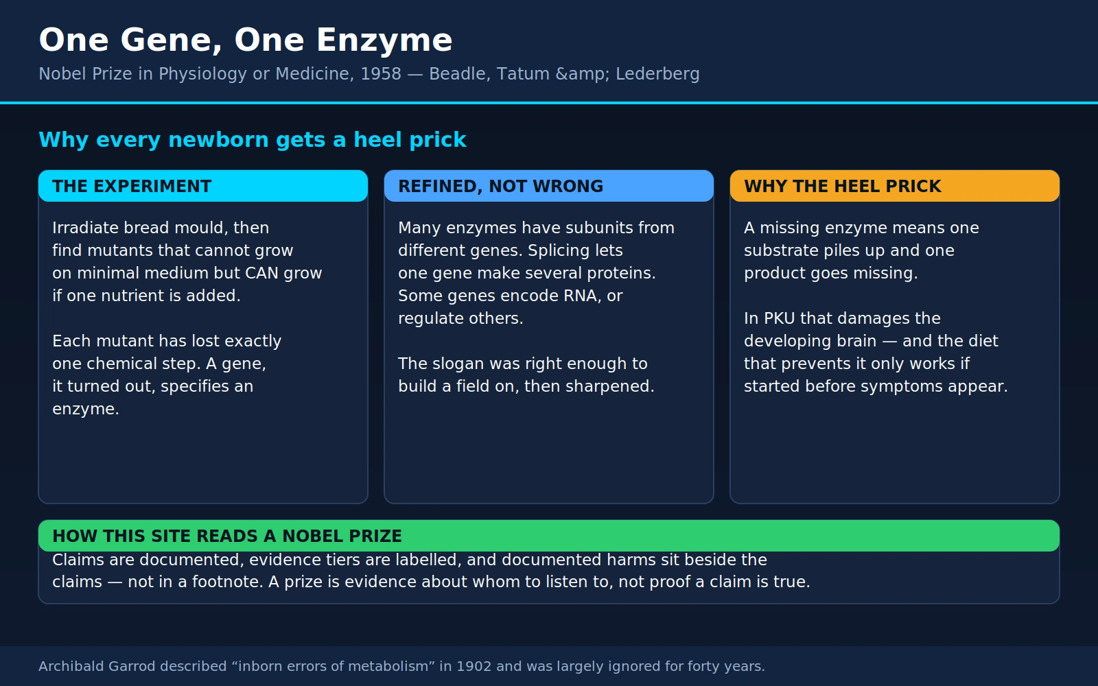
Table of Contents
- The Prize and the Three Men
- The Question: What Does a Gene Actually Do?
- Neurospora, 1941: Running the Experiment Backwards
- One Gene, One Enzyme — and How the Slogan Was Refined
- Garrod Got There First
- Lederberg's Bacteria: Sex, Phage, and Antibiotic Resistance
- Inborn Errors of Metabolism: The Practical Heart
- Newborn Screening: Why the Heel Prick Exists
- Living With PKU
- What This Does Not License
- Where Mainstream Medicine Agrees — and What Remains Debated
- Key Research Papers
- Connections
- Featured Videos
1. The Prize and the Three Men
In December 1958 the Nobel Prize in Physiology or Medicine was divided between an idea and a technique. One half went jointly to George Wells Beadle (1903–1989) and Edward Lawrie Tatum (1909–1975) “for their discovery that genes act by regulating definite chemical events.” The other half went to Joshua Lederberg (1925–2008) “for his discoveries concerning genetic recombination and the organization of the genetic material of bacteria.” The official record is at nobelprize.org — 1958 Prize in Physiology or Medicine.
Between them, those two sentences describe the moment biology stopped being a science of what is inherited and became a science of how inheritance works. Beadle and Tatum showed that a gene's job is to control a specific chemical step. Lederberg showed that bacteria — organisms nobody thought had genetics at all — swap genes with each other, which turned the bacterium into the laboratory workhorse on which nearly everything else in molecular biology was subsequently built.
The three were not peers in the ordinary sense. Beadle was a Nebraska farm boy turned Caltech geneticist; Tatum was a biochemist, the son of a pharmacologist, and the man who could actually run the chemistry Beadle's genetics required. Their collaboration is a textbook case of a discovery that neither discipline could have made alone: Beadle knew what question to ask, Tatum knew how to ask it in a flask.
Lederberg was something else again. He arrived in Tatum's laboratory at Yale in 1946 as a medical student on leave, having interrupted his training at Columbia to spend a year trying an experiment most senior scientists thought was doomed. He was 21. The experiment worked, he never went back to finish medicine, and he was 33 years old when he collected the Nobel Prize — still one of the youngest laureates in the history of the medicine prize. He went on to found the department of genetics at Stanford, then to the presidency of Rockefeller University, and to a long second career advising the United States government on infectious disease and biological weapons.
Esther Lederberg, who is not on the medal
There is a fourth name in this story, and this site has now recorded enough of these to call the pattern what it is.
Esther Miriam Zimmer Lederberg (1922–2006) was a microbial geneticist who worked alongside Joshua Lederberg for two decades and was married to him from 1946 to 1966. Three of the most consequential contributions to come out of that laboratory are hers or jointly hers:
- Replica plating — the technique of pressing a plate of bacterial colonies onto sterile velvet and stamping the pattern onto fresh plates, so that thousands of colonies can be screened for a trait in their exact original positions. It is published as Lederberg J and Lederberg EM, J Bacteriol 1952;63(3):399-406, and it is the tool that made large-scale bacterial genetics practical. Nearly every mutant-hunting screen since owes it something.
- The discovery of bacteriophage lambda — the temperate virus lurking inside the standard laboratory strain of Escherichia coli, which she found in 1951. Lambda became one of the central objects of molecular biology: a model for how a virus decides between lying dormant and killing its host, the vehicle for the transduction experiments described below, and, decades later, a routine tool for moving DNA around. The genetic analysis appeared with her as first author: Lederberg EM and Lederberg J, Genetics 1953;38(1):51-64.
- The F fertility factor — work on the plasmid that determines which bacterial cell acts as donor in the conjugation Joshua was awarded for.
She did not share the 1958 prize. She spent much of her career in non-tenured research positions at Stanford, and by most accounts had to fight for the title of research professor. Nothing here requires deciding what the Nobel committee should have done in 1958; the useful thing is simply to say what happened, and to notice that it keeps happening. Readers who want the pattern rather than the anecdote will find it running through this site's coverage of Watson, Crick and Wilkins — where Rosalind Franklin's diffraction data did the decisive work — and of the medicine prize as a whole, whose omissions are as informative as its awards.
2. The Question: What Does a Gene Actually Do?
It is genuinely hard, now, to feel how blank this question was in 1940.
By then a great deal was settled. Gregor Mendel's ratios had been rediscovered in 1900 and had held up. Thomas Hunt Morgan's fruit-fly group had shown that genes sit in a linear order along chromosomes and can be mapped by how often they are separated during reproduction. Hermann Muller had shown in 1927 that X-rays cause mutations, which meant genes were physical things that radiation could damage. Geneticists could predict inheritance patterns with real precision.
What nobody could say was what a gene did. You could trace a factor for white eyes through ten generations of flies without having the faintest idea how a stretch of chromosome makes an eye white. Between the gene and the trait lay a gap that genetics had no instrument to cross. Some biologists suspected genes were enzymes themselves. Others thought each gene somehow governed the whole organism's development in a way that could not be broken into steps. The chemical identity of the gene was itself unsettled — most biochemists in 1940 assumed the hereditary material was protein, since DNA looked far too monotonous to carry information. Oswald Avery's demonstration that DNA was the transforming principle was still four years away in 1944, and the Hershey–Chase confirmation eight years away in 1952.
So the field faced a problem of method, not just of ignorance. The classical approach ran from trait to gene: notice something visible — wing shape, eye color, flower position — then breed for it and map the responsible locus. That approach can only ever find genes for things you already noticed. Worse, the traits it finds are the endpoints of long developmental cascades, arriving at the observer's eye a hundred chemical steps downstream of whatever the gene actually did. It is like trying to understand an engine by studying the exhaust.
Beadle had already run at this problem from the classical direction. Working with Boris Ephrussi in Paris in the mid-1930s, he had transplanted eye tissue between fruit-fly larvae of different genotypes and shown that eye pigment depended on diffusible substances made elsewhere in the body — strong evidence that genes were acting through chemistry. But the chemistry of fly pigments was a nightmare, and after several years the project had produced more frustration than answers. What Beadle needed was an organism whose chemistry was simple enough to read directly, and a way of running the whole enquiry backwards.
3. Neurospora, 1941: Running the Experiment Backwards
The organism was Neurospora crassa: the orange-pink bread mould that grows on stale bread and, in the tropics, on burnt sugarcane fields. Beadle and Tatum, then at Stanford, chose it for a set of extremely practical reasons.
Neurospora grows fast — days, not weeks. It has a well-understood sexual cycle, so crosses can be made and progeny analysed. Crucially, it spends most of its life as a haploid organism with only one copy of each gene, so a recessive mutation shows itself immediately instead of hiding behind a functional partner as it would in a fly or a human. And — this is the decisive property — wild-type Neurospora grows happily on a minimal medium: sugar, a few inorganic salts, and the single vitamin biotin. From that austere diet the mould builds every amino acid, every vitamin, every nucleotide it needs. It is, in effect, a small chemical factory whose entire product list is known.
The design
That last property is what let Beadle and Tatum reverse the direction of the whole enquiry. Their procedure, reported in Proc Natl Acad Sci U S A 1941;27(11):499-506, ran like this:
- Damage genes at random. Irradiate Neurospora spores — Muller had shown that radiation mutates genes, and nobody needed to know which gene was hit.
- Grow the survivors on a rich, complete medium supplemented with a broad mixture of vitamins and amino acids. This is the subtle step. A mould that has lost the ability to make its own vitamin B6 would simply die on minimal medium and never be seen. On rich medium it survives, because the missing substance is handed to it.
- Test each survivor on minimal medium. Most grow. A rare few do not — they are alive on the rich diet and dead on the austere one. Each of those has lost the ability to manufacture something.
- Find out what. Take the failing strain and offer it minimal medium plus one added substance at a time: this vitamin, that amino acid. When it grows again, you have identified the exact compound the organism can no longer make.
- Cross it and count. Mate the mutant with a normal strain and follow the defect through the progeny. If the requirement segregates cleanly — half the offspring needing the supplement, half not — it behaves as a single Mendelian gene.
Read that sequence again and notice what it does. The classical geneticist starts with a visible trait and hunts for the gene. Beadle and Tatum started with a gene lesion of unknown identity and read out the chemistry it broke. They did not need to guess in advance which biochemical step to study; the mould told them, by starving until they supplied the one thing it could no longer make. They had converted an invisible genetic change into a specific, nameable chemical deficiency — and the answer arrived as a bottle off the shelf.
The strains they generated became famous as auxotrophs: organisms that require a specific nutrient their wild relatives synthesise for themselves. In the follow-up work the naming convention was almost comically direct — a strain that could not make para-aminobenzoic acid was reported as an “aminobenzoicless” mutant (Tatum EL, Beadle GW, Proc Natl Acad Sci U S A 1942;28(6):234-43). Within a few years the Stanford group and others had accumulated hundreds of such mutants, and the pattern held every time: one mutation, one blocked chemical step.
The logic that fell out of it
Something more than a catalogue emerged. When several different mutants all failed to make the same end product, you could work out the order of the steps by seeing which intermediates rescued which mutants. A mutant blocked early in a pathway is rescued by any intermediate downstream of its block; a mutant blocked late is rescued only by the final compounds. Line up which strain is rescued by which substance and the sequence of the biochemical pathway falls out — without ever purifying an enzyme.
Metabolism, in other words, could now be mapped by genetics. That is the quiet revolution inside the famous one. The pathways drawn on the walls of every biochemistry department were reconstructed in large part by this method, and its logic is still what a modern geneticist means by a “genetic screen.”
4. One Gene, One Enzyme — and How the Slogan Was Refined
The conclusion crystallised into four words that dominated biology for a generation: one gene, one enzyme. Each gene, on this view, specifies one enzyme; each enzyme catalyses one chemical step; a broken gene means a broken step, and the substrate of that step piles up behind the blockage while the product goes missing downstream.
It is now known to be too simple. That fact deserves more than a footnote, because how it was too simple is one of the better illustrations available of what a scientific model is for.
The four corrections
Many enzymes are built from several different proteins, each with its own gene. This was the first and most important correction, and it produced the revised slogan one gene, one polypeptide. The cleanest demonstration came from tryptophan synthase, the bacterial enzyme that makes the amino acid tryptophan: it is assembled from two distinct kinds of subunit, alpha and beta, encoded by two separate adjacent genes. Charles Yanofsky, who did that work and used it to nail down the relationship between gene sequence and protein sequence, later wrote up exactly why this enzyme was such a good test case — Genetics 2005;169(2):511-6. Hemoglobin makes the same point in a form patients meet: it has alpha and beta chains from different genes, which is why sickle cell disease and beta-thalassemia are diseases of the beta gene while alpha-thalassemia is a different disease of a different gene.
One gene can make several different proteins. Most human genes are interrupted by non-coding stretches (introns) that are cut out of the RNA transcript before it is translated. Which pieces get kept can vary by tissue, by developmental stage, and by circumstance — alternative splicing. A single gene may therefore yield a family of related proteins with different properties. The roughly 20,000 protein-coding genes in the human genome specify a considerably larger number of distinct proteins.
Many genes do not encode enzymes at all — and many do not encode proteins. Structural proteins such as collagen and keratin are not enzymes. Neither are antibodies, ion channels, receptors, or transporters. And a large fraction of the genome's transcribed output is functional RNA that is never translated: transfer RNAs, ribosomal RNAs, microRNAs that tune how much of a protein gets made, long non-coding RNAs whose jobs are still being catalogued.
Some genes exist to control other genes. This was the discovery that earned the 1965 prize for François Jacob, André Lwoff and Jacques Monod: regulatory genes whose products switch other genes on and off in response to conditions. A cell does not simply possess a fixed set of enzymes; it decides which to make, when, and how much. The heart cell and the liver cell in the same body carry the same genes and behave completely differently, and gene regulation is the reason. Barbara McClintock's transposable elements — genes that physically move and thereby change the expression of their neighbours — had already put a crack in the tidy picture in the 1940s, though it took the field decades to accept it.
Right enough to build a field on
None of that makes Beadle and Tatum wrong in any useful sense. Their hypothesis was a simplification that was accurate in the domain where it was tested — single-subunit metabolic enzymes in a haploid mould — and it was productive precisely because it was sharp enough to be pushed until it broke. It gave biochemistry a way to find genes, gave genetics a way to find chemistry, and gave medicine the concept that a heritable disease can be a single missing catalytic step. Every one of the corrections above was discovered by people using their method.
Compare Newton's mechanics, which is wrong at high velocity and near large masses, and which still lands spacecraft. A model earns its keep by being useful and by being specific enough to fail informatively. “One gene, one enzyme” did both. The honest description of what happened is not that Beadle and Tatum made an error; it is that they built the first floor, and the people who added the second floor could only do so by standing on it.
5. Garrod Got There First
Nearly four decades before the bread mould, an English physician had the whole idea and could not get anyone to care.
Archibald Edward Garrod (1857–1936) was a London consultant with an unfashionable interest in chemical pathology. His subject was alkaptonuria, a rare condition whose most arresting sign is that the patient's urine turns black on standing. Alkaptonuric urine contains homogentisic acid, an intermediate in the breakdown of the amino acids phenylalanine and tyrosine, which normally never accumulates because it is destroyed as fast as it is made. Over decades the pigment deposits in cartilage and connective tissue, darkening the ears and the whites of the eyes and producing a degenerative arthritis of the spine and large joints called ochronosis.
Garrod noticed two things that nobody else had put together. First, alkaptonuria was frequently present from birth, ran in families, and was strikingly common among children of first-cousin marriages. Working with the geneticist William Bateson, he recognised that this was exactly the pattern Mendel's newly rediscovered rules predicted for a recessive trait. Second — and this is the leap — he proposed that the underlying defect was not an organ or a tissue but a single missing chemical step: the affected person simply lacked whatever agent normally destroys homogentisic acid, so the substance backed up and spilled into the urine.
He published this in 1902. His paper on the subject in the Medico-Chirurgical Transactions is indexed as Med Chir Trans 1902;85:69-78. His better-known statement of the argument, “The incidence of alkaptonuria: a study in chemical individuality,” appeared in The Lancet the same year. That Lancet paper is not indexed in PubMed as an original record; what PubMed carries is a republication issued a century later as a classical article, Yale J Biol Med 2002;75(4):221-31 — we flag the distinction because a modern date on a hundred-year-old idea is exactly the kind of thing that gets miscited.
In 1908 Garrod delivered the Croonian Lectures under the title Inborn Errors of Metabolism, expanded into a book the following year, in which he set out four such conditions — alkaptonuria, cystinuria, pentosuria and albinism — as a class of disorders in which an inherited factor blocks a specific chemical conversion. The phrase he coined is still the name of the field.
And then, essentially, nothing. Garrod's argument required both Mendelian genetics and enzyme chemistry, and in 1908 those were separate worlds with separate journals and few shared readers. Geneticists were absorbed in breeding experiments; clinicians saw four rare curiosities of no obvious general importance. The synthesis he offered had no audience prepared to receive it. Beadle himself, once the Neurospora work was published, was candid that Garrod had reached the concept first — and it is worth being clear about what he added: Garrod inferred a missing chemical step from patients he could observe but could not experiment on; Beadle and Tatum could manufacture such lesions to order and prove the general rule.
The Coris close the loop in a human disease
The bridge between mould genetics and human medicine was built by Carl and Gerty Cori, who took the 1947 Nobel Prize for their work on how glycogen is made and broken down. In the early 1950s they examined the livers of children with von Gierke disease — glycogen storage disease type I, in which glycogen accumulates catastrophically and blood sugar collapses between meals — and found the specific enzyme glucose-6-phosphatase missing. That was the first time a human inherited disease was traced to the absence of one identified enzyme: Garrod's inference, finally demonstrated at the bench. Gerty Cori did the work while dying of a bone marrow disease; she is the third woman to have won a Nobel Prize in a science category and the first American woman to take one.
6. Lederberg's Bacteria: Sex, Phage, and Antibiotic Resistance
The other half of the 1958 prize was, on the face of it, a much narrower result: bacteria have sex. Its consequences were anything but narrow.
Conjugation, 1946
Until the mid-1940s bacteria were widely thought to lie outside genetics altogether. They reproduce by splitting in two, so every daughter is a copy; with no sexual process there is no recombination, and with no recombination there is nothing for a geneticist to map. Bacteriology and genetics were separate trades.
Joshua Lederberg, then 21 and on leave from medical school, went to Tatum's laboratory at Yale to test the assumption — and to do it he used exactly the tool Tatum had just invented. Tatum had applied the Neurospora approach to E. coli and generated bacterial auxotrophs: strains unable to make particular nutrients. Lederberg took two such strains, each crippled in different ways, and mixed them.
Call one strain unable to make nutrients A and B, and the other unable to make C and D. Neither can grow on minimal medium. Mix them, plate the mixture on minimal medium, and if nothing happens, bacteria do not exchange genes. But colonies appeared — cells that could make all four nutrients, and therefore had to have acquired functional copies of A and B from one parent and C and D from the other. The result was published as a short note: Lederberg J, Tatum EL, Nature 1946;158(4016):558, with the full account following as J Bacteriol 1947;53(6):673-84.
The design of that experiment is worth pausing on, because it is doing something clever. Requiring two defects in each parent was the safeguard: a single strain reverting spontaneously to health would have to reverse two independent mutations at once, which essentially never happens, whereas a genuine exchange produces prototrophs readily. The rare event is made to stand out against a background that has been engineered to be silent.
Bernard Davis then showed in 1950 that the exchange required the cells to touch: two cultures separated by a filter that passes liquid but not bacteria produced no recombinants (J Bacteriol 1950;60(4):507-8). The process, conjugation, involves a donor cell extending a pilus to a recipient and passing DNA through the contact. Which cell plays donor is determined by the F fertility factor, a plasmid — a small, independently replicating loop of DNA separate from the chromosome — work in which Esther Lederberg was centrally involved.
Transduction, 1952
Six years later Lederberg and his graduate student Norton Zinder repeated the mixing experiment in Salmonella and got recombinants again — but this time the filter experiment gave the opposite answer. Genes moved between cultures that could not touch. Something passed through the filter and carried genetic material with it.
That something turned out to be a bacteriophage: a virus that infects bacteria, and which occasionally packages a fragment of its host's DNA by mistake, then injects that stolen fragment into the next cell it infects. Zinder ND, Lederberg J, J Bacteriol 1952;64(5):679-99, named the process transduction. It is a second, entirely different mechanism for moving genes between bacteria — and it needs no cell contact at all.
Why this mattered far beyond bacteria
It made bacteria a genetic system. This is the point that justifies half a Nobel Prize for what looks like a technical curiosity. A bacterium divides every twenty minutes; a billion of them fit in a millilitre of broth; a single plate can hold more individuals than every fruit fly ever bred. Once you can cross bacteria, map their genes, and select rare events out of enormous populations, you have an experimental system of a completely different order of power from anything in classical genetics.
Essentially all of early molecular biology ran on that system. The genetic code was cracked in bacterial extracts — the work of Nirenberg, Khorana and Holley. Gene regulation was worked out in E. coli by Jacob and Monod. Messenger RNA, DNA replication, restriction enzymes, and eventually the whole toolkit of genetic engineering came out of bacterial and phage genetics. It is not an exaggeration to say that the recombinant insulin in a diabetic's pen traces its lineage to a 21-year-old medical student mixing two crippled cultures of E. coli in 1946.
And it explains antibiotic resistance. This is the part that reaches an ordinary reader's life directly, so we will state it plainly.
Resistance to an antibiotic is not only something a bacterium evolves for itself by mutation. It is something a bacterium can be handed, ready-made, by another bacterium of an entirely different species. The mechanisms Lederberg's group discovered are precisely the mechanisms by which that happens:
- Conjugation transfers plasmids, and resistance genes are frequently carried on plasmids. A single plasmid may carry resistance to several drug classes at once, so one transfer event can convert a susceptible organism into a multidrug-resistant one in a step.
- Transduction moves resistance genes inside phage particles, with no cell contact required.
- Transformation — the uptake of naked DNA from the environment, the process Avery used to identify DNA as the hereditary material — is a third route.
Collectively this is horizontal gene transfer, and it is why resistance spreads through hospitals, sewage, farm slurry and the human gut far faster than mutation alone could explain. A recent review of the transfer systems in Gram-negative bacteria is Microbiol Immunol 2025;69(7):367-376. It also supplies the practical argument behind the advice everyone has heard and few follow: an unnecessary antibiotic course does not merely fail to help you, it applies selection pressure to a population of organisms that is fully capable of trading the resulting resistance genes onward. Selman Waksman's streptomycin — the first drug effective against tuberculosis — met resistant strains within a couple of years of entering the clinic. Our Bacteria section covers the individual organisms and what actually works against them.
A third experiment worth knowing about
Replica plating did not merely save labour. The Lederbergs used it to settle a genuine argument about how bacteria acquire resistance in the first place, in the same 1952 paper that introduced the technique.
The question was whether resistant bacteria arise because of exposure to the killing agent — the organism adapting in response to the threat — or whether resistant mutants are already present in the population by chance, and exposure merely reveals them by killing everything else. The experiment: replica-plate a master plate of colonies onto a plate laced with bacteriophage, note the positions of the few colonies that survive, then go back to the original, never-exposed master plate and pick from those same positions. Those colonies turn out to be resistant already, despite having never met the phage.
Mutation, therefore, comes first; selection follows. This is Darwin in a petri dish over an afternoon, and it is also the correct mental model for what happens when a person takes an antibiotic: the drug does not teach bacteria to resist it, it clears the field for the ones that already could.
7. Inborn Errors of Metabolism: The Practical Heart
Here is where a 1941 experiment on bread mould reaches into an ordinary hospital, and it does so through a piece of logic simple enough to hold in your head.
The general logic
Metabolism is an assembly line. A substance arrives, an enzyme converts it into the next substance, another enzyme converts that, and so on to the finished product. Take out one worker and two things happen at once:
- The substrate piles up. Material arrives at the missing step and stops. It accumulates in blood, in tissue, in urine — and if it is toxic at high concentration, that accumulation is the disease.
- The product goes missing. Everything downstream of the block is not made. If the body needs it, that shortage is also the disease.
Which of the two does the damage varies by condition, and it determines the treatment. From that logic, three strategies follow directly — and essentially every treatment for an inborn error of metabolism is one of them, or a combination:
- Restrict the substrate. If you cannot process a substance, stop putting it in. This is dietary therapy, and it is the mainstay for the amino-acid disorders.
- Replace the product. If the missing product is needed, supply it directly. In phenylketonuria, tyrosine — normally made from phenylalanine — becomes an essential nutrient that has to come from the diet or the formula.
- Replace the enzyme. Manufacture the missing protein and infuse it. This is enzyme replacement therapy, and it works best where the enzyme's normal address is a compartment the bloodstream can deliver to.
A fourth strategy has joined them more recently: stabilise the crippled enzyme. Some disease-causing mutations do not abolish an enzyme, they make it unstable or clumsy. Supplying a large excess of the enzyme's cofactor can coax residual activity out of it — a mechanism with real clinical consequences, described below for both PKU and homocystinuria.
Phenylketonuria — the one everybody's newborn is tested for
PKU is deficiency of phenylalanine hydroxylase, the liver enzyme that converts the amino acid phenylalanine into tyrosine. It is inherited recessively: two carrier parents, one-in-four risk per pregnancy, and carriers are entirely healthy. Across Europe the average prevalence is roughly 1 in 10,000 newborns, with notably higher rates in Ireland and Turkey and a very low rate in Finland — figures set out in the European guidelines, Orphanet J Rare Dis 2017;12(1):162.
Phenylalanine is in every protein, so it arrives with every meal. Without the enzyme it accumulates in the blood and crosses into the brain, where high concentrations interfere with the transport of other amino acids and with the synthesis of neurotransmitters. Untreated, the consequence is severe, permanent intellectual disability, often with seizures and behavioural disturbance. Treated from the first weeks of life, the consequence is a person with normal intelligence who has to think about food.
That gap — between the untreated and the treated outcome of the identical genotype — is as stark as anything in medicine, and it is entirely a function of when treatment starts. The European guideline cites work showing that every four weeks of delay in starting treatment costs roughly four IQ points. A newborn with PKU looks completely normal. By the time anything is visibly wrong, the damage is done and is not reversible. There is no way to find these babies except to test all of them — which is the entire argument for newborn screening, and the subject of the next section. Our clinical page is Phenylketonuria, and the amino acid itself is covered at Phenylalanine and Tyrosine.
Galactosemia
Classical galactosemia is deficiency of galactose-1-phosphate uridyltransferase, an enzyme in the pathway that processes galactose — half of the lactose molecule in milk. The substrate that accumulates is toxic to liver, kidney and brain, and unlike PKU the presentation is fast: a newborn who is feeding on breast milk or standard formula can develop jaundice, liver failure, poor feeding and a dangerous E. coli sepsis within the first week or two of life.
Treatment is immediate and lifelong removal of galactose and lactose from the diet, which is life-saving. It is important to be honest about what it does not do: even with excellent dietary control from birth, a substantial proportion of people with classical galactosemia experience long-term difficulties — speech and language problems, learning difficulties, and primary ovarian insufficiency in girls and women. This is one reason galactosemia is a good corrective to any tidy sense that identifying an enzyme solves a disease. Our page is Galactosemia.
MCAD deficiency — the fasting disorder
Medium-chain acyl-CoA dehydrogenase deficiency works on an entirely different principle from the two above, and it is worth understanding because it explains why the disorder is so treacherous.
Between meals, and especially overnight or during an illness that stops a child eating, the body switches from burning sugar to burning fat. Fatty acids are dismantled in a repeating cycle, each turn requiring a dehydrogenase enzyme matched to the length of the chain. In MCADD the medium-chain enzyme is missing, so fat-burning stalls halfway. The child cannot make enough energy, blood sugar falls, and partly processed fatty acids accumulate.
The result is a child who appears completely healthy — until an ordinary childhood illness with vomiting produces a metabolic crisis: profound hypoglycaemia, lethargy, seizures, coma, sometimes death, sometimes brain injury in the survivors. Historically some of these deaths were classified as sudden infant death syndrome or Reye syndrome. And the management is almost absurdly simple once the diagnosis is known: don't let the child go without food for long, and treat any illness that prevents eating as an emergency requiring glucose. An emergency regimen is issued to the family. The condition is essentially benign when known about and lethal when not, which is exactly the profile that makes newborn screening worth doing.
Homocystinuria
Classical homocystinuria is deficiency of cystathionine beta-synthase, an enzyme in the pathway that disposes of the amino acid methionine. Homocysteine accumulates, and the consequences are dislocation of the lenses of the eyes, a Marfan-like tall thin build with long limbs, osteoporosis, learning difficulties, and — the dangerous one — a high risk of blood clots and stroke, sometimes in childhood.
It illustrates the cofactor strategy well. Cystathionine beta-synthase uses vitamin B6 (pyridoxine) as a cofactor, and a substantial minority of patients carry mutations that respond to pharmacological doses of B6 with a large drop in homocysteine. Those who respond are managed very differently from those who do not, who need a methionine-restricted diet plus betaine to route homocysteine down an alternative path. Folate and vitamin B12 support the same pathway. Our page is Homocystinuria; see also Methionine.
Two adjacent cautions belong here. First, the drastically raised homocysteine of this rare genetic disease is a different quantity from the mildly raised homocysteine sometimes reported on general wellness panels; the first is unambiguously dangerous, the second is a much weaker and more contested signal. Second, another amino-acid disorder on the same screening panels — maple syrup urine disease — is one of the fastest-moving of all, capable of causing brain injury within days of birth.
Lysosomal storage diseases and enzyme replacement
The lysosome is the cell's recycling compartment, packed with enzymes that break down worn-out molecules. Lose one of those enzymes and its particular substrate accumulates inside the lysosome year after year, swelling the cell and eventually wrecking whatever tissue is worst affected. This is a large family: Gaucher disease, Fabry disease, Pompe disease, Tay-Sachs disease, and dozens more.
These are the conditions where enzyme replacement therapy works, and the reason is anatomical: cells actively take up enzymes from the bloodstream and deliver them to the lysosome, so an infused enzyme can reach the compartment where it is needed.
Pompe disease is the sharpest illustration, and it closes a circle in this story. Pompe is glycogen storage disease type II — a disorder of the same glycogen metabolism the Coris mapped, caused by absence of the lysosomal enzyme acid alpha-glucosidase. The infantile form was, until this century, uniformly fatal: an enlarged heart, floppy weak muscles, and death usually in the first year. In the pivotal trial of recombinant human acid alpha-glucosidase in 18 rapidly progressing infants, all 18 survived to 18 months of age, and against an untreated historical control group the treatment reduced the risk of death by 99% and the risk of death or invasive ventilation by 92% (Neurology 2007;68(2):99-109). Eleven of the eighteen had infusion-associated reactions, all mild or moderate.
Two caveats keep that honest. Comparison against a historical control group is weaker evidence than a randomised comparison — though for a uniformly fatal infantile disease, no ethics committee would approve the randomised version. And enzyme replacement does not cross the blood–brain barrier well, so conditions whose damage is primarily neurological — Tay-Sachs is the standard example — are not helped by it. The success is real and it is bounded.
8. Newborn Screening: Why the Heel Prick Exists
Somewhere between 24 and 48 hours after a baby is born, a nurse warms the heel, pricks it, and blots a few spots of blood onto a card of filter paper. It is the most widely applied genetic test in the world, most parents never think about it again, and it exists because of a specific chain of reasoning that runs directly out of everything above.
Robert Guthrie and the bacterial trick
By the late 1950s PKU was understood and the low-phenylalanine diet was known to work — if started early enough. The problem was purely one of finding the babies. Blood phenylalanine could be measured, but only by laborious chemistry on a decent volume of blood, one sample at a time. Screening a whole population that way was out of the question.
Robert Guthrie, an American physician with a son with intellectual disability and a niece with PKU, solved it with a piece of lateral thinking that belongs squarely in the Beadle–Tatum tradition: he used a bacterium as the measuring instrument.
His assay takes a strain of Bacillus subtilis and an inhibitor that prevents it growing. Phenylalanine overcomes the inhibition. Punch a small disc out of a dried blood spot, lay it on an agar plate seeded with the bacteria and the inhibitor, incubate overnight, and a ring of bacterial growth appears around the disc in proportion to how much phenylalanine the blood contained. The method is called a bacterial inhibition assay, and its virtues were exactly the ones population screening needs: it works on a dried spot of blood that can be posted through the mail, it costs pennies, and hundreds of discs fit on a single plate. Guthrie R and Susi A published it as Pediatrics 1963;32:338-43.
The filter-paper card is still called a Guthrie card in many countries, sixty years later, even though the assay on it has changed completely.
Why this is one of public health's clearest wins
Screening a whole population of healthy people is usually a fraught proposition, because most screening finds things that would never have hurt anyone. Newborn screening for treatable metabolic disease is the case where the argument is close to airtight, and it is worth spelling out why:
- The condition is invisible at birth. A newborn with PKU or MCADD looks entirely well. There is no clinical sign to prompt testing.
- The damage is done before symptoms appear — and in PKU it is irreversible. Waiting for a sign means waiting past the point where treatment helps.
- Effective treatment exists and is often cheap and simple: a diet, a feeding rule, an emergency plan.
- The window is narrow. Four weeks of delay in PKU costs about four IQ points. There is no second chance later.
Every one of Wilson and Jungner's classic criteria for a screening programme is satisfied at once, which is rare. The intervention is not a drug with side effects weighed against benefits; for most of these conditions it is information the family did not otherwise have, delivered in time to be useful.
Tandem mass spectrometry: from one condition to dozens
The transformation came in the 1990s with tandem mass spectrometry, which sorts molecules by mass with enough precision to measure dozens of amino acids and acylcarnitines simultaneously — from the same dried blood spot, in a couple of minutes per sample. One test, one card, many conditions.
The consequences were measured directly in Australia, where screening, diagnosis and clinical services were centralised enough to permit a clean comparison. Among 362,000 newborns screened by tandem mass spectrometry, the prevalence of inborn errors excluding PKU was 15.7 per 100,000 births (95% CI 11.9 to 20.4), against adjusted rates of 8.6 to 9.5 per 100,000 in the four preceding four-year cohorts diagnosed clinically (N Engl J Med 2003;348(23):2304-12). Roughly speaking, screening was finding about twice as many affected children as clinical diagnosis had.
The same group followed those children to age six and reported outcomes across more than two million births (Pediatrics 2009;124(2):e241-8). Excluding MCADD, 21 unscreened children with metabolic disorders diagnosed after the first five days of life had died or had significant intellectual or physical disability (1.35 per 100,000), compared with 2 in the screened cohort (0.43 per 100,000). We should note honestly what that comparison does and does not establish: the odds ratio was 3.1 with a 95% confidence interval of 0.73 to 13.32, which crosses 1 and is therefore not statistically significant on its own. The authors' argument for a real benefit rests on accounting additionally for the children who were never diagnosed at all in the unscreened cohort, and their conclusion is that screening produced fewer deaths and fewer clinically significant disabilities at six years. It is good evidence. It is not a knockdown randomised trial, and there will not be one.
The panel is not the same everywhere
A parent moving between countries — or between American states — is often surprised to learn that their new baby was screened for a different list of conditions than their previous one. This is not an oversight; it reflects genuinely different judgements about which conditions clear the bar, plus different budgets and laboratory capacity. Modern screening systems are collectively capable of detecting more than 50 different conditions, and which ones any given programme actually reports varies substantially by region; a survey covering five world regions is Semin Perinatol 2015;39(3):171-87. In the United States there is a federally recommended uniform panel, but individual states decide what to adopt, and they do not all decide the same way.
If you want to know what your own baby was tested for, the answer is held by your state or national screening programme, and it is a reasonable thing to ask.
A positive screen is not a diagnosis — and this is genuinely distressing
This deserves to be stated as plainly as possible, because parents meet it at the worst possible moment and the system communicates it badly.
A screening test is deliberately tuned to miss nothing, which guarantees it will flag babies who turn out to be fine. That is not a malfunction; it is the design. The cost of missing a real case is a permanently damaged child, so the threshold is set low, and the price of setting it low is false positives. It is a trade the programme makes on purpose.
Take PKU. The screen looks for hyperphenylalaninemia, defined as any blood phenylalanine above 120 µmol/L. A raised value has several possible explanations besides PKU: a defect in the tetrahydrobiopterin (BH4) cofactor rather than the enzyme itself, a high protein intake, liver disease, prematurity or a sample taken too early, or a mild elevation that will never need treatment at all. Distinguishing these is exactly what the confirmatory testing is for. The same is true across the panel — a flagged acylcarnitine can reflect the mother's diet or a medication as easily as the baby's genes.
Meanwhile, the family gets a phone call saying the newborn screen was abnormal and further tests are needed. The literature on what that does to parents is unambiguous: false-positive results are associated with increased anxiety and stress, and that distress can persist even after the child's good health is confirmed (J Inherit Metab Dis 2006;29(5):677-82). The same review found that the distress is substantially reduced by better explanation at the point of recall — which is to say the harm is largely a communication failure, not an inevitable feature of screening.
So, for a parent reading this after a phone call: an abnormal newborn screen means a number was outside the expected range and needs checking. It does not mean your child has the condition, and most flagged babies do not. The confirmatory tests exist precisely because the screen is not the answer. Ask the person who calls you three questions — which condition is being considered, what the confirmatory test is, and when you will have the result — and ask to be put in touch with the metabolic team directly rather than waiting for letters.
9. Living With PKU
This section is for people who actually have PKU in their lives, and it tries to be specific rather than reassuring.
Diet for life — the advice that got reversed
When dietary treatment began, the expectation was that the critical period was early brain development and that the diet could be relaxed once the child was grown. Through the 1970s and into the 1980s, many clinics let adolescents come off the diet. That advice has been reversed, and the reversal is one of the more instructive u-turns in modern medicine.
What changed the picture was longer follow-up. Studies through the 1980s and 1990s established first that stopping treatment during childhood and pre-adolescence was unsafe, and then that adults who had come off the diet were not simply fine. Early-treated adults with poor phenylalanine control show measurable deficits in executive function, attention, verbal memory, expressive naming and verbal fluency, along with social and emotional difficulties — effects that are subtle enough to have been missed by short studies and real enough to matter to the person living with them. Some of it improves when control is regained.
Both major guidelines now recommend treatment for life. The American College of Medical Genetics guideline (Genet Med 2014;16(2):188-200) describes the emergence of neurodevelopmental and psychological problems in people treated from birth, together with the discovery of the maternal PKU effect, as having produced “a general call in the field for treatment for life.” The European guideline states the position with unusual candour: there is currently no strong evidence that it is safe to discontinue dietary treatment in adults, therefore treatment for life is recommended — “even though it is acknowledged that dietary management is associated with significant patient burden.”
Note what that sentence is: a guideline committee conceding that it is asking something hard on evidence that is not conclusive, and saying so out loud rather than pretending the case is stronger than it is. If you were told as a teenager that you could come off the diet, you were given the best advice available at the time. It has changed.
The European guideline also recommends lifelong follow-up regardless of whether a person is adhering to treatment or has chosen not to treat — explicitly so that people who left the system as teenagers are not left without care. If that describes you, the door is open; clinics expect returners and are not there to scold.
What the numbers mean
Blood phenylalanine is reported in micromoles per litre, and the targets differ by age and by which guideline your clinic follows:
- Under 12 years: the European guideline calls the evidence strong for an upper target of 360 µmol/L.
- Twelve and over: the European guideline recommends aiming for 120–600 µmol/L — and states plainly that for this age group it could find no study supporting a tighter upper target of 360.
- Pregnancy and pre-conception: 120–360 µmol/L. This is the tightest range in the whole of PKU care, and the reason is in the next subsection.
- There is also a floor. Levels below 120 µmol/L should be avoided — phenylalanine is an essential amino acid, and driving it too low causes its own problems, including restricted fetal growth in pregnancy.
North American practice has generally used a tighter ceiling above age 12 than the European 600. If your numbers do not match a figure you read online, that difference is very likely the explanation, and it is a fair question to put to your own clinic: what upper target are we aiming for, and why that one?
The diet itself
People without PKU eat a great deal of phenylalanine without noticing: the third US national nutrition survey put mean daily intake as high as 3,400 mg across life stages. A child with classic PKU typically tolerates 200 to 500 mg of phenylalanine a day. People with milder variants often tolerate 500 mg or more.
That constraint means almost all natural protein is out: meat, fish, eggs, dairy, ordinary bread and pasta, nuts, beans and lentils. What remains is a measured allowance of low-protein foods, specially manufactured low-protein staples, and — the cornerstone — a phenylalanine-free amino acid formula supplying the protein the diet cannot, plus tyrosine, which is now an essential nutrient because the body cannot make it. These formulas also carry the vitamins and minerals the restricted diet omits; when intake of the formula slips, deficiencies of iron, zinc, selenium and vitamin B12 follow, and the formula is therefore not optional.
Aspartame is worth its own line, because it is the one hidden source people meet daily. Aspartame is a dipeptide about half phenylalanine by weight, used in diet drinks, sugar-free gum, sweets, desserts and tabletop sweeteners. The European guideline's figures: roughly 130 mg of aspartame in a 360 mL can of diet cola, about 40 mg in a portion of aspartame-flavoured jelly, 15–20 mg per teaspoon of artificial sweetener, and about 5 mg in a stick of sugar-free gum. This is why every product containing aspartame carries the warning “Contains a source of phenylalanine” — a label most shoppers have read a thousand times without knowing it was put there for a rare metabolic condition.
Being honest about the burden
The clinical literature does not soft-pedal this, and neither will we. Weighing food, calculating exchanges, drinking a formula many people find genuinely unpleasant, explaining yourself at every restaurant and birthday party, and doing it every single day for a lifetime is a heavy load. Adherence deteriorates in adolescence, reliably and predictably, and this is documented as a normal feature of the condition rather than a personal failing. Guidelines note the association between life-long severe dietary restriction and disturbed eating attitudes. Returning to the diet after years away is described as very challenging, partly because the formula tastes worse to a palate accustomed to ordinary food.
Cost is real too. Medical foods and low-protein staples are expensive, and insurance coverage is inconsistent — in some systems formula is covered but low-protein foods are not, which is a strange place to draw a line given that the two only work together. Patient organisations in most countries campaign on exactly this and know the local rules better than anyone; they are worth contacting early rather than late.
Maternal PKU — the one thing that cannot wait
If there is a single fact on this page that a reader with PKU needs to carry away, it is this one.
High maternal phenylalanine damages a developing fetus regardless of whether the baby has inherited PKU. Phenylalanine crosses the placenta and concentrates on the fetal side; a baby who is a healthy carrier can still be seriously harmed. The consequences are microcephaly, intellectual disability, restricted growth, and congenital heart disease.
Lenke and Levy documented this in an international survey of 524 pregnancies in 155 women, finding greatly increased rates of intellectual disability, microcephaly and congenital heart disease, correlated with the mother's blood phenylalanine: 95% of mothers with blood phenylalanine of 20 mg/dL or higher had at least one child with intellectual disability (N Engl J Med 1980;303(21):1202-8).
The dose-response for heart defects, from the later Maternal PKU Collaborative Study as summarised in the European guideline, is about as clear as clinical data gets. Where maternal blood phenylalanine was 120–360 µmol/L during the first eight weeks of gestation, there were no cases of congenital heart disease. At 360–600, one case. At 600–900, five. Above 900, twenty-six.
The first eight weeks is the critical window — and that is often before a woman knows she is pregnant. This is why the advice is not “tighten control when you are pregnant” but “be in range before you conceive.” Any woman with PKU or with mild hyperphenylalaninemia who might become pregnant should be in contact with a metabolic clinic about it in advance, and contraception is part of that conversation until control is established. Women whose own levels are low enough that they need no treatment for themselves may still need treatment for a pregnancy.
There is a hard-edged corollary. Women who were treated in childhood and lost to follow-up in their teens may reach their twenties not knowing any of this. Tracing them is an active concern of every metabolic service. If you have PKU, or if you were on “a special diet as a child” and never learned why, this is a reason to make a phone call.
Sapropterin and pegvaliase
Two drug treatments now sit alongside the diet, and neither replaces it for everyone.
Sapropterin (a synthetic form of tetrahydrobiopterin, BH4) is the cofactor phenylalanine hydroxylase needs. In patients whose mutations leave some residual enzyme, flooding the system with cofactor coaxes more activity out of it. In the phase III randomised placebo-controlled trial, 88 patients received treatment: those given sapropterin at 10 mg/kg daily had a mean fall in blood phenylalanine of 236 µmol/L at six weeks, against a 3 µmol/L rise on placebo (p<0.0001). Reaching a 30% or greater reduction: 18 of 41 (44%) on sapropterin versus 4 of 47 (9%) on placebo (Lancet 2007;370(9586):504-10). Note what that says as much as what it does not: most patients did not respond. Responsiveness depends on genotype, has to be established by a formal trial of the drug, and where it exists it typically increases natural protein tolerance rather than abolishing dietary restriction — a real improvement in daily life, not a cure.
Pegvaliase is a different idea entirely: a pegylated form of the bacterial enzyme phenylalanine ammonia lyase, injected under the skin, which destroys phenylalanine in the bloodstream by a route the human body does not possess. It is approved for adults with uncontrolled blood phenylalanine above 600 µmol/L despite prior management. In the final results of the phase 3 programme, across 261 adults with a mean of 36.6 months of treatment, 71.3% reached blood phenylalanine at or below 600 µmol/L, 65.1% at or below 360, and 59.4% at or below 120 (Mol Genet Metab Rep 2024;39:101084).
Two honest qualifications. It is slow: median time to first reaching 600 was 4.4 months, to 360 was 8.0 months, and to 120 was 11.6 months — with individual ranges running to several years. And because it introduces a bacterial protein into the body, the immune response is the central safety issue; the commonest adverse events were joint pain, injection-site reactions and headache, and hypersensitivity events were concentrated in the first six months, with the safety profile improving thereafter. Anaphylaxis is a recognised risk, which is why the drug is dispensed with adrenaline and a formal induction schedule. For adults who cannot achieve control any other way, it can be transformative. It is not a casual option.
10. What This Does Not License
This site takes the view that the most useful thing we can do with a genuinely powerful idea is mark the edge of it, because the edge is where the selling happens.
The intuition “your genes determine your enzymes, therefore your genes determine what you should eat” is the sales pitch behind an entire industry: “genetic metabolic typing,” nutrigenomic diet plans, DNA-based meal kits, and personalised supplement subscriptions keyed to a cheek swab. The pitch borrows the authority of Beadle, Tatum and Garrod. It does not inherit their evidence.
Four reasons the analogy fails
Real inborn errors are rare, severe, and unmistakable. PKU affects roughly 1 in 10,000. These are not conditions that produce vague fatigue or trouble losing weight; untreated, they produce intellectual disability, metabolic crises, organ failure and death. If you are an adult reading a nutrigenomics report, you do not have an undiagnosed classical inborn error of metabolism. It would have declared itself.
They are diagnosed by measuring metabolites, not by genotyping. This is the crucial technical point and it is the exact inversion of the consumer model. The newborn screen does not read the baby's DNA. It measures the chemical consequence — how much phenylalanine, which acylcarnitines, at what concentration. Genotyping typically comes afterwards, to refine prognosis and predict drug responsiveness. Metabolic medicine reads the output because the output is what actually matters, and two people with the same variant can have quite different enzyme activity. A consumer test that reads variants and infers your metabolism has the logic backwards.
Ordinary variation in how people respond to food is polygenic and weakly predictive. Whether you gain more weight on a low-fat or low-carbohydrate diet, how your cholesterol moves with saturated fat, how you handle caffeine — these are influenced by many variants of individually tiny effect, interacting with each other and with everything else in your life. A handful of single-nucleotide polymorphisms explains a small fraction of the variation. This is a completely different genetic architecture from a single enzyme knocked out, and the same reasoning does not transfer between them.
The market itself does not meet the standard it implies. A recent scoping review examined 104 companies offering 204 nutrition-related testing panels, at a mean cost of about US$234. Only 54% publicly disclosed which genes their recommendations were based on, and across the panels 3,309 different genes were invoked — a number that should provoke scepticism by itself (Adv Nutr 2026;17(8):100687). An earlier analysis of 45 such companies reached a similar conclusion and called for minimum quality standards (Nutrients 2020;12(2):566). If nearly half of a market will not say what its advice is based on, that is the finding.
How we tier it
- 🟢 Well established. Single-gene inborn errors of metabolism; diagnosis by metabolite measurement with genetic confirmation; dietary, cofactor and enzyme-replacement treatment under specialist supervision. This is real medicine with decades of outcome data.
- 🟢 Well established. Pharmacogenetics in specific, validated situations — a small number of gene–drug pairs where a variant genuinely changes dosing or drug choice, used in clinical practice with published guidelines.
- 🟡 Plausible, weak, actively researched. Common genetic variation influencing nutrient handling — lactase persistence is the textbook clear case, and there are others. Real biology; typically small effects; rarely enough on its own to change what a person should eat.
- 🔴 Not supported. Consumer “genetic metabolic typing,” DNA-matched meal plans and genotype-targeted supplement subscriptions sold as precision nutrition. The tests measure real variants; the leap to a confident personal diet prescription is not supported by evidence, and in about half the market you cannot even inspect the reasoning.
The practical version: if you have a symptom, measure the chemistry. A blood test that tells you what your body is actually doing beats a genotype that tells you what it might be inclined to do. That is not an anti-genetics position — it is the position the field that invented this whole way of thinking arrived at, and it is why the heel-prick card measures phenylalanine rather than sequencing the PAH gene.
11. Where Mainstream Medicine Agrees — and What Remains Debated
Settled
- Genes act by specifying proteins; a mutation that disables an enzyme blocks a specific chemical step, and the resulting substrate build-up or product shortage causes disease.
- Bacteria exchange genetic material by conjugation, transduction and transformation, and this is a principal route by which antibiotic-resistance genes spread between species.
- Resistant mutants generally arise before exposure to the drug; the drug selects them rather than creating them.
- Screening newborns for PKU and treating from the first weeks of life prevents intellectual disability. This is not seriously contested anywhere.
- Treatment for PKU should continue for life, and blood phenylalanine control before conception and throughout pregnancy is essential to protect the fetus.
- A positive newborn screen requires confirmatory testing and is not a diagnosis.
Genuinely debated
Which conditions belong on a newborn panel. Tandem mass spectrometry can report far more than the panel does; the constraint is judgement, not technology. Every addition brings more true cases, more false positives, more anxious families, and more laboratory and follow-up cost. Reasonable programmes weigh these differently, which is why the panels differ between countries and between American states.
Screening for conditions with no proven treatment. This is the sharpest of the arguments and it has no clean answer. Against: the classical purpose of screening is to enable treatment, and a diagnosis you cannot act on converts a healthy-seeming infancy into a period of dread, with no compensating benefit. For: families are spared the “diagnostic odyssey” of years of tests; they can make informed reproductive decisions; the child can enter research and gene-therapy trials early, which is often precisely when such treatments must be given to work at all; and some families are simply clear that they would rather know. This argument has been running for two decades and is likely to keep running, because it is a genuine conflict of values rather than a gap in the data.
Mild variants: where is the line for treatment? The European guideline recommends treating untreated phenylalanine concentrations between 360 and 600 µmol/L during the first 12 years, while noting the evidence is inconsistent, and states that patients aged 12 and over with untreated levels below 600 do not require treatment. Reasonable people disagree about where mild hyperphenylalaninemia stops being a disease.
The adult target range. As above: the European guideline settled on 600 µmol/L as the upper target above age 12 and said explicitly that it could find no study supporting a target of 360 for that age group. North American practice has been tighter. Since the tighter target imposes a substantially heavier daily burden, this is not an academic disagreement.
Long-term outcomes in well-treated adults. Even people treated from birth with good control show, as a group, some measurable differences in executive function and attention. How much of that is residual phenylalanine effect, how much is the nutritional profile of the diet itself, and how much reflects growing up with a demanding chronic condition, is not resolved. The European guideline is candid that there are no large controlled longitudinal studies to settle the optimal adult target.
The quality of the evidence base generally. The complete European PKU guidelines comprise 70 statements developed by specialists from ten countries using formal appraisal methods — and the authors state that the level of evidence for most recommendations is C or D, with sub-optimal study designs and patient numbers. That is not a criticism of the guideline; it is what rare disease looks like. The conditions are too uncommon for large randomised trials, and withholding an effective treatment to create a control arm is not ethical. Treat it as a general lesson: in rare disease, expert consensus over accumulated cohort data is frequently the best that exists, and saying so openly is a mark of a good guideline rather than a bad one.
12. Key Research Papers
- Beadle GW, Tatum EL. Genetic control of biochemical reactions in Neurospora. Proc Natl Acad Sci U S A 1941;27(11):499-506 — the founding paper; also indexed at doi:10.1073/pnas.27.11.499.
- Tatum EL, Beadle GW. Genetic control of biochemical reactions in Neurospora: an “aminobenzoicless” mutant. Proc Natl Acad Sci U S A 1942;28(6):234-43 — the method applied to a named biochemical step.
- Garrod AE. About alkaptonuria. Med Chir Trans 1902;85:69-78. Garrod's better-known Lancet paper of the same year, “The incidence of alkaptonuria: a study in chemical individuality,” is not indexed in PubMed as an original; PubMed carries a later republication as a classical article, Yale J Biol Med 2002;75(4):221-31.
- Lederberg J, Tatum EL. Gene recombination in Escherichia coli. Nature 1946;158(4016):558 — the one-page note that made bacteria a genetic system; the full account is J Bacteriol 1947;53(6):673-84.
- Davis BD. Nonfiltrability of the agents of genetic recombination in Escherichia coli. J Bacteriol 1950;60(4):507-8 — the U-tube experiment showing conjugation requires cell contact.
- Lederberg J, Lederberg EM. Replica plating and indirect selection of bacterial mutants. J Bacteriol 1952;63(3):399-406 — Esther Lederberg's technique, and the demonstration that resistance mutations precede exposure.
- Zinder ND, Lederberg J. Genetic exchange in Salmonella. J Bacteriol 1952;64(5):679-99 — the discovery of transduction.
- Lederberg EM, Lederberg J. Genetic studies of lysogenicity in Escherichia coli. Genetics 1953;38(1):51-64 — the genetics of bacteriophage lambda, with Esther Lederberg as first author.
- Yanofsky C. The favorable features of tryptophan synthase for proving Beadle and Tatum's one gene-one enzyme hypothesis. Genetics 2005;169(2):511-6 — how the hypothesis was tested, and why it became one gene, one polypeptide.
- Guthrie R, Susi A. A simple phenylalanine method for detecting phenylketonuria in large populations of newborn infants. Pediatrics 1963;32:338-43 — the bacterial inhibition assay that created newborn screening.
- Wilcken B, Wiley V, Hammond J, Carpenter K. Screening newborns for inborn errors of metabolism by tandem mass spectrometry. N Engl J Med 2003;348(23):2304-12 — 362,000 screened newborns; roughly double the detection rate of clinical diagnosis, with the authors' own caveat that it was not yet clear which detected cases would have become symptomatic.
- Wilcken B, Haas M, Joy P, et al. Expanded newborn screening: outcome in screened and unscreened patients at age 6 years. Pediatrics 2009;124(2):e241-8 — more than two million births; note that the headline sub-group comparison gave an odds ratio of 3.1 with a 95% CI of 0.73–13.32 and was not significant on its own.
- Therrell BL, Padilla CD, Loeber JG, et al. Current status of newborn screening worldwide: 2015. Semin Perinatol 2015;39(3):171-87 — how far panels vary across five world regions.
- Hewlett J, Waisbren SE. A review of the psychosocial effects of false-positive results on parents and current communication practices in newborn screening. J Inherit Metab Dis 2006;29(5):677-82 — anxiety persisting after the child is confirmed well, and how better communication reduces it.
- Vockley J, Andersson HC, Antshel KM, et al. Phenylalanine hydroxylase deficiency: diagnosis and management guideline. Genet Med 2014;16(2):188-200 — the American College of Medical Genetics guideline; the source of “treatment for life.”
- van Wegberg AMJ, MacDonald A, Ahring K, et al. The complete European guidelines on phenylketonuria: diagnosis and treatment. Orphanet J Rare Dis 2017;12(1):162 — 70 statements from ten countries; the authors note most recommendations rest on evidence of level C or D.
- Lenke RR, Levy HL. Maternal phenylketonuria and hyperphenylalaninemia. An international survey of the outcome of untreated and treated pregnancies. N Engl J Med 1980;303(21):1202-8 — 524 pregnancies in 155 women; the paper that established maternal PKU syndrome.
- Levy HL, Milanowski A, Chakrapani A, et al. Efficacy of sapropterin dihydrochloride (tetrahydrobiopterin, 6R-BH4) for reduction of phenylalanine concentration in patients with phenylketonuria: a phase III randomised placebo-controlled study. Lancet 2007;370(9586):504-10 — 44% versus 9% reached a 30% reduction, which also means most patients did not respond.
- Harding CO, Longo N, Northrup H, et al. Pegvaliase for the treatment of phenylketonuria: final results of a long-term phase 3 clinical trial program. Mol Genet Metab Rep 2024;39:101084 — 261 adults, mean 36.6 months of treatment, with response times measured in months to years.
- Kishnani PS, Corzo D, Nicolino M, et al. Recombinant human acid alpha-glucosidase: major clinical benefits in infantile-onset Pompe disease. Neurology 2007;68(2):99-109 — enzyme replacement in a previously uniformly fatal disease; compared against a historical control group, not a randomised one.
- Wachino JI. Horizontal gene transfer systems for spread of antibiotic resistance in Gram-negative bacteria. Microbiol Immunol 2025;69(7):367-376 — conjugation, transformation and transduction as the routes resistance travels today.
- McCartney C, Day K, Adamski M, et al. A scoping review of direct-to-consumer nutrigenetic testing: mapping genes and associated nutrition recommendations. Adv Nutr 2026;17(8):100687 — 104 companies, 204 panels, mean US$234, and only 54% disclosing the genes behind their advice.
- Floris M, Cano A, Porru L, et al. Direct-to-consumer nutrigenetics testing: an overview. Nutrients 2020;12(2):566 — an earlier analysis of 45 companies, concluding that minimum quality standards are needed.
Live PubMed Searches
- One gene one enzyme Neurospora
- Newborn screening tandem mass spectrometry
- Phenylketonuria diet for life
- Bacterial conjugation antibiotic resistance transfer
- Nutrigenomics personalized diet evidence
External Resources
- Nobel Prize in Physiology or Medicine 1958 — official summary
- George Beadle — Nobel Lecture
- Edward Tatum — Nobel Lecture
- Joshua Lederberg — Nobel Lecture
Connections
- All Notable Doctors
- Nobel Prize in Physiology or Medicine — the full roll, and the recurring pattern of who gets left off it
- Carl & Gerty Cori — who proved a human disease was one missing enzyme, and whose glycogen work underlies Pompe disease
- Jacob, Lwoff & Monod — gene regulation: the correction that showed genes also switch other genes on and off
- Barbara McClintock — transposable elements, and a genome far less static than the 1941 picture allowed
- Watson, Crick & Wilkins — the structure of the molecule these genes turned out to be made of, and another prize with a missing name
- Nirenberg, Khorana & Holley — cracking the genetic code, using the bacterial systems Lederberg's work made possible
- Selman Waksman — streptomycin, and the resistance that followed it: conjugation and transduction are how those genes travel
- Robert Koch — who made bacteria visible and cultivable; Lederberg made them genetic
- Genetic Diseases — the full section, including the inborn errors described on this page
- Phenylketonuria — the condition behind the heel prick, in clinical detail
- Homocystinuria — and the vitamin B6-responsive subgroup that shows the cofactor strategy working
- Galactosemia — where treatment is life-saving but not curative
- Pompe Disease — enzyme replacement therapy in a previously fatal infantile disease
- Maple Syrup Urine Disease — another amino-acid disorder on the newborn panel, and one of the fastest-moving
- Gaucher Disease and Fabry Disease — lysosomal storage diseases where infused enzyme reaches its target
- Tay-Sachs Disease — and why enzyme replacement does not help a disease of the brain
- Bacteria — the organisms whose genetics Lederberg opened up, and whose resistance genes travel by his mechanisms
- Phenylalanine and Tyrosine — the two ends of the reaction PKU breaks
- Methionine — the amino acid whose disposal pathway fails in homocystinuria