Chronic Obstructive Pulmonary Disease (COPD)
Interactive Visualization Breathing & Gas Exchange — move the diaphragm yourself Fill the alveoli and watch oxygen diffuse into the blood while carbon dioxide leaves, with live pO₂/pCO₂ meters and an exercise mode. Launch → Interactive Visualization Blood pH — hold the acid–base balance yourself Keep blood pH between 7.35 and 7.45 — blow off CO₂ with the lungs in minutes, dump acid through the kidneys over hours, then break it with hyperventilation or diabetic ketoacidosis. Launch → Interactive Visualization Hemoglobin & the O₂ Curve — bend the oxygen curve Load oxygen onto hemoglobin one cooperative step at a time to draw the S-curve — then shift it with exercise, watch carbon monoxide lock it shut, or switch to fetal hemoglobin. Launch → Interactive Visualization Lung Surfactant — keep the air-sacs open See why 300 million wet air-sacs don't collapse from surface tension — a soapy surfactant holds them open — then take it away, as in a premature baby, and watch the lungs stiffen and collapse. Launch → Interactive Visualization Asthma — squeeze the airway shut, then open it again Narrow a bronchiole and watch resistance climb with the fourth power of the radius — then fire a rescue inhaler and pull the peak flow back out of the red zone. Launch →
Table of Contents
- What is COPD?
- What Actually Goes Wrong
- Types of COPD
- Symptoms of COPD
- How It Varies Between People
- Risk Factors
- Diagnosis: The Tests and the Numbers
- Staging and the ABE Groups
- Treatment Options
- Oxygen, Ventilation and Surgery
- Exacerbations: What Guidelines and Trials Report
- What the Evidence Does Not Support
- Prevention Strategies
- Practical Detail: Nutrition and Daily Life
- Warning Signs Listed in the Guidelines
- Complications of COPD
- Key Research Papers
- Connections
- Featured Videos
What is COPD?
The 2023 report of the Global Initiative for Chronic Obstructive Lung Disease (GOLD) defines chronic obstructive pulmonary disease (COPD) as a heterogeneous lung condition with chronic respiratory symptoms — breathlessness, cough, sputum and/or exacerbations — caused by abnormalities of the airways (bronchitis, bronchiolitis) and/or the air sacs (emphysema) that produce persistent, often progressive, airflow obstruction. The World Health Organization (WHO) notes that the condition is sometimes called emphysema or chronic bronchitis, that it is not curable, and that its symptoms can improve with treatment.
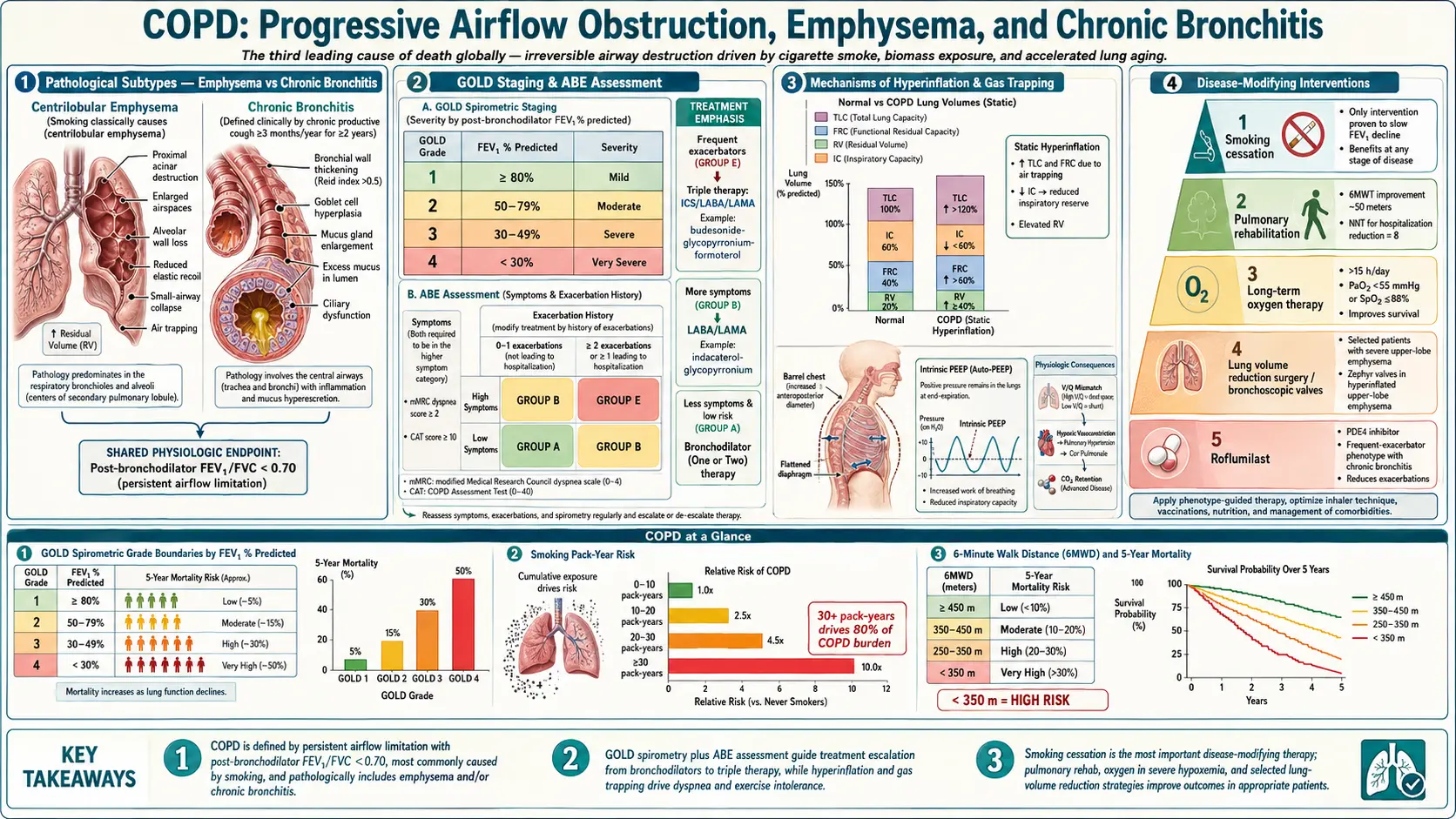
A healthy adult lung holds hundreds of millions of air sacs: a 2004 count of six adult human lungs found a mean of 480 million alveoli (range 274–790 million). In emphysema, the WHO describes destruction of the tiny air sacs at the end of the airways. Air that cannot get out leaves the lungs over-inflated — hyperinflation — and the GOLD report lists a flattened diaphragm and an enlarged air space behind the breastbone among its signs on a chest X-ray.
Hyperinflation helps explain why bronchodilators can ease breathlessness. In a 2004 randomised trial in 187 people with COPD, the long-acting bronchodilator tiotropium produced sustained reductions in lung hyperinflation at rest and during exercise; the rise in inspiratory capacity let each breath expand further, and exertional breathlessness fell while exercise endurance rose by 21% compared with placebo.
The WHO lists COPD as the third leading cause of death worldwide, with 3.4 million deaths in 2023 (about 6% of all deaths); nearly 90% of COPD deaths in people under 70 occur in low- and middle-income countries. The condition is widely under-diagnosed: in population surveys of 30,874 adults aged 40 or over at 44 sites in 27 countries, 81.4% of people with spirometry-defined COPD had never been given the diagnosis.
What Actually Goes Wrong
The WHO and GOLD list tobacco smoke, household smoke from biomass fuel, outdoor air pollution, and occupational dusts, chemical agents and fumes as the main inhaled exposures. Three processes are described:
- Protease–antiprotease imbalance. Alpha-1 antitrypsin’s main function is to protect the lung against proteolytic damage from neutrophil elastase. Hydrogen peroxide, a component of cigarette smoke, is known to be essential for inactivating alpha-1 antitrypsin; a 2000 laboratory study found that oxidation of either of two methionines in the protein removes its anti-elastase activity. People born with alpha-1 antitrypsin deficiency develop early-onset emphysema, and the 2017 European Respiratory Society statement notes that even a proportion of never-smokers with the deficiency develop progressive lung disease.
- Small-airway narrowing and loss. The major sites of obstruction in COPD are the small airways, under 2 mm across. In a 2011 CT and micro-CT study, the number of airways 2.0–2.5 mm across was already reduced in GOLD stage 1 disease; lungs removed at transplantation for GOLD stage 4 disease had 72–89% fewer terminal bronchioles; and the narrowing and disappearance of terminal bronchioles preceded emphysematous destruction.
- Mucus hypersecretion. Chronic bronchitis is present in 27–35% of people with COPD, according to the 2023 GOLD report, which links it to faster lung-function decline, more exacerbations and higher mortality.
Two further findings matter. First, stopping smoking does not restore lost lung function, but in Fletcher and Peto’s 1977 study of London working men the average further rate of FEV1 loss reverted to normal once a susceptible smoker stopped. In the Lung Health Study (Anthonisen et al., Am J Respir Crit Care Med 2002), men who quit at the start declined by 30.2 mL a year over 11 years, against 66.1 mL a year in men who kept smoking (women: 21.5 versus 54.2 mL a year); at 11 years, 38% of continuing smokers had an FEV1 below 60% of predicted, compared with 10% of sustained quitters. Second, the GOLD report states that COPD almost always coexists with other diseases: cardiovascular disease is found in 20–70% of people with COPD, and osteoporosis, anxiety and depression are common and under-diagnosed.
Types of COPD
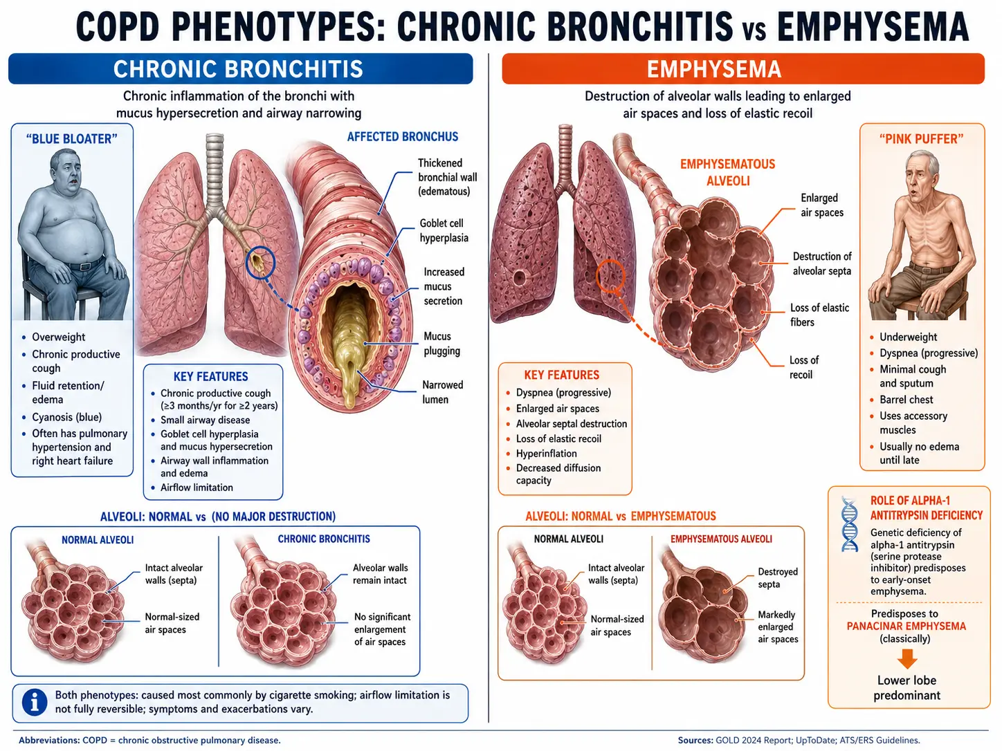
The GOLD definition covers abnormalities of the airways and/or the air sacs, and the two classic patterns below often occur together.
1. Chronic bronchitis
- Definition: the traditional definition quoted by GOLD is cough and sputum production for at least three months a year for two consecutive years.
- Who has it: 27–35% of people with COPD in the 2023 GOLD report, more often men, younger patients, people with more pack-years of smoking, more severe obstruction, rural residence or occupational exposure.
- What it is linked to: faster lung-function decline, exacerbations and mortality.
2. Emphysema
- Definition: destruction of the tiny air sacs at the end of the airways (WHO).
- On CT: GOLD notes that the presence, severity and distribution of emphysema bear on surgical or bronchoscopic lung volume reduction, and that emphysema on CT is associated with faster FEV1 decline, higher mortality and an increased risk of lung cancer.
3. Overlaps worth naming
- COPD with asthma — the 2023 GOLD report recommends that people with COPD and concomitant asthma be treated as for asthma.
- Alpha-1 antitrypsin deficiency (AATD) — an inherited cause of early-onset emphysema. In a Swedish series of 246 adults with the severe Pi*Z form, breathlessness began at a median age of 40 in smokers and 53 in non-smokers. GOLD reports the Pi*ZZ genotype in 0.12% of people with COPD. The 2003 American Thoracic Society/European Respiratory Society statement made a strong recommendation to test all symptomatic adults with COPD for AATD, and the 2016 Alpha-1 Foundation guideline continues to recommend testing all adults with symptomatic fixed airflow obstruction, whether labelled COPD or asthma; it describes testing of first-degree relatives as currently the most efficient way of finding new cases.
- Bronchiectasis with COPD — GOLD reports bronchiectasis on CT in about 30% of people with COPD, marked by more sputum, recurrent infections and more frequent exacerbations, and associated with higher mortality; GOLD recommends a chest CT when it is suspected.
Symptoms of COPD
The WHO lists difficulty breathing, chronic cough (sometimes with phlegm) and tiredness as the most common symptoms, usually developing from mid-life onwards.
- Breathlessness — as COPD progresses, the WHO notes, people find it harder to carry out normal daily activities, often because of breathlessness.
- Chronic cough, sometimes with phlegm.
- Wheezing, a symptom COPD shares with asthma; the WHO notes that people may have both conditions.
- Tiredness.
- Flare-ups (exacerbations) — symptoms can get worse quickly; the WHO describes flare-ups that usually last a few days and often need additional medicine. GOLD describes worsening breathlessness with cough and purulent sputum as the typical pattern, while noting that other conditions can mimic it.
- Low body weight — a Cochrane review notes that people with COPD and low body weight have lower exercise capacity and higher mortality than those who are adequately nourished.
How It Varies Between People
- Exacerbation frequency behaves like a trait. In the ECLIPSE cohort of 2,138 patients, frequent exacerbations (two or more in the first year) occurred in 22% of people with GOLD stage 2 disease, 33% with stage 3 and 47% with stage 4. The single best predictor of exacerbations at every stage was a history of exacerbations, and the frequent-exacerbation pattern was relatively stable over three years.
- Symptoms and lung function are assessed separately. Since its 2017 report, GOLD has separated the spirometric grade from symptom evaluation, deriving its treatment groups from symptoms and exacerbation history alone. In the BODE study, an index combining body-mass index, airflow obstruction, breathlessness and six-minute-walk distance predicted death better than FEV1 alone (C statistic 0.74 versus 0.65).
- Blood eosinophils and inhaled steroids. The 2023 GOLD report states that adding an inhaled corticosteroid has little or no effect at a blood eosinophil count below 100 cells/µL, that counts of 300 cells/µL or more identify people with a strong likelihood of benefit, and that the effect is graded in between.
- Never-smokers get COPD too. A 2009 Lancet review estimated that 25–45% of people with COPD have never smoked. GOLD reports that in low- and middle-income countries COPD in non-smokers may account for up to 60–70% of cases, and the WHO that tobacco smoking accounts for over 70% of cases in high-income countries but 30–40% in low- and middle-income countries.
- Sex. GOLD reports that COPD prevalence in developed countries is now almost equal in men and women, and that women report more breathlessness, worse health status and more exacerbations than men at similar severity of airflow limitation.
Risk Factors
- Tobacco smoking — GOLD reports that smokers have more respiratory symptoms, faster FEV1 decline and higher COPD mortality than non-smokers, yet fewer than 50% of heavy smokers develop COPD. Passive smoke exposure, pipes, cigars, water pipes and marijuana are also listed as risk factors.
- Biomass fuel smoke — wood, animal dung, crop residues and coal burned in poorly functioning stoves (GOLD). The 2009 Lancet review estimated that about 3 billion people are exposed to biomass smoke, compared with 1.01 billion who smoke tobacco, and suggested biomass smoke might be the biggest risk factor for COPD globally.
- Occupational dusts, chemical agents and fumes — described by GOLD as an under-appreciated risk factor. An estimate from the US NHANES III survey quoted by GOLD attributed 19.2% of COPD to workplace exposures overall and 31.1% among never-smokers.
- Alpha-1 antitrypsin deficiency — the most relevant genetic risk factor identified, according to GOLD, which also reports no increased COPD risk in heterozygous carriers (MZ and SZ) in the absence of smoking.
- Impaired lung growth — GOLD reports that about half of patients develop COPD through accelerated FEV1 decline and about half through abnormal lung growth and development with a normal rate of decline. Smoking during pregnancy and severe respiratory infections in childhood are linked to reduced lung function later.
- Outdoor air pollution — described by GOLD as a major worldwide cause of COPD that also increases exacerbations, hospitalisations and mortality.
- Asthma — GOLD notes that many studies report asthma and atopy in infancy as a possible risk factor for COPD in adulthood.
Diagnosis: The Tests and the Numbers
Spirometry: required for the diagnosis
The GOLD reports state that spirometry is required for the clinical diagnosis of COPD, and that a post-bronchodilator FEV1/FVC ratio below 0.7 is needed to establish it; a chest X-ray cannot confirm the diagnosis. GOLD uses the fixed 0.7 ratio because it is simple, independent of reference values and was used in the clinical trials behind its treatment recommendations. It acknowledges that, compared with the lower limit of normal (LLN) for the ratio, the fixed cut-off may under-diagnose young adults and over-diagnose the elderly, especially in mild disease. GOLD also notes that when the first post-bronchodilator ratio is below 0.6 it is very unlikely to rise spontaneously above 0.7.
The GOLD grades of airflow obstruction use post-bronchodilator FEV1 as a percentage of predicted, in people who meet the ratio criterion:
- GOLD 1 (mild) — FEV1 80% predicted or above.
- GOLD 2 (moderate) — 50% or above and below 80%.
- GOLD 3 (severe) — 30% or above and below 50%.
- GOLD 4 (very severe) — below 30%.
Other tests, and what each adds
- Blood eosinophil count — together with exacerbation history, the main factor in GOLD’s guidance on inhaled corticosteroids (see Treatment Options).
- Alpha-1 antitrypsin testing — recommended for all symptomatic adults with COPD by the 2003 ATS/ERS statement and for all adults with symptomatic fixed airflow obstruction by the 2016 Alpha-1 Foundation guideline. A serum level below the protective threshold of 11 µmol/L increases the risk of emphysema, and the 2017 European Respiratory Society statement notes that more accurate and less expensive genetic diagnostic methods have been developed since 2003.
- Pulse oximetry and arterial blood gases — GOLD notes that pulse oximetry is not as accurate as arterial blood gas measurement, and that blood gases are measured to decide on ventilatory support during an exacerbation.
- Chest CT — the 2023 GOLD report recommends chest CT for people with persistent exacerbations, symptoms out of proportion to their airflow limitation, severe obstruction with significant hyperinflation and gas trapping, or who meet lung-cancer screening criteria. It notes that most people with COPD meet those criteria, and recommends annual low-dose CT screening for lung cancer in COPD due to smoking; for COPD not due to smoking, it finds the data insufficient to establish benefit over harm.
- Six-minute walk test — part of the BODE index (Body-mass index, airflow Obstruction, Dyspnoea, Exercise capacity), which predicted death from any cause and from respiratory causes better than FEV1 alone.
Staging and the ABE Groups
Since 2017, GOLD has kept how obstructed the airways are (the numerical grade above) separate from how a person is doing, which drives its treatment groups. Symptoms are scored with the modified Medical Research Council (mMRC) breathlessness questionnaire or the COPD Assessment Test (CAT), and exacerbations over the previous year are counted. The 2023 report replaced groups C and D with a single group E:
- Group A — no exacerbation, or one that did not lead to hospital admission, with an mMRC score of 0–1 or a CAT score below 10.
- Group B — the same exacerbation history, with an mMRC score of 2 or more or a CAT score of 10 or more.
- Group E — a heavier exacerbation history (two or more moderate exacerbations, or one leading to hospital admission), whatever the symptom score.
Treatment Options
Treatments with trial evidence on survival
Most COPD treatments in the trials below improved symptoms, exacerbations or quality of life rather than survival. Those with a reported survival effect include a smoking-cessation programme (the Lung Health Study, in 5,887 middle-aged volunteers with asymptomatic airway obstruction: all-cause mortality 8.83 versus 10.38 per 1,000 person-years over up to 14.5 years, P = 0.03, even though only 21.7% of the intervention group had stopped smoking at five years); long-term oxygen in severe chronic hypoxaemia (the MRC and NOTT trials, below); and lung volume reduction surgery in one subgroup (NETT, below). A 2024 review of the GOLD 2023 report notes that the report also discusses new findings suggesting that triple inhaled therapy can reduce mortality.
Inhaled bronchodilators
- Long-acting muscarinic antagonists (LAMA) — tiotropium, glycopyrronium (glycopyrrolate), umeclidinium. The UPLIFT trial (Tashkin et al., NEJM 2008) randomised 5,993 people to tiotropium or placebo for four years: tiotropium improved lung function, quality of life and exacerbations, and reduced exacerbation-related hospitalisations and respiratory failure, but did not significantly reduce the rate of FEV1 decline, which was the co-primary end point.
- Long-acting beta-2 agonists (LABA) — salmeterol, formoterol, indacaterol, vilanterol.
- LAMA + LABA combination — the 2023 GOLD report states that dual bronchodilation is more effective than monotherapy with similar side effects, recommends it as initial therapy for groups B and E (except people in group E with eosinophils of 300 cells/µL or more, in whom triple therapy can be considered), and no longer encourages LABA plus inhaled corticosteroid. In FLAME (Wedzicha et al., NEJM 2016), among people with at least one exacerbation in the previous year (1,680 on indacaterol–glycopyrronium, 1,682 on salmeterol–fluticasone), the dual bronchodilator was not only non-inferior but superior: the annual rate of all exacerbations was 11% lower (3.59 versus 4.03), moderate or severe exacerbations were 0.98 versus 1.19 a year, pneumonia occurred in 3.2% versus 4.8%, and the effect did not depend on the baseline eosinophil count.
- Short-acting bronchodilators — short-acting beta-2 agonists, with or without short-acting anticholinergics, which GOLD lists as the initial bronchodilators for treating an exacerbation.
Inhaled corticosteroids: benefit depends on eosinophils and exacerbations
TORCH (Calverley et al., NEJM 2007) compared salmeterol–fluticasone with placebo, either drug alone, for three years in 6,112 people. All-cause mortality was 12.6% with the combination and 15.2% with placebo (hazard ratio 0.825, P = 0.052), which did not reach the predetermined level of statistical significance. The combination reduced exacerbations from 1.13 to 0.85 a year, while pneumonia was reported in 19.6% of the combination group and 18.3% of the fluticasone group, against 12.3% with placebo.
Later trials tested single-inhaler triple therapy (inhaled corticosteroid + LAMA + LABA). In IMPACT (Lipson et al., NEJM 2018; 10,355 patients), moderate or severe exacerbations were 0.91 a year with fluticasone furoate–umeclidinium–vilanterol, against 1.07 with fluticasone furoate–vilanterol and 1.21 with umeclidinium–vilanterol, and severe exacerbations leading to hospital admission were 0.13 versus 0.19 a year compared with umeclidinium–vilanterol; clinician-diagnosed pneumonia was more frequent with triple therapy than with umeclidinium–vilanterol (hazard ratio 1.53). In ETHOS (Rabe et al., NEJM 2020; 8,509 patients with at least one exacerbation in the past year), the annual rate was 1.08 and 1.07 with triple therapy at two budesonide doses, against 1.42 with glycopyrrolate–formoterol and 1.24 with budesonide–formoterol; confirmed pneumonia occurred in 3.5–4.5% of the groups taking an inhaled corticosteroid and 2.3% of the glycopyrrolate–formoterol group.
The 2023 GOLD report bases the decision to add an inhaled corticosteroid mainly on exacerbation history and the blood eosinophil count:
- 300 cells/µL or more — a strong likelihood of benefit; triple therapy can be considered from the start in group E, and stopping the steroid is more likely to be followed by exacerbations.
- 100–300 cells/µL — a graded effect, so some people are likely to benefit; for persistent exacerbations on LAMA + LABA, escalation to triple therapy is recommended at 100 cells/µL or more.
- Below 100 cells/µL — little or no effect.
Other drug options
- Roflumilast — an oral PDE4 inhibitor. In REACT (Martinez et al., Lancet 2015), 1,945 people with severe COPD, chronic bronchitis and at least two exacerbations in the previous year despite an inhaled corticosteroid–LABA combination were randomised to roflumilast or placebo for a year. In the primary analysis, moderate-to-severe exacerbations were 13.2% lower (rate ratio 0.868, p = 0.0529, not statistically significant); a predefined sensitivity analysis found them 14.2% lower (p = 0.0424). Adverse events (67% versus 59%) and withdrawals because of them (11% versus 5%) were more common with roflumilast. A 2020 Cochrane review of PDE4 inhibitors found a reduced likelihood of exacerbation (odds ratio 0.78), more diarrhoea (odds ratio 3.20), more psychiatric adverse events with roflumilast 500 µg (odds ratio 2.13), weight loss, insomnia and depressive mood symptoms, and no effect on mortality. GOLD lists roflumilast as an option for continued exacerbations, particularly in people with chronic bronchitis and an FEV1 below 50% predicted.
- Azithromycin — Albert et al. (NEJM 2011) randomised 1,142 people at increased risk of exacerbations to azithromycin 250 mg daily or placebo for a year, excluding people with hearing impairment, resting tachycardia or an apparent risk of QT prolongation. Exacerbations were 1.48 versus 1.83 per patient-year and the median time to the first was 266 versus 174 days; hearing decrements were more common with azithromycin (25% versus 20%), and the authors noted that the effect on microbial resistance patterns is not known. GOLD lists a macrolide as an option for continued exacerbations, particularly in people who do not currently smoke.
- Dupilumab — in BOREAS (Bhatt et al., NEJM 2023), 939 people with blood eosinophils of at least 300 cells/µL and an elevated exacerbation risk despite triple therapy received dupilumab 300 mg or placebo every two weeks. Moderate or severe exacerbations were 0.78 versus 1.10 a year (rate ratio 0.70), and lung function and quality of life improved more with dupilumab.
- Mucolytics — a 2019 Cochrane review (38 trials, 10,377 participants; N-acetylcysteine, carbocysteine, erdosteine and ambroxol) found people on mucolytics more likely to remain exacerbation-free (Peto odds ratio 1.73; eight people treated for about nine months for one more to stay exacerbation-free). More recent and longer trials showed smaller effects, the type or dose did not seem to change the effect, impact on lung function and quality of life was limited, and the effect on mortality was uncertain.
- Vaccination — the 2023 GOLD report lists vaccination against influenza, pneumococcus, COVID-19, pertussis and herpes zoster, depending on local guidelines. A 2018 Cochrane review found that inactivated influenza vaccine reduced exacerbations in people with COPD (two trials, 180 participants, low-quality evidence), mainly those occurring three or more weeks after vaccination, with mild, transient local reactions; the trials were too small to detect an effect on mortality.
Pulmonary rehabilitation
A 2015 Cochrane review of 65 randomised trials (3,822 participants) defined pulmonary rehabilitation as exercise training for at least four weeks, with or without education or psychological support; most programmes ran for eight or twelve weeks. Rehabilitation improved breathlessness, fatigue, emotional function and sense of control by more than the minimal clinically important difference, improved the St George’s Respiratory Questionnaire total score by 6.89 units, and increased six-minute walk distance by 43.93 m compared with usual care; the authors judged further trials against usual care unnecessary. After an exacerbation, a 2016 Cochrane review (20 trials, 1,477 participants) found moderate-quality evidence that rehabilitation reduced hospital readmissions (odds ratio 0.44, with marked heterogeneity), no statistically significant effect on mortality, and high-quality evidence of better quality of life and a 62 m longer walk distance. The 2023 GOLD report recommends a formal programme for people in groups B and E, and notes that trials suggest tele-rehabilitation is safe, with benefits similar to centre-based programmes across a range of outcomes.
Oxygen, Ventilation and Surgery
- Long-term oxygen therapy (LTOT) — two trials in severe chronic hypoxaemia found longer survival. In the Medical Research Council trial (Lancet 1981), 87 people under 70 with chronic bronchitis or emphysema, severe arterial hypoxaemia, carbon dioxide retention and a history of heart failure were randomised to oxygen for at least 15 hours a day or no oxygen: 19 of 42 treated patients and 30 of 45 controls died over five years. In the Nocturnal Oxygen Therapy Trial (Ann Intern Med 1980), 203 hypoxaemic patients were randomised to continuous or 12-hour nocturnal oxygen; mortality with nocturnal-only oxygen was 1.94 times that with continuous oxygen. The 2023 GOLD executive summary states that its criteria for prescribing long-term oxygen are unchanged and are set out in the full report.
- Moderate desaturation — the Long-Term Oxygen Treatment Trial (LOTT, NEJM 2016) followed 738 people with moderate resting desaturation (SpO2 89–93%) or desaturation on exercise for 1–6 years. Long-term oxygen did not lengthen the time to death or first hospitalisation (hazard ratio 0.94) and gave no sustained benefit in hospitalisations, exacerbations, quality of life, lung function or six-minute walk distance.
- Home non-invasive ventilation (NIV) — in the HOT-HMV trial (JAMA 2017), 116 people with persistent hypercapnia (PaCO2 above 53 mmHg) two to four weeks after an exacerbation were randomised to home oxygen with or without home NIV. Median time to readmission or death was 4.3 months with NIV versus 1.4 months without (adjusted hazard ratio 0.49); the 12-month risk of readmission or death was 63.4% versus 80.4%, and 16 versus 19 patients had died at 12 months.
- Lung volume reduction — in NETT (Fishman et al., NEJM 2003; 1,218 patients with severe emphysema), surgery did not improve overall survival (0.11 deaths per person-year in both groups) but improved exercise capacity more often. Among people with predominantly upper-lobe emphysema and low baseline exercise capacity, mortality was lower with surgery (risk ratio 0.47); among those with non-upper-lobe emphysema and high exercise capacity, it was higher (risk ratio 2.06). The 2023 GOLD report states that surgical or bronchoscopic lung volume reduction may be considered in selected people with heterogeneous or homogeneous emphysema and significant hyperinflation despite optimised care, and bullectomy in people with a large bulla.
- Lung transplantation — GOLD states that it may be considered in selected people with very severe COPD and no relevant contraindications.
Exacerbations: What Guidelines and Trials Report
The 2023 GOLD report describes exacerbations as harming health status, disease progression and prognosis, and reports a five-year mortality of about 50% after a hospital admission for one.
- Bronchodilators — GOLD lists short-acting beta-2 agonists, with or without short-acting anticholinergics, as the initial treatment, given by metered-dose inhaler (with a spacer if necessary) or by nebuliser; it describes air-driven nebulisation as preferable to oxygen-driven, to avoid raising carbon dioxide levels.
- Systemic corticosteroids — GOLD recommends 40 mg of prednisone-equivalent a day for 5 days and reports that longer courses increase the risk of pneumonia and mortality. In the REDUCE trial (JAMA 2013; 314 patients), 5 days of prednisone 40 mg was non-inferior to 14 days for re-exacerbation within six months (37.2% versus 38.4%) with a lower cumulative dose (379 versus 793 mg). GOLD also notes that steroids may be less effective in people with lower blood eosinophils.
- Antibiotics — GOLD recommends them for people with increased sputum volume and purulence and for most of those needing mechanical ventilation, for 5–7 days, with the choice based on local resistance patterns.
- Written action plans — a 2016 Cochrane review (7 trials, 1,550 participants) found that an action plan with brief education reduced the likelihood of hospital admission (odds ratio 0.69) and of emergency department visits (odds ratio 0.55) over 12 months, with no difference in mortality; people with action plans used more courses of oral steroids and antibiotics.
- Controlled oxygen — GOLD recommends titrating supplemental oxygen to a saturation of 88–92%. In a 2010 ambulance trial of 405 patients with a presumed exacerbation, mortality was 9% with high-flow oxygen and 4% with titrated oxygen (9% versus 2% in confirmed COPD), and titrated oxygen was associated with less respiratory acidosis and hypercapnia.
What the Evidence Does Not Support
- Inhaled steroids for everyone with COPD. GOLD reports little or no effect below 100 eosinophils/µL, and pneumonia was more frequent in the steroid-containing arms of TORCH, IMPACT and ETHOS. GOLD states that stopping the steroid can be considered if pneumonia or other important side effects occur, noting that exacerbations are more likely to follow when eosinophils are 300 cells/µL or more.
- Long-term oxygen for moderate desaturation. LOTT found no benefit in survival, hospitalisation, quality of life or walking distance.
- Tiotropium as a disease-modifying drug. UPLIFT tested whether it slows FEV1 decline; it did not, although it improved lung function, quality of life and exacerbations.
- Beta-blockers to prevent exacerbations. BLOCK COPD (NEJM 2019) randomised 532 people with moderate or severe COPD and no established indication for a beta-blocker to metoprolol or placebo. The trial stopped early for futility and safety concerns: time to first exacerbation did not differ (202 versus 222 days), exacerbations leading to hospital admission were more common with metoprolol (hazard ratio 1.91), and there were 11 deaths with metoprolol versus 5 with placebo.
- Low-dose morphine for chronic breathlessness. In the MABEL trial (Lancet Respir Med 2025; 143 people with severe breathlessness from heart or lung conditions), long-acting oral morphine 5–10 mg twice daily did not improve worst breathlessness at day 28 compared with placebo, and caused more adverse events (251 versus 162) and serious adverse events (15 versus 3).
- Long-term azithromycin beyond the studied group. The benefit was shown in selected people at increased exacerbation risk with no hearing impairment, resting tachycardia or apparent QT risk; the trial authors noted that its effect on microbial resistance is not known.
Prevention Strategies
- Stopping smoking. Stopping does not restore lost lung function but returns its rate of loss towards normal (Fletcher and Peto, 1977), and the Lung Health Study smoking-cessation programme lowered all-cause mortality. A 2016 Cochrane review of smokers with COPD found that varenicline (risk ratio 3.34), sublingual nicotine tablets (2.60) and bupropion (2.03) increased quit rates over placebo, that pharmacotherapy plus intensive behavioural support beat placebo plus the same support (risk ratio 2.53), and no convincing evidence for preferring any particular form of treatment.
- Household air pollution — GOLD states that research is needed on how reducing household air pollution affects COPD risk.
- Occupational exposure — GOLD describes workplace dusts, chemical agents and fumes as an under-appreciated risk factor (see Risk Factors).
- Vaccination — influenza, pneumococcus, COVID-19, pertussis and herpes zoster, depending on local guidelines, in the 2023 GOLD report.
- Early life — GOLD links smoking during pregnancy, prematurity, early-life hospitalisations and severe childhood respiratory infections to reduced lung function in adulthood.
- Case-finding spirometry. The 2023 GOLD report states that screening spirometry is probably not indicated in people without symptoms or significant exposures, while in people with symptoms and/or risk factors (for example more than 20 pack-years of smoking, recurrent chest infections, or prematurity) the diagnostic yield is relatively high and spirometry should be considered for case finding. Fletcher and Peto concluded in 1977 that severe obstructive lung disease could be prevented by screening smokers’ lung function in early middle age, if those with reduced function could be induced to stop smoking.
Practical Detail: Nutrition and Daily Life
- Inhaler devices. The 2023 GOLD report states that the choice of device depends on availability, patient preference and the ability to perform the correct inhalation manoeuvre, and that correct use requires education and training and regular reassessment at follow-up.
- Nutrition. A 2012 Cochrane review (17 trials, 632 participants) found moderate-quality evidence that nutritional supplementation promotes significant weight gain in people with COPD, especially if malnourished, with improvements in fat-free mass and six-minute walk distance; respiratory muscle strength and quality of life improved in malnourished patients. GOLD lists lower body-mass index among the factors associated with a poor outcome after a hospital admission. Protein-rich whole foods include eggs, fish such as salmon and sardines, poultry, meat, lentils, beans, nuts, whole-milk dairy and Greek yogurt, eaten alongside vegetables, fruit, olive oil and whole grains such as brown rice, oats, barley and buckwheat.
- Physical activity. GOLD reports that physical activity is decreased in COPD and that maintaining it is the main challenge.
- Handheld fan. A 2026 meta-analysis of 12 randomised trials (463 people with advanced disease, including COPD) found that face-directed fan therapy reduced immediate breathlessness, with high heterogeneity between trials, but showed no significant benefit for cumulative short-term breathlessness; long-term benefits were unclear.
- Assessments discussed in the guidelines and trials on this page: post-bronchodilator spirometry; a blood eosinophil count; alpha-1 antitrypsin testing; pulmonary rehabilitation; a written exacerbation action plan; inhaler-technique review; assessment of blood oxygen; and, in COPD due to smoking, low-dose CT lung-cancer screening. Decisions about tests and treatment belong with a clinician.
Warning Signs Listed in the Guidelines
The 2023 GOLD report lists these as indications for hospital care during an exacerbation:
- Severe symptoms — sudden worsening of breathlessness at rest, a high breathing rate, falling oxygen saturation, confusion or drowsiness.
- Acute respiratory failure.
- New physical signs such as cyanosis (blue discoloration) or peripheral oedema (swelling).
- Failure to respond to initial treatment.
- Serious coexisting illness, such as heart failure or a newly occurring arrhythmia.
GOLD also notes that people with COPD are at increased risk of decompensated heart failure, pneumonia and pulmonary embolism, which can mimic or aggravate an exacerbation.
Interactions and cautions
- High-flow oxygen — in the 2010 ambulance trial, high-flow oxygen was associated with higher mortality, more acidosis and more hypercapnia than titrated oxygen; GOLD’s target saturation in an exacerbation is 88–92%.
- Opioids — in MABEL, low-dose morphine caused more adverse events than placebo without improving breathlessness.
- Azithromycin — the long-term trial excluded people with hearing impairment, resting tachycardia or an apparent risk of QT prolongation, and hearing decrements were more common on the drug.
- Beta-blockers — GOLD states that coexisting conditions are treated according to the usual standards regardless of COPD. In people with no established indication, BLOCK COPD found more exacerbations leading to hospital admission with metoprolol.
- Roflumilast — associated with weight loss, insomnia, depressive mood symptoms and psychiatric adverse events in the Cochrane review.
- Repeated systemic steroids — GOLD reports that recurrent use increases the risk of osteoporosis.
Complications of COPD
- Respiratory failure — low blood oxygen, with carbon dioxide retention in some people; GOLD lists acute respiratory failure among the indications for hospital care.
- Cor pulmonale — the MRC oxygen trial enrolled people with chronic hypoxic cor pulmonale complicating chronic bronchitis and emphysema.
- Pneumonia — more frequent with inhaled corticosteroids in TORCH, IMPACT and ETHOS.
- Lung cancer — emphysema on CT is associated with an increased risk (GOLD); lung cancer caused 33% of the 731 deaths over 14.5 years in the Lung Health Study.
- Cardiovascular disease — present in 20–70% of people with COPD, from heart failure and ischaemic heart disease to arrhythmias, peripheral vascular disease and hypertension (GOLD); cardiovascular disease caused 22% of deaths in the Lung Health Study.
- Bronchiectasis — in about 30% of people with COPD (GOLD).
- Sleep apnoea — in about 14% of people with COPD; GOLD reports more frequent oxygen desaturation and longer sleep time with low oxygen and high carbon dioxide than in sleep apnoea without COPD.
- Osteoporosis, often under-diagnosed and associated with poor health status and prognosis (GOLD).
- Anxiety and depression, which GOLD describes as important, under-diagnosed and associated with poor prognosis; cognitive impairment is reported in 32% of people with COPD.
- Frailty — weakness, fatigue, low physical activity and unintentional weight loss, reported as more prevalent in COPD (GOLD).
Key Research Papers
Historical background
Charles Fletcher and Richard Peto’s prospective study of London working men, published in 1977, found that FEV1 falls gradually over a lifetime, that clinically significant airflow obstruction never develops in most non-smokers and many smokers, and that in susceptible people smoking causes irreversible obstructive changes; if a susceptible smoker stops, lost function is not recovered, but the average further rate of loss reverts to normal. They also concluded that infections and chronic mucus hypersecretion do not make airflow obstruction progress faster. The earlier history of the condition is covered on the COPD History page.
Key research papers
Each citation below was checked against its PubMed record; the linked DOI resolves to the paper named.
- Christenson SA, Smith BM, Bafadhel M, Putcha N. Chronic obstructive pulmonary disease. Lancet. 2022;399(10342):2227–2242. — doi:10.1016/S0140-6736(22)00470-6 · PubMed PMID: 35533707
- Vestbo J, Hurd SS, Agustí AG, et al. Global strategy for the diagnosis, management, and prevention of chronic obstructive pulmonary disease: GOLD executive summary. Am J Respir Crit Care Med. 2013;187(4):347–365. — doi:10.1164/rccm.201204-0596PP · PubMed PMID: 22878278
- Vogelmeier CF, Criner GJ, Martinez FJ, et al. Global strategy for the diagnosis, management, and prevention of chronic obstructive lung disease 2017 report: GOLD executive summary. Am J Respir Crit Care Med. 2017;195(5):557–582. — doi:10.1164/rccm.201701-0218PP · PubMed PMID: 28128970
- Anthonisen NR, Connett JE, Murray RP. Smoking and lung function of Lung Health Study participants after 11 years. Am J Respir Crit Care Med. 2002;166(5):675–679. — doi:10.1164/rccm.2112096 · PubMed PMID: 12204864
- Calverley PM, Anderson JA, Celli B, et al. Salmeterol and fluticasone propionate and survival in chronic obstructive pulmonary disease (TORCH). N Engl J Med. 2007;356(8):775–789. — doi:10.1056/NEJMoa063070 · PubMed PMID: 17314337 — the mortality difference did not reach statistical significance.
- Tashkin DP, Celli B, Senn S, et al. A 4-year trial of tiotropium in chronic obstructive pulmonary disease (UPLIFT). N Engl J Med. 2008;359(15):1543–1554. — doi:10.1056/NEJMoa0805800 · PubMed PMID: 18836213 — no effect on the rate of FEV1 decline.
- Albert RK, Connett J, Bailey WC, et al. Azithromycin for prevention of exacerbations of COPD. N Engl J Med. 2011;365(8):689–698. — doi:10.1056/NEJMoa1104623 · PubMed PMID: 21864166
- Martinez FJ, Calverley PM, Goehring UM, et al. Effect of roflumilast on exacerbations in patients with severe chronic obstructive pulmonary disease uncontrolled by combination therapy (REACT). Lancet. 2015;385(9971):857–866. — doi:10.1016/S0140-6736(14)62410-7 · PubMed PMID: 25684586
- Wedzicha JA, Banerji D, Chapman KR, et al. Indacaterol–glycopyrronium versus salmeterol–fluticasone for COPD (FLAME). N Engl J Med. 2016;374(23):2222–2234. — doi:10.1056/NEJMoa1516385 · PubMed PMID: 27181606
- Lipson DA, Barnhart F, Brealey N, et al. Once-daily single-inhaler triple versus dual therapy in patients with COPD (IMPACT). N Engl J Med. 2018;378(18):1671–1680. — doi:10.1056/NEJMoa1713901 · PubMed PMID: 29668352
- Rabe KF, Martinez FJ, Ferguson GT, et al. Triple inhaled therapy at two glucocorticoid doses in moderate-to-very-severe COPD (ETHOS). N Engl J Med. 2020;383(1):35–48. — doi:10.1056/NEJMoa1916046 · PubMed PMID: 32579807
- Bhatt SP, Rabe KF, Hanania NA, et al. Dupilumab for COPD with type 2 inflammation indicated by eosinophil counts (BOREAS). N Engl J Med. 2023;389(3):205–214. — doi:10.1056/NEJMoa2303951 · PubMed PMID: 37272521
- Nocturnal Oxygen Therapy Trial Group. Continuous or nocturnal oxygen therapy in hypoxemic chronic obstructive lung disease: a clinical trial. Ann Intern Med. 1980;93(3):391–398. — doi:10.7326/0003-4819-93-3-391 · PubMed PMID: 6776858
- Fishman A, Martinez F, Naunheim K, et al. A randomized trial comparing lung-volume-reduction surgery with medical therapy for severe emphysema (NETT). N Engl J Med. 2003;348(21):2059–2073. — doi:10.1056/NEJMoa030287 · PubMed PMID: 12759479
- World Health Organization. Chronic obstructive pulmonary disease (COPD). Fact sheet. — who.int fact sheet
- Agustí A, Celli BR, Criner GJ, et al. Global Initiative for Chronic Obstructive Lung Disease 2023 Report: GOLD Executive Summary. Eur Respir J. 2023;61(4):2300239. — doi:10.1183/13993003.00239-2023 · PubMed PMID: 36858443
- Sharma M, Joshi S, Banjade P, et al. Global Initiative for Chronic Obstructive Lung Disease (GOLD) 2023 Guidelines Reviewed. Open Respir Med J. 2024;18:e18743064279064. — doi:10.2174/0118743064279064231227070344 · PubMed PMID: 38660684
- Lamprecht B, Soriano JB, Studnicka M, et al. Determinants of underdiagnosis of COPD in national and international surveys. Chest. 2015;148(4):971–985. — doi:10.1378/chest.14-2535 · PubMed PMID: 25950276
- Ochs M, Nyengaard JR, Jung A, et al. The number of alveoli in the human lung. Am J Respir Crit Care Med. 2004;169(1):120–124. — doi:10.1164/rccm.200308-1107OC · PubMed PMID: 14512270
- O’Donnell DE, Flüge T, Gerken F, et al. Effects of tiotropium on lung hyperinflation, dyspnoea and exercise tolerance in COPD. Eur Respir J. 2004;23(6):832–840. — doi:10.1183/09031936.04.00116004 · PubMed PMID: 15218994
- McDonough JE, Yuan R, Suzuki M, et al. Small-airway obstruction and emphysema in chronic obstructive pulmonary disease. N Engl J Med. 2011;365(17):1567–1575. — doi:10.1056/NEJMoa1106955 · PubMed PMID: 22029978
- Fletcher C, Peto R. The natural history of chronic airflow obstruction. Br Med J. 1977;1(6077):1645–1648. — doi:10.1136/bmj.1.6077.1645 · PubMed PMID: 871704
- Salvi SS, Barnes PJ. Chronic obstructive pulmonary disease in non-smokers. Lancet. 2009;374(9691):733–743. — doi:10.1016/S0140-6736(09)61303-9 · PubMed PMID: 19716966
- Hurst JR, Vestbo J, Anzueto A, et al. Susceptibility to exacerbation in chronic obstructive pulmonary disease (ECLIPSE). N Engl J Med. 2010;363(12):1128–1138. — doi:10.1056/NEJMoa0909883 · PubMed PMID: 20843247
- Celli BR, Cote CG, Marin JM, et al. The body-mass index, airflow obstruction, dyspnea, and exercise capacity index in chronic obstructive pulmonary disease. N Engl J Med. 2004;350(10):1005–1012. — doi:10.1056/NEJMoa021322 · PubMed PMID: 14999112
- Stoller JK, Aboussouan LS. Alpha1-antitrypsin deficiency. Lancet. 2005;365(9478):2225–2236. — doi:10.1016/S0140-6736(05)66781-5 · PubMed PMID: 15978931
- Miravitlles M, Dirksen A, Ferrarotti I, et al. European Respiratory Society statement: diagnosis and treatment of pulmonary disease in α1-antitrypsin deficiency. Eur Respir J. 2017;50(5):1700610. — doi:10.1183/13993003.00610-2017 · PubMed PMID: 29191952
- Sandhaus RA, Turino G, Brantly ML, et al. The diagnosis and management of alpha-1 antitrypsin deficiency in the adult. Chronic Obstr Pulm Dis. 2016;3(3):668–682. — doi:10.15326/jcopdf.3.3.2015.0182 · PubMed PMID: 28848891
- Larsson C. Natural history and life expectancy in severe alpha1-antitrypsin deficiency, Pi Z. Acta Med Scand. 1978;204(5):345–351. — doi:10.1111/j.0954-6820.1978.tb08452.x · PubMed PMID: 309708
- Taggart C, Cervantes-Laurean D, Kim G, et al. Oxidation of either methionine 351 or methionine 358 in alpha 1-antitrypsin causes loss of anti-neutrophil elastase activity. J Biol Chem. 2000;275(35):27258–27265. — doi:10.1074/jbc.M004850200 · PubMed PMID: 10867014
- Anthonisen NR, Skeans MA, Wise RA, et al. The effects of a smoking cessation intervention on 14.5-year mortality: a randomized clinical trial. Ann Intern Med. 2005;142(4):233–239. — doi:10.7326/0003-4819-142-4-200502150-00005 · PubMed PMID: 15710956
- Medical Research Council Working Party. Long term domiciliary oxygen therapy in chronic hypoxic cor pulmonale complicating chronic bronchitis and emphysema. Lancet. 1981;1(8222):681–686. — PubMed PMID: 6110912
- Long-Term Oxygen Treatment Trial Research Group; Albert RK, Au DH, Blackford AL, et al. A randomized trial of long-term oxygen for COPD with moderate desaturation (LOTT). N Engl J Med. 2016;375(17):1617–1627. — doi:10.1056/NEJMoa1604344 · PubMed PMID: 27783918
- Murphy PB, Rehal S, Arbane G, et al. Effect of home noninvasive ventilation with oxygen therapy vs oxygen therapy alone on hospital readmission or death after an acute COPD exacerbation: a randomized clinical trial (HOT-HMV). JAMA. 2017;317(21):2177–2186. — doi:10.1001/jama.2017.4451 · PubMed PMID: 28528348
- McCarthy B, Casey D, Devane D, et al. Pulmonary rehabilitation for chronic obstructive pulmonary disease. Cochrane Database Syst Rev. 2015;(2):CD003793. — doi:10.1002/14651858.CD003793.pub3 · PubMed PMID: 25705944
- Puhan MA, Gimeno-Santos E, Cates CJ, et al. Pulmonary rehabilitation following exacerbations of chronic obstructive pulmonary disease. Cochrane Database Syst Rev. 2016;12(12):CD005305. — doi:10.1002/14651858.CD005305.pub4 · PubMed PMID: 27930803
- Kopsaftis Z, Wood-Baker R, Poole P. Influenza vaccine for chronic obstructive pulmonary disease (COPD). Cochrane Database Syst Rev. 2018;6(6):CD002733. — doi:10.1002/14651858.CD002733.pub3 · PubMed PMID: 29943802
- Poole P, Sathananthan K, Fortescue R. Mucolytic agents versus placebo for chronic bronchitis or chronic obstructive pulmonary disease. Cochrane Database Syst Rev. 2019;5(5):CD001287. — doi:10.1002/14651858.CD001287.pub6 · PubMed PMID: 31107966
- Janjua S, Fortescue R, Poole P. Phosphodiesterase-4 inhibitors for chronic obstructive pulmonary disease. Cochrane Database Syst Rev. 2020;5(5):CD002309. — doi:10.1002/14651858.CD002309.pub6 · PubMed PMID: 32356609
- Leuppi JD, Schuetz P, Bingisser R, et al. Short-term vs conventional glucocorticoid therapy in acute exacerbations of chronic obstructive pulmonary disease: the REDUCE randomized clinical trial. JAMA. 2013;309(21):2223–2231. — doi:10.1001/jama.2013.5023 · PubMed PMID: 23695200
- Austin MA, Wills KE, Blizzard L, et al. Effect of high flow oxygen on mortality in chronic obstructive pulmonary disease patients in prehospital setting: randomised controlled trial. BMJ. 2010;341:c5462. — doi:10.1136/bmj.c5462 · PubMed PMID: 20959284
- Howcroft M, Walters EH, Wood-Baker R, et al. Action plans with brief patient education for exacerbations in chronic obstructive pulmonary disease. Cochrane Database Syst Rev. 2016;12(12):CD005074. — doi:10.1002/14651858.CD005074.pub4 · PubMed PMID: 27990628
- van Eerd EA, van der Meer RM, van Schayck OC, et al. Smoking cessation for people with chronic obstructive pulmonary disease. Cochrane Database Syst Rev. 2016;(8):CD010744. — doi:10.1002/14651858.CD010744.pub2 · PubMed PMID: 27545342
- Dransfield MT, Voelker H, Bhatt SP, et al. Metoprolol for the prevention of acute exacerbations of COPD (BLOCK COPD). N Engl J Med. 2019;381(24):2304–2314. — doi:10.1056/NEJMoa1908142 · PubMed PMID: 31633896
- Johnson MJ, Williams B, Keerie C, et al. Morphine for chronic breathlessness (MABEL) in the UK: a multi-site, parallel-group, dose titration, double-blind, randomised, placebo-controlled trial. Lancet Respir Med. 2025;13(11):967–977. — doi:10.1016/S2213-2600(25)00205-X · PubMed PMID: 41033333
- Pereira RA, Barcellos G, Lenz G, et al. Fan therapy for dyspnea in palliative and supportive care: a systematic review and meta-analysis of randomized controlled trials. Respir Care. 2026;71(5):527–535. — doi:10.1177/19433654261424868 · PubMed PMID: 41761933
- Ferreira IM, Brooks D, White J, et al. Nutritional supplementation for stable chronic obstructive pulmonary disease. Cochrane Database Syst Rev. 2012;12:CD000998. — doi:10.1002/14651858.CD000998.pub3 · PubMed PMID: 23235577
PubMed Topic Searches
The following PubMed topic searches surface the current peer-reviewed literature on COPD. Each link opens a live query; results update as new papers are indexed.
- PubMed: Search: chronic obstructive pulmonary disease
- PubMed: Search: COPD GOLD guidelines
- PubMed: Search: COPD exacerbation
- PubMed: Search: COPD blood eosinophil inhaled corticosteroid
- PubMed: Search: emphysema
- PubMed: Search: chronic bronchitis
- PubMed: Search: pulmonary rehabilitation
- PubMed: Search: alpha 1 antitrypsin deficiency
- PubMed: Search: COPD smoking cessation
- PubMed: Search: long term oxygen therapy COPD
- PubMed: Search: endobronchial valve emphysema
- PubMed: Search: lung volume reduction surgery
- PubMed: Search: COPD non invasive ventilation hypercapnia
- PubMed: Search: COPD cardiovascular comorbidity
Connections
- Pulmonology
- Asthma: What Happens in an Attack — interactive animation
- Surfactant: Why Your Lungs Don’t Collapse — interactive animation
- Hemoglobin & the Oxygen Dissociation Curve — interactive animation
- Blood pH: The Acid–Base Balancing Act — interactive animation
- Breathing & Gas Exchange — interactive animation
- Pneumonia
- Shortness of Breath
- Chronic Cough
- Asthma
- Pulmonary Hypertension
- Vitamin D3
- Eucalyptus
- Breathwork
- Interstitial Lung Disease
- Obstructive Sleep Apnea
- Pulmonary Embolism
- Fatigue
- Depression
- Lead Toxicity
- Magnesium
- Hypertension
- Chest Pain
- Cancer
- Mullein
- Lung Cancer
- RSV
- Herbs covered on this site that discuss this condition: Elecampane · Boswellia · Horehound · Savory · White Turmeric / Zedoary (Curcuma zedoaria)
- Influenza — a common trigger of exacerbations, and why annual vaccination is part of COPD care
- Dr. Bryan Ardis: Tobacco and Nicotine Bans
- Toxins