Acanthamoeba Treatment — Why It's a Siege, Not a Course
Conventional Medical Treatment
The proven therapy in detail: topical biguanides (PHMB, chlorhexidine) plus a diamidine, the punishing every-hour dosing schedule, the slow taper, and oral miltefosine for severe disease.
Ivermectin — What the Evidence Shows
People search for ivermectin against Acanthamoeba. We review the actual evidence honestly — and explain why it is not an established treatment for this infection.
Castor Oil — Folk Remedy vs Evidence
A popular folk remedy meets the clinical record. Why putting castor oil in an eye with an active amoebic infection is not a treatment — and can be dangerous.
Table of Contents
- Why Acanthamoeba Is So Hard to Treat
- The Standard of Care, in Brief
- Treatment Differs by Disease
- When Surgery Is Needed
- Investigational and Alternative Approaches — Read This Carefully
- Cost and Access
- What Recovery Looks Like
- Key Research Papers
- Featured Videos
1. Why Acanthamoeba Is So Hard to Treat
Acanthamoeba is not a typical infection, and it cannot be cleared with a typical antibiotic course. The organism lives in two forms. The active, feeding form is the trophozoite. When conditions turn hostile — including the moment a drug is introduced — it can convert into a cyst: a dormant cell wrapped in a tough, double-walled shell. That cyst wall is the heart of the problem. It resists antiseptics, drying out, heat, and the chlorine levels used in tap water and swimming pools. A drug that kills every trophozoite but leaves cysts behind has not cured anything; the survivors simply wait, re-emerge weeks later, and the infection relapses.
The eye compounds the difficulty. The cornea — the clear front window of the eye where Acanthamoeba keratitis takes hold — is avascular, meaning it has no blood vessels of its own. Blood vessels are how the immune system and many medicines reach infected tissue, so an avascular cornea is a place where the body's defenses arrive late and weakly, and where drugs delivered by mouth or vein penetrate poorly. That is why eye disease is treated mainly with drops applied directly to the surface, and why those drops have to be given relentlessly.
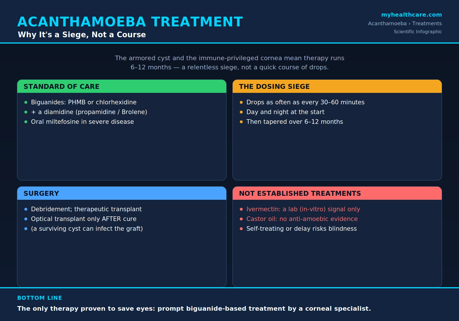
Put together, these facts explain the defining feature of Acanthamoeba therapy: it is long. Treatment that must kill cysts as well as trophozoites, in a tissue the immune system struggles to reach, routinely runs six to twelve months. It is best understood as a siege rather than a short course — a sustained campaign to outlast an organism built to wait you out. This is also why early, specialist-supervised treatment matters so much: the longer the amoeba is left to dig in and encyst, the harder and longer the siege becomes.
2. The Standard of Care, in Brief
For Acanthamoeba keratitis, the cornerstone of treatment is a topical biguanide. Two are used: polyhexamethylene biguanide (PHMB) and chlorhexidine. Their crucial advantage is that they are cysticidal as well as trophocidal — they damage the dormant cyst, not just the active form — which is exactly what is required to break the relapse cycle. A biguanide is usually combined with a diamidine, most often propamidine isethionate (sold as Brolene) or hexamidine, to broaden the attack on the parasite.
The dosing is intense. In the opening phase, drops are often given as frequently as every 30 to 60 minutes around the clock, including overnight, for the first days. As the eye responds, the frequency is slowly tapered over weeks and months — never stopped abruptly, because stopping too soon invites the surviving cysts to reactivate. In severe or treatment-resistant (refractory) cases, specialists may add oral miltefosine, a systemic anti-amoebic drug used as an adjunct rather than a substitute for the topical regimen.
This page is a map, not the full route. The complete regimen — specific agents, concentrations, the taper schedule, and how miltefosine is added — is covered on the dedicated Conventional Medical Treatment page. All of it belongs in the hands of a corneal specialist (ophthalmologist), not a self-care routine.
3. Treatment Differs by Disease
Acanthamoeba does not cause a single illness, and the treatment changes depending on where in the body the infection sits. The three main presentations are managed very differently.
Keratitis (the eye)
Acanthamoeba keratitis is the most common form, especially in contact lens wearers. It is fought at the corneal surface with the topical biguanide siege described above — PHMB or chlorhexidine, usually plus a diamidine, dosed hourly at first and tapered over many months. See Symptoms & Diagnosis for how keratitis is recognized and confirmed.
Granulomatous amebic encephalitis (GAE, the brain)
GAE is a rare but extremely serious infection of the central nervous system that occurs mainly in people with weakened immune systems. Because it is a deep, systemic infection, it cannot be treated with eye drops. Management is multi-drug and systemic — individualized combinations of anti-amoebic agents given by mouth or vein, frequently including miltefosine — and is handled by infectious-disease specialists, often in consultation with public-health authorities.
Cutaneous and disseminated disease (skin and beyond)
In immunocompromised patients, Acanthamoeba can cause skin lesions or spread (disseminate) to multiple organs. Treatment combines topical and systemic agents and, critically, depends on restoring the patient's immune function wherever possible, since the immune system does much of the work of containing the parasite.
4. When Surgery Is Needed
Medicine is the foundation of Acanthamoeba treatment, but surgery has specific, limited roles — usually when the infection has damaged the cornea or refuses to clear with drops alone.
Epithelial debridement is the simplest procedure: the infected surface layer of the cornea (the epithelium) is gently scraped away. This removes a reservoir of organisms and lets the drops penetrate the tissue beneath more effectively. It is often done early.
Therapeutic corneal transplant is performed as a rescue when the cornea is melting (progressively thinning and dissolving) or has perforated. Here the goal is to save the structural integrity of the eye in the face of damage the drugs cannot reverse quickly enough.
Optical corneal transplant — a graft performed to restore clear vision after the infection is over — carries a vital caveat: it should be attempted only after the infection is proven to be cured. A single surviving cyst left in the eye can seed the new donor cornea and reinfect the graft, undoing the surgery. This is one more reason confirmation of cure, not just symptom relief, governs the timeline.
Enucleation — surgical removal of the eye — is a genuine last resort, reserved for an eye that is blind, intractably painful, or a source of uncontrolled infection that threatens the rest of the body. It is rare, but it is part of the honest picture of how severe untreated or late-treated disease can become.
5. Investigational and Alternative Approaches — Read This Carefully
Let us be plain, because eyesight is on the line: the only treatment proven to save eyes from Acanthamoeba keratitis is prompt, biguanide-based therapy supervised by a corneal specialist. Everything else discussed below is either investigational or unproven for this infection.
People understandably search for other options, and two names come up often: ivermectin and castor oil. This site does not pretend those searches do not happen — instead it reviews the evidence for each, honestly, on dedicated pages. But the bottom line is the same for both: neither is an established Acanthamoeba treatment.
- Ivermectin — What the Evidence Shows: ivermectin is an important antiparasitic for several human diseases, but it is not a recognized therapy for Acanthamoeba, and there is no good clinical evidence that it cures Acanthamoeba keratitis.
- Castor Oil — Folk Remedy vs Evidence: castor oil is a traditional remedy with no evidence base as an Acanthamoeba treatment, and putting any unproven oil into an actively infected eye carries real risk.
The danger is not only that these substances do not work. It is the delay they cause. Acanthamoeba rewards early treatment and punishes lateness; the cyst digs in, the cornea scars, and what might have been a months-long medical siege becomes surgery — or permanent loss of vision. Self-treating an active eye infection, or putting off real care to try a home remedy, risks permanent blindness. If you have eye pain, light sensitivity, redness, or blurred vision — especially as a contact lens wearer — the safe move is to be seen by an eye specialist urgently, not to experiment.
6. Cost and Access
Because the therapy is so long, it is also expensive — a burden that is part of the real-world story of this disease. The figures below are reported values that have appeared in case reports and patient accounts; individual costs vary widely by country, insurance, and severity.
A frequently cited example is oral miltefosine (Impavido), which has been reported at around $16,000 for a single 28-capsule course. For a severe case requiring extended, multi-drug treatment, the total medication cost has been reported near $62,000. These are headline-grabbing numbers, but they reflect a genuine reality: specialty anti-amoebic drugs are scarce, and months of compounded biguanide and diamidine drops add up.
Access can also be a hurdle because some of these drugs are not routinely stocked. In the United States, miltefosine for free-living amoebae has been distributed through the U.S. Centers for Disease Control and Prevention (CDC), which has historically served as a route to obtain the drug for these rare infections. A corneal or infectious-disease specialist is the person who navigates this on a patient's behalf — another reason early specialist involvement matters.
7. What Recovery Looks Like
It helps to know, honestly, what the months of treatment feel like — both to prepare and to take the threat seriously enough to seek care early.
Recovery from Acanthamoeba keratitis is slow and demanding. In the early weeks it means putting in drops every hour or two, day and night, then slowly tapering as the eye allows. Many patients endure severe light sensitivity (photophobia) — some describe living in a darkened room for weeks because daylight is unbearable. The pain and the relentless dosing schedule frequently make it impossible to work during the worst of it.
For those whose corneas are too damaged to recover with medicine alone and who need an optical transplant, the timeline stretches further. Because surgeons must first confirm the amoeba is truly gone before grafting — a surviving cyst could infect the new cornea — patients may face up to a year of waiting to confirm cure before a vision-restoring transplant can even be attempted. The reward of patience is a real chance to save the eye; the cost is time, discomfort, and perseverance. None of this is meant to frighten — it is meant to make the case for the one thing that shortens it all: getting to a specialist early.
Key Research Papers
Peer-reviewed studies underpinning the points on this page — the biology that makes Acanthamoeba so hard to treat, the biguanide and diamidine regimens that define the standard of care, the role of oral miltefosine and surgical adjuncts, and the risk factors behind keratitis. Each citation links to the full text via DOI. Treatment of an active infection should always be directed by a corneal or infectious-disease specialist.
- Lorenzo-Morales J, Khan NA, Walochnik J. An Update on Acanthamoeba Keratitis: Diagnosis, Pathogenesis and Treatment. Parasite. 2015;22:10.
- Marciano-Cabral F, Cabral G. Acanthamoeba spp. as Agents of Disease in Humans. Clinical Microbiology Reviews. 2003;16(2):273–307.
- Larkin DFP, Kilvington S, Dart JKG. Treatment of Acanthamoeba Keratitis with Polyhexamethylene Biguanide. Ophthalmology. 1992;99(2):185–191.
- Seal D, Hay J, Kirkness C, et al. Successful Medical Therapy of Acanthamoeba Keratitis with Topical Chlorhexidine and Propamidine. Eye. 1996;10(4):413–421.
- Kosrirukvongs P, Wanachiwanawin D, Visvesvara GS. Treatment of Acanthamoeba Keratitis with Chlorhexidine. Ophthalmology. 1999;106(4):798–802.
- Dewan N, Ming W, Holland SP, Yeung SN, Iovieno A. Oral Miltefosine as Adjunctive Treatment for Recalcitrant Acanthamoeba Keratitis. Cornea. 2019;38(7):914–917.
- Richoz O, Gatzioufas Z, Hafezi F. Corneal Collagen Cross-Linking for the Treatment of Acanthamoeba Keratitis. Cornea. 2013;32(10):e189.
- Carnt N, Hoffman JJ, Verma S, et al. Acanthamoeba Keratitis: Confirmation of the UK Outbreak and a Prospective Case-Control Study Identifying Contributing Risk Factors. British Journal of Ophthalmology. 2018;102(12):1621–1628.
- Carnt N, Minassian DC, Dart JKG. Acanthamoeba Keratitis Risk Factors for Daily Wear Contact Lens Users: A Case-Control Study. Ophthalmology. 2023;130(1):48–55.
Live PubMed Searches
Each link opens a live PubMed query so results stay current as new papers are indexed.
- Acanthamoeba keratitis treatment
- Polyhexamethylene biguanide (PHMB) Acanthamoeba
- Chlorhexidine Acanthamoeba keratitis
- Miltefosine Acanthamoeba
- Acanthamoeba keratitis corneal transplant
- Granulomatous amebic encephalitis treatment
- Acanthamoeba cyst resistance
- Acanthamoeba keratitis contact lens risk factors
Connections
- Conventional Medical Treatment
- Ivermectin — What the Evidence Shows
- Castor Oil — Folk Remedy vs Evidence
- Acanthamoeba
- Symptoms & Diagnosis
- Acanthamoeba Keratitis
- Granulomatous Amebic Encephalitis
- Parasites
- Castor Oil (Remedy)
- Ophthalmology
- Infectious Disease
- Silver Nanoparticles