Cutaneous and Disseminated Acanthamoeba Infection
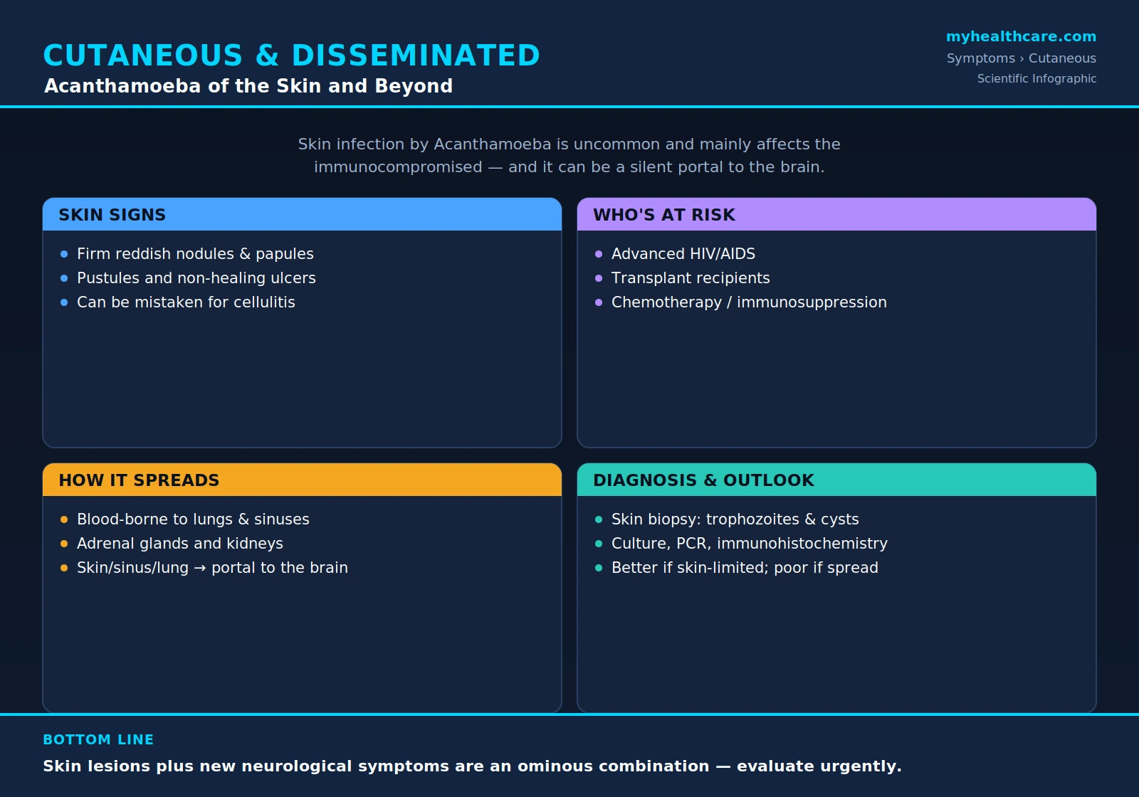
Table of Contents
- What Is Cutaneous Acanthamoeba Infection?
- Who Is at Risk
- Skin Symptoms
- Disseminated Disease
- The Link to Brain Disease
- Diagnosis
- Treatment
- Prognosis
- Key Research Papers
- Featured Videos
1. What Is Cutaneous Acanthamoeba Infection?
Cutaneous Acanthamoeba infection is an infection of the skin caused by Acanthamoeba, a free-living amoeba found widely in soil, dust, and fresh and brackish water. The same organism that causes the eye infection Acanthamoeba keratitis in contact-lens wearers can, under very different circumstances, invade and persist in the skin. When it does, it produces chronic, often non-healing nodules, ulcers, and abscesses rather than the brief sores caused by ordinary bacterial skin infections.
Cutaneous Acanthamoeba infection is uncommon. Unlike keratitis, which strikes otherwise healthy people, skin and disseminated disease occur almost entirely in people whose immune defenses are weakened. The amoeba is an opportunist: in a person with a healthy immune system, the body's macrophages and other defenses usually clear it before it can establish itself, so the skin form is rare in the general population. The combination of its rarity and its resemblance to far more common conditions means it is frequently overlooked at first, and a diagnosis is often reached only after a skin biopsy.
Two related Acanthamoeba syndromes affect the body beyond the eye. The first is the cutaneous (skin) form described on this page. The second is granulomatous amebic encephalitis (GAE), a slowly progressive brain infection. The two are connected: skin involvement and brain involvement frequently occur together in the same severely immunocompromised patient, and skin lesions can precede or accompany the neurological disease.
2. Who Is at Risk
Cutaneous and disseminated Acanthamoeba infection is predominantly a disease of the immunocompromised. The people most affected share a common thread: their cell-mediated immunity — the arm of the immune system that controls organisms living inside or attacking tissue — is impaired. The major risk groups are:
- Advanced HIV/AIDS. People with very low CD4 T-cell counts have historically accounted for a large share of reported cutaneous Acanthamoeba cases. The skin lesions may be the first sign of disease.
- Organ- and stem-cell-transplant recipients. The same drugs that prevent transplant rejection also suppress the defenses that would otherwise hold the amoeba in check. Cases have been reported after liver, kidney, lung, heart, and bone-marrow transplantation.
- People on chemotherapy or immunosuppressive drugs. Patients receiving cancer chemotherapy, high-dose corticosteroids, or biologic immunosuppressants — for example for leukemia, lymphoma, or graft-versus-host disease — are also vulnerable.
Other conditions that weaken immunity, such as poorly controlled diabetes, kidney failure, and chronic illness, may add to the risk. The unifying feature is reduced immune surveillance, which lets an organism that healthy people brush off take hold and spread.
3. Skin Symptoms
The skin findings of cutaneous Acanthamoeba infection are varied, which is part of why they are so often mistaken for something else. The most common lesions are:
- Firm reddish papules and nodules — raised, solid bumps under or within the skin, sometimes tender, that may be a few millimeters to several centimeters across.
- Pustules — pus-filled lesions that can be mistaken for ordinary bacterial folliculitis or acne.
- Non-healing ulcers — open sores with ragged edges that fail to close over weeks to months despite standard wound care or antibiotics.
The lesions most often appear on the face, trunk, arms, and legs, and there may be one lesion or many. Early on, the appearance can closely mimic cellulitis (a spreading bacterial skin infection), a deep fungal infection, an atypical mycobacterial infection, vasculitis, or even a skin cancer. A telling clue is the behavior of the lesions over time: rather than coming to a head and healing the way a boil or a cut would, Acanthamoeba skin lesions tend to persist and slowly progress, enlarging or multiplying and resisting the antibiotics that would resolve an ordinary bacterial infection. A chronic skin lesion that will not heal in a person with weakened immunity should raise the question of an unusual organism such as Acanthamoeba.
4. Disseminated Disease
Disseminated infection means the amoeba has spread beyond its original site and is seeding organs throughout the body. Acanthamoeba can enter the body through several portals — broken or diseased skin, the sinuses (through the nose), or the lungs (by inhaling contaminated dust or aerosols). From any of these entry points the organism can invade small blood vessels and travel through the bloodstream to distant sites.
Once disseminated, Acanthamoeba can affect:
- Lungs — producing pneumonitis (lung inflammation) that may look like an ordinary or opportunistic pneumonia on a chest X-ray or CT scan.
- Sinuses — causing a chronic sinusitis that fails to clear with usual treatment, sometimes with destruction of the surrounding bone and tissue.
- Adrenal glands and kidneys — the organism has been found in these organs at autopsy in disseminated cases.
- Skin — widespread skin nodules and ulcers are themselves often a manifestation of disseminated, bloodstream-spread disease rather than a purely local problem.
Because the lungs, sinuses, and skin can serve as both entry points and targets, disseminated Acanthamoeba infection is best thought of as a whole-body illness in a severely immunocompromised host, not an isolated organ infection. Its non-specific symptoms — fever, weight loss, cough, persistent skin lesions — overlap with many other infections that affect such patients, which again delays recognition.
5. The Link to Brain Disease
The most serious aspect of cutaneous and disseminated Acanthamoeba infection is its connection to the brain. A skin, sinus, or lung infection can act as a silent portal of entry that later seeds the central nervous system, producing granulomatous amebic encephalitis (GAE) — a slowly progressive, frequently fatal brain infection.
In many reported cases, skin lesions appeared weeks to months before any neurological symptoms, meaning the skin can give an early warning that the same organism is present and capable of reaching the brain. For this reason, the combination of chronic skin lesions plus new neurological symptoms — headache, confusion, personality change, seizures, weakness on one side, or visual disturbance — in an immunocompromised person is an ominous combination that warrants urgent evaluation. A skin biopsy that identifies Acanthamoeba can establish the diagnosis far more easily and safely than a brain biopsy, so recognizing the skin form early may shorten the path to treating the brain infection before it becomes overwhelming.
6. Diagnosis
Because cutaneous Acanthamoeba infection is rare and imitates common conditions, the diagnosis is frequently missed at first and is usually made only when a sample of affected tissue is examined directly. The key tests are:
- Skin biopsy with histology. The single most important step. Under the microscope, a pathologist looks for Acanthamoeba trophozoites (the active, feeding form) and cysts (the dormant, double-walled form) within the tissue, often surrounded by inflammation. Recognizing these forms is what distinguishes the infection from a bacterial, fungal, or malignant process.
- Culture. Tissue can be grown on special agar plates seeded with bacteria as a food source, on which the amoebae leave characteristic tracks as they feed.
- PCR. Molecular testing detects Acanthamoeba DNA and can confirm the organism and identify its genotype.
- Immunohistochemistry. Antibody-based staining highlights the amoebae in tissue sections, helping confirm an uncertain microscopic diagnosis.
- Imaging. Chest CT, sinus imaging, and brain MRI are used to look for spread to the lungs, sinuses, and central nervous system once cutaneous infection is found.
Specialized confirmation, including genotyping and reference testing, is available through public-health reference laboratories such as the U.S. Centers for Disease Control and Prevention. Because so few clinicians have seen a case, a high index of suspicion in the right patient — a non-healing skin lesion in someone immunocompromised — is often what prompts the biopsy that finally yields the answer.
7. Treatment
There is no single drug reliably curative for Acanthamoeba infection, so treatment is built around combinations of agents used together, applied both topically to the skin and systemically to reach internal organs. Detailed regimens are covered on the Conventional Medical Treatment page; the principles are:
- Topical agents for skin lesions — antiseptic and antifungal preparations such as chlorhexidine or ketoconazole applied directly to accessible lesions to reduce the local burden of organisms.
- Systemic combination therapy — drugs given by mouth or vein to treat disseminated disease, typically several used together. Agents reported in the literature include miltefosine, pentamidine, azole antifungals (such as fluconazole, itraconazole, or voriconazole), flucytosine, and sulfadiazine. The exact combination is individualized and guided by specialist advice.
- Restoring immune function — perhaps the most decisive factor. Wherever possible, reducing immunosuppression, treating underlying HIV with antiretroviral therapy to rebuild the CD4 count, or otherwise improving the patient's immune defenses gives the drugs a far better chance of working.
Treatment is typically prolonged and is best managed by clinicians experienced with free-living amoebae, often in consultation with infectious-disease specialists and reference laboratories.
8. Prognosis
The outlook depends heavily on how far the infection has spread and on whether the immune system can be restored. The prognosis is better when the disease is limited to the skin, is caught and biopsied early, and the patient's immune function can be improved — for example by starting effective HIV therapy or by easing transplant immunosuppression where safe to do so. Localized cutaneous disease in a patient whose immunity recovers offers the best chance of cure.
The prognosis is poor once the infection has disseminated widely through the bloodstream or, especially, once it has reached the brain as granulomatous amebic encephalitis, which carries a very high mortality. This stark difference in outcome is the central reason that recognizing the skin form early — before the organism reaches the brain — matters so much, and why a non-healing skin lesion in an immunocompromised person should never be dismissed.
Key Research Papers
Peer-reviewed studies and case reports on cutaneous and disseminated Acanthamoeba infection — spanning the biology and pathogenesis of the organism, primary skin disease in AIDS, fatal disseminated infection after transplantation, and successful treatment of disseminated disease. Each citation links to the full text via DOI.
- Marciano-Cabral F, Cabral G. Acanthamoeba spp. as Agents of Disease in Humans. Clinical Microbiology Reviews. 2003;16(2):273–307.
- Visvesvara GS, Moura H, Schuster FL. Pathogenic and Opportunistic Free-Living Amoebae: Acanthamoeba spp., Balamuthia mandrillaris, Naegleria fowleri, and Sappinia diploidea. FEMS Immunology & Medical Microbiology. 2007;50(1):1–26.
- Siddiqui R, Khan NA. Biology and Pathogenesis of Acanthamoeba. Parasites & Vectors. 2012;5:6.
- Khan NA. Pathogenesis of Acanthamoeba Infections. Microbial Pathogenesis. 2003;34(6):277–285.
- Migueles S, Kumar P. Primary Cutaneous Acanthamoeba Infection in a Patient with AIDS. Clinical Infectious Diseases. 1998;27(6):1547–1548.
- Helton J, Loveless M, White CR. Cutaneous Acanthamoeba Infection Associated with Leukocytoclastic Vasculitis in an AIDS Patient. The American Journal of Dermatopathology. 1993;15(2):146–149.
- Young AL, LeBoeuf NR, Tsiouris SJ, Husain S, Grossman ME. Fatal Disseminated Acanthamoeba Infection in a Liver Transplant Recipient Immunocompromised by Combination Therapies for Graft-Versus-Host Disease. Transplant Infectious Disease. 2010;12(6):529–537.
- Slater CA, Sickel JZ, Visvesvara GS, Pabico RC, Gaspari AA. Successful Treatment of Disseminated Acanthamoeba Infection in an Immunocompromised Patient. New England Journal of Medicine. 1994;331(2):85–87.
- Sell JJ, Rupp FW, Orrison WW Jr. Granulomatous Amebic Encephalitis Caused by Acanthamoeba. Neuroradiology. 1997;39(6):434–436.
Live PubMed Searches
Each link opens a live PubMed query so results stay current as new papers are indexed.
- Cutaneous Acanthamoeba infection
- Disseminated Acanthamoeba
- Acanthamoeba skin lesions in AIDS
- Acanthamoeba in transplant recipients
- Acanthamoeba miltefosine treatment
- Acanthamoeba in immunocompromised hosts
- Granulomatous amebic encephalitis and skin
- Free-living amoebae human infection
Connections
- Acanthamoeba
- Symptoms & Diagnosis
- Acanthamoeba Keratitis (Eye)
- Granulomatous Amebic Encephalitis (Brain)
- Conventional Medical Treatment
- Parasitic Diseases
- Dermatology
- Sepsis
- Meningitis
- HIV / AIDS
- Pulmonology (Lungs)
- Neurology
- Kidney Disease
- Endocrinology (Adrenal Glands)