Conventional Medical Treatment of Acanthamoeba Infection
Acanthamoeba is a free-living amoeba found in tap water, soil, swimming pools, and contact-lens solutions. In people it causes two very different problems: a sight-threatening eye infection called Acanthamoeba keratitis, which most often strikes contact-lens wearers, and rare but frequently fatal infections of the brain and skin (granulomatous amebic encephalitis and cutaneous disease) that mostly affect people with weakened immune systems. The standard, allopathic approach to all of these is built around one stubborn biological fact: the amoeba can wrap itself in a tough, dormant cyst that ordinary antimicrobials cannot reach. Conventional treatment is therefore long, intensive, and demanding — and it must be supervised by a corneal specialist or an infectious-disease team.
Important: the medicines, concentrations, and dosing intervals described below are typical ranges reported in the medical literature, presented to help patients understand what care involves. They are not a self-treatment guide. Several of the most important agents are unlicensed, compounded by specialist pharmacies, or supplied only through public-health channels. Any treatment must be directed and monitored by a qualified specialist.
Table of Contents
- The Goal: Kill Both the Trophozoite and the Cyst
- Biguanides — the Backbone
- Diamidines
- The Dosing Siege
- Miltefosine (Impavido)
- Adjuncts and Controversies
- Surgery
- Treating GAE and Cutaneous Disease
- Monitoring and Duration
- Key Research Papers
- Featured Videos
1. The Goal: Kill Both the Trophozoite and the Cyst
Acanthamoeba exists in two forms. The active, feeding, dividing form is the trophozoite. When the environment turns hostile — whether from dryness, starvation, or the very drugs used to treat it — the trophozoite transforms into a cyst: a rounded, double-walled, metabolically dormant capsule. The inner wall (the endocyst) is rich in cellulose, and the outer wall (the ectocyst) is wrinkled and resistant. This cyst is the central problem of treatment. It can survive desiccation, disinfectants, freezing, and many antimicrobial drugs, and it can remain viable in human tissue for long periods before re-emerging as a trophozoite weeks or months later.
This is why a drug that merely kills the active amoeba is not enough. Conventional therapy aims to be cysticidal — capable of killing the encysted form, not just the trophozoite — and it must be continued long enough to catch amoebae that encyst, hide, and later wake up. A relapse weeks after symptoms improve is usually a sign that surviving cysts have re-activated rather than a brand-new infection. The combination of cyst resistance and the risk of late re-activation is the single biggest reason treatment runs for many months rather than the days or weeks that bacterial eye infections require.
2. Biguanides — the Backbone
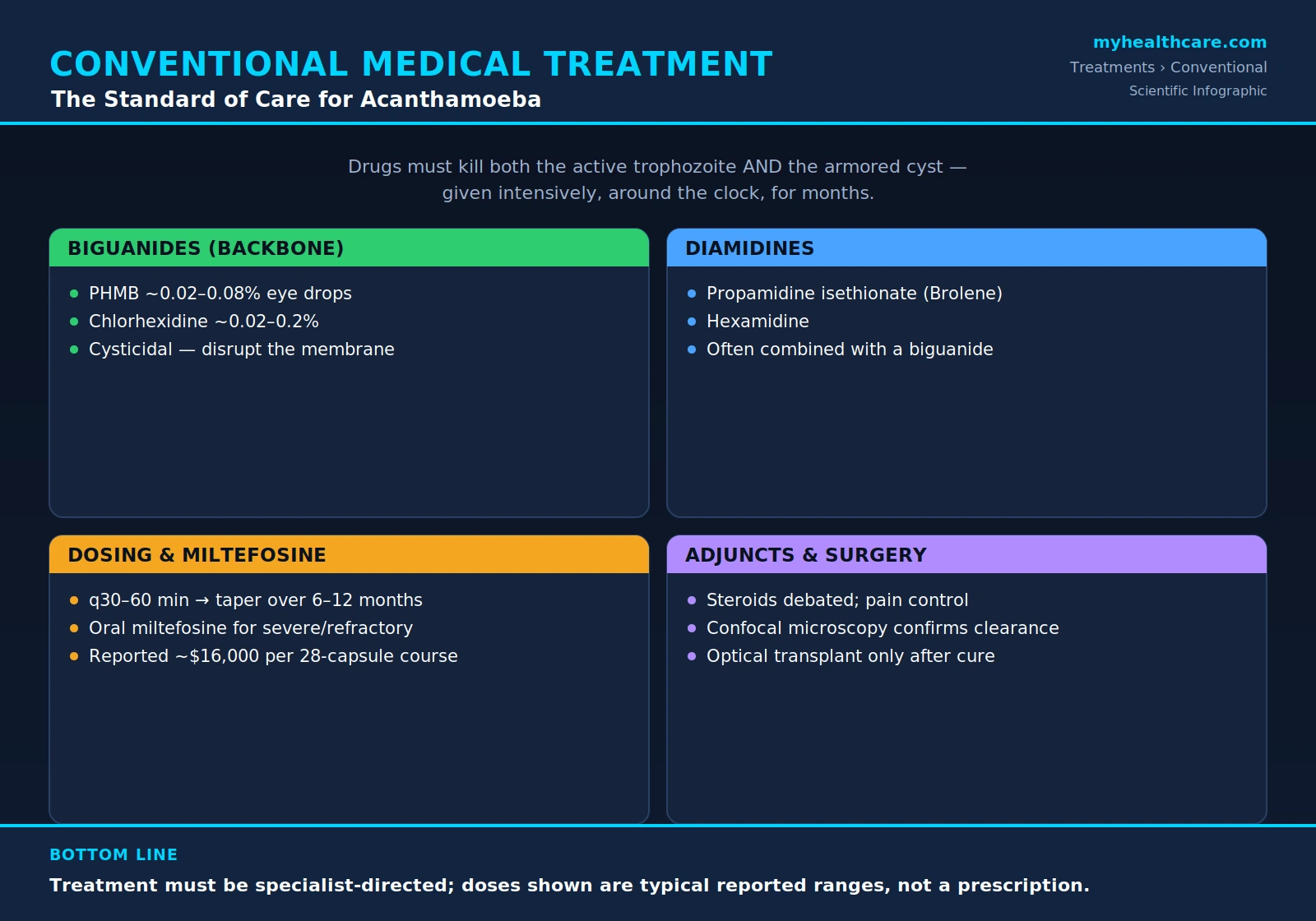
The foundation of Acanthamoeba keratitis treatment is a class of antiseptic compounds called biguanides, given as frequent topical eye drops. The two used in practice are polyhexamethylene biguanide (PHMB), also called polihexanide, and chlorhexidine. Biguanides are cationic (positively charged) molecules that are attracted to the negatively charged surface of the amoeba. They bind to and disrupt the lipid membrane and the cyst wall, increasing permeability so the cell leaks its contents and dies. Crucially, biguanides are among the few topically tolerated agents that are active against the cyst, not just the trophozoite, which is why they anchor the regimen.
PHMB is typically compounded as a topical drop in the range of about 0.02% to 0.08%, and chlorhexidine in the range of roughly 0.02% to 0.2%. These are unlicensed (off-label, pharmacy-compounded) eye drops in most countries rather than commercial products. Historically PHMB 0.02% was standard, but interest has grown in a stronger single-agent approach: PHMB 0.08% has become a leading monotherapy, supported by clinical research into higher-concentration polihexanide for keratitis. Even at these low percentages the drops can be irritating to the ocular surface, and the prescribing specialist balances killing power against toxicity to the cornea.
Laboratory susceptibility testing has shown that both PHMB and chlorhexidine retain activity against cysts of multiple Acanthamoeba species, which is the practical basis for choosing them as the backbone of therapy. Some specialists use a biguanide alone; others combine it with a diamidine (next section) for added effect, particularly in severe or slow-responding cases.
3. Diamidines
The second topical class is the diamidines. The two used for the eye are propamidine isethionate — sold in some countries as the over-the-counter eye preparation Brolene — and hexamidine. Diamidines interfere with the amoeba's DNA and disrupt its membrane and metabolism. They are good at killing trophozoites and have some activity against cysts, but on their own they are generally considered less reliably cysticidal than the biguanides.
For this reason diamidines are most often used in combination with a biguanide rather than alone. A common conventional regimen pairs PHMB (or chlorhexidine) with propamidine or hexamidine, so that the two drug classes attack the amoeba through different mechanisms at the same time. Because propamidine is available without prescription in some regions, patients occasionally start it themselves before a diagnosis is confirmed; while it can help, it is not a substitute for specialist-directed biguanide therapy, and prolonged diamidine use can itself be toxic to the corneal surface.
4. The Dosing Siege
What makes Acanthamoeba keratitis treatment so grueling is not just the drugs but the schedule. To overwhelm the amoeba and reach cysts before they can hide, the topical drops are given extremely frequently at the start of therapy. A typical reported induction phase involves instilling the drops roughly every 30 to 60 minutes, around the clock — including waking through the night — for the first several days. This intensity is then gradually reduced.
As the eye responds, the specialist slowly tapers the frequency: stepping down to hourly during waking hours, then every few hours, then a handful of times a day over the following weeks and months. The full course commonly continues for 6 to 12 months, and the taper is deliberately cautious because stopping too soon allows surviving cysts to re-activate. Patients describe this relentless regimen — setting alarms every hour through the night for days, then sustaining drops for the better part of a year — as one of the hardest parts of the whole illness, both physically exhausting and emotionally draining. Understanding why the schedule is so punishing (the cyst, again) helps people stick with it.
5. Miltefosine (Impavido)
Miltefosine (brand name Impavido) is an oral antiparasitic drug, originally developed against leishmaniasis, that has become an important option for the most difficult Acanthamoeba infections. It is an alkylphosphocholine that damages the parasite's cell membrane and disturbs its lipid metabolism. In the eye, miltefosine is used as an adjunctive (add-on) or salvage therapy for severe or refractory keratitis that is not responding to topical biguanides and diamidines alone. It is also a mainstay of treatment for the dangerous non-eye forms of disease — granulomatous amebic encephalitis (GAE) and disseminated infection — where case reports document successful outcomes when miltefosine was added to a multi-drug regimen.
In the United States, miltefosine for free-living amoeba infections has historically been supplied through the Centers for Disease Control and Prevention (CDC), reflecting how rare these infections are and how restricted the drug's availability has been. Access and cost are real obstacles: a reported course of 28 capsules has cost on the order of $16,000, and the drug carries its own toxicities, including gastrointestinal upset and reproductive concerns (it is contraindicated in pregnancy). Miltefosine is a specialist-only, often institution-coordinated medication — not something a patient can simply fill at a pharmacy — and it is reserved for the situations where it is genuinely needed.
6. Adjuncts and Controversies
Beyond the amoeba-killing drugs, several supportive measures are used — and one of them is genuinely controversial. Topical corticosteroids reduce the intense inflammation and pain that Acanthamoeba keratitis causes, and they can make a suffering patient much more comfortable. But there is a real concern that steroids, by dampening the immune response, may allow the amoeba to persist or even encourage encystment, potentially prolonging the infection. The medical literature reflects an honest, unresolved debate: some specialists avoid steroids entirely until the infection is well controlled on anti-amoebic therapy, while others use them carefully alongside continued biguanide drops. The safe summary is that steroids are used selectively, under specialist judgment, and not as a routine first step.
Other adjuncts are less contentious. Pain control is important because this infection is notoriously painful, often out of proportion to how the eye looks. Cycloplegic drops (which paralyze the focusing muscle) ease the deep aching caused by spasm of the ciliary body and reduce the risk of inflammatory adhesions inside the eye. Epithelial debridement — gently scraping away infected surface tissue early on — can lower the amoebic load and help the drops penetrate. Finally, corneal cross-linking (PACK-CXL), in which riboflavin and ultraviolet-A light are used to stiffen and sterilize the cornea, has been explored as an add-on for infectious keratitis; for Acanthamoeba specifically it remains investigational, with mixed results, and is not a standard part of treatment.
7. Surgery
Most cases are managed medically, but surgery becomes necessary when the cornea is severely damaged or the infection cannot be controlled with drops. Therapeutic keratoplasty — a corneal transplant performed to remove infected, melting, or perforating tissue — is used as a rescue when the eye is failing despite maximal medical therapy. This is different from an optical corneal transplant, which is done purely to restore vision through a scarred but quiet cornea.
The timing of an optical transplant matters enormously. Surgeons generally wait to perform a vision-restoring graft only after the infection is confirmed cured, because a single surviving cyst left in or near the transplant bed can re-activate and reinfect the new graft, undoing the surgery and threatening the eye again. This caution is one more reason the overall course of care can stretch toward a year. In the worst outcomes — an eye that is blind, painful, and beyond saving despite every measure — enucleation (surgical removal of the eye) is considered as a last resort to relieve suffering and remove the source of infection.
8. Treating GAE and Cutaneous Disease
When Acanthamoeba spreads beyond the eye — into the brain as granulomatous amebic encephalitis (GAE), or into the skin as cutaneous disease, usually in people with compromised immune systems — there is no single proven cure and outcomes are often poor. Because no one drug works reliably, specialists assemble individualized, multi-drug systemic regimens, frequently combining several agents reported in successful case reports. These have included miltefosine, flucytosine, pentamidine, sulfadiazine, an azole antifungal such as fluconazole or voriconazole, and azithromycin, used in various combinations.
Just as important as the drugs is addressing the host. Many GAE and cutaneous cases occur in transplant recipients, people with HIV/AIDS, or others on immunosuppressive medication. Wherever it is medically possible, the team works to reduce immunosuppression, giving the patient's own immune system a chance to help fight the infection — a delicate balance that must be weighed against the reason the immunosuppression was being given in the first place. These cases are managed by infectious-disease specialists, often with input from public-health authorities such as the CDC.
9. Monitoring and Duration
Because cysts are invisible to the naked eye and can lurk in the cornea, specialists rely on careful monitoring to judge whether treatment is working and when it is safe to stop. In vivo confocal microscopy is a non-invasive imaging technique that lets clinicians look directly into the living cornea at high magnification and watch for the bright, round cyst forms. Seeing cysts clear over time helps confirm that therapy is succeeding; seeing them persist signals that treatment must continue. Repeat cultures and clinical examination round out the picture.
Even with good monitoring, relapses occur, which is why drops are tapered slowly rather than stopped abruptly and why the total course commonly runs up to a year. The same caution governs the wait before any vision-restoring corneal transplant: surgeons want strong evidence that the cysts are truly gone before placing a new graft that a surviving amoeba could reinfect. The overarching theme of conventional Acanthamoeba care — long therapy, slow tapers, patient monitoring, and a deliberate wait before reconstructive surgery — all traces back to the resilience of that one dormant cyst.
Key Research Papers
Peer-reviewed studies and reviews underpinning the conventional management of Acanthamoeba infection — covering the biology of the cyst, the biguanide and diamidine drops that anchor topical therapy, the role of miltefosine in severe and disseminated disease, and the long-term outcomes of treated keratitis. Each citation links to the full text via DOI.
- Marciano-Cabral F, Cabral G. Acanthamoeba spp. as Agents of Disease in Humans. Clinical Microbiology Reviews. 2003;16(2):273–307.
- Lorenzo-Morales J, Khan NA, Walochnik J. An Update on Acanthamoeba Keratitis: Diagnosis, Pathogenesis and Treatment. Parasite. 2015;22:10.
- Dart JKG, Saw VPJ, Kilvington S. Acanthamoeba Keratitis: Diagnosis and Treatment Update 2009. American Journal of Ophthalmology. 2009;148(4):487–499.
- Khan NA. Acanthamoeba: Biology and Increasing Importance in Human Health. FEMS Microbiology Reviews. 2006;30(4):564–595.
- Visvesvara GS, Moura H, Schuster FL. Pathogenic and Opportunistic Free-Living Amoebae: Acanthamoeba spp., Balamuthia mandrillaris, Naegleria fowleri, and Sappinia diploidea. FEMS Immunology & Medical Microbiology. 2007;50(1):1–26.
- Larkin DFP, Kilvington S, Dart JKG. Treatment of Acanthamoeba Keratitis with Polyhexamethylene Biguanide. Ophthalmology. 1992;99(2):185–191.
- Seal DV, Hay J, Kirkness CM. Chlorhexidine or Polyhexamethylene Biguanide for Acanthamoeba Keratitis. The Lancet. 1995;345(8942):136.
- Narasimhan S, Madhavan HN, Therese KL. Development and Application of an In Vitro Susceptibility Test for Acanthamoeba Species Isolated from Keratitis to Polyhexamethylene Biguanide and Chlorhexidine. Cornea. 2002;21(2):203–205.
- Dewan N, Ming W, Holland SP, et al. Oral Miltefosine as Adjunctive Treatment for Recalcitrant Acanthamoeba Keratitis. Cornea. 2019;38(7):914–917.
- Aichelburg AC, Walochnik J, Assadian O, et al. Successful Treatment of Disseminated Acanthamoeba sp. Infection with Miltefosine. Emerging Infectious Diseases. 2008;14(11):1743–1746.
- Page MA, Mathers WD. Acanthamoeba Keratitis: A 12-Year Experience Covering a Wide Spectrum of Presentations, Diagnoses, and Outcomes. Journal of Ophthalmology. 2013;2013:670242.
- Carnt N, Hoffman JJ, Verma S, et al. Acanthamoeba Keratitis: Confirmation of the UK Outbreak and a Prospective Case-Control Study Identifying Contributing Risk Factors. British Journal of Ophthalmology. 2018;102(12):1621–1628.
Live PubMed Searches
Each link opens a live PubMed query so results stay current as new papers are indexed.
- Acanthamoeba keratitis treatment
- Polyhexamethylene biguanide Acanthamoeba
- Chlorhexidine Acanthamoeba keratitis
- Propamidine isethionate Acanthamoeba
- Miltefosine Acanthamoeba
- Granulomatous amebic encephalitis treatment
- Acanthamoeba keratoplasty corneal transplant
- Acanthamoeba cyst cysticidal drug
- Confocal microscopy Acanthamoeba keratitis
- Corneal crosslinking infectious keratitis
Connections
- Acanthamoeba
- Acanthamoeba Treatments
- Ivermectin
- Castor Oil
- Acanthamoeba Keratitis
- Acanthamoeba Symptoms
- Ophthalmology
- Parasites
- Meningitis
- Sepsis
- Silver Nanoparticles
- Goldenseal
- Oregano
- Escherichia coli
- All Conditions
- Lab Tests