Granulomatous Amebic Encephalitis (GAE) — Acanthamoeba Brain Infection
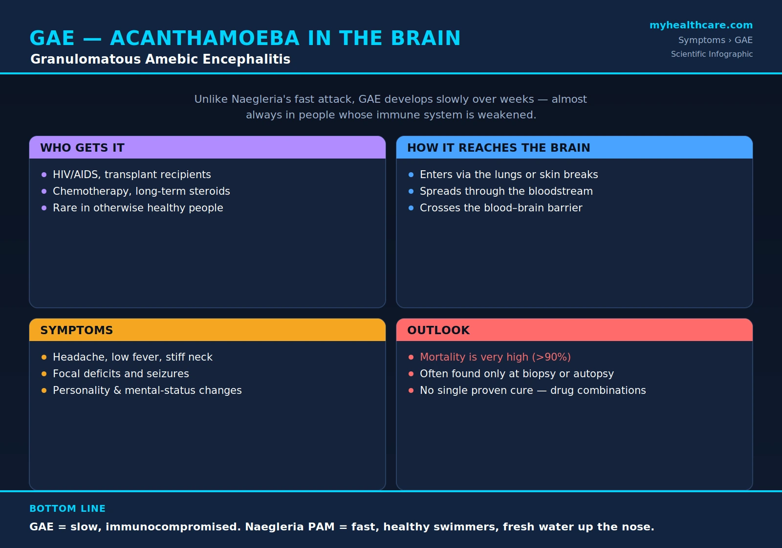
Granulomatous amebic encephalitis (GAE) is a rare but devastating infection of the brain caused by free-living amoebae — chiefly Acanthamoeba species and the closely related Balamuthia mandrillaris. Unlike the lightning-fast brain infection caused by Naegleria fowleri, GAE creeps in over weeks to months, almost always in people whose immune defenses are weakened. Because its early symptoms mimic so many other neurological conditions, it is frequently recognized only late in its course — and it is fatal in the large majority of reported cases. This page explains what GAE is, who is at risk, how the amoeba reaches the brain, why it is so hard to diagnose, and how it differs from the other amebic infections of the central nervous system.
Table of Contents
- What Is GAE?
- Who Gets It
- How It Reaches the Brain
- Symptoms
- Why It Is So Hard to Diagnose
- Prognosis
- Treatment
- GAE vs. Other Amebic Brain Infections
- Key Research Papers
- Featured Videos
1. What Is GAE?
Granulomatous amebic encephalitis is a rare, subacute-to-chronic, and usually fatal infection of the brain caused by free-living amoebae. The principal agents are Acanthamoeba species (such as A. castellanii, A. culbertsoni, and A. polyphaga) and the related amoeba Balamuthia mandrillaris. These are not parasites that depend on a human host to survive — they live freely in soil, dust, fresh and brackish water, swimming pools, hot tubs, air-conditioning units, and even tap water and contact-lens solutions. Humans are accidental hosts.
The word "granulomatous" refers to the body's immune response. When the amoeba invades brain tissue, the immune system attempts to wall it off by forming granulomas — small nodules of inflammatory cells. These granulomas, along with areas of tissue death (necrosis) and inflammation of the small blood vessels, are the pathological hallmark of the disease. Notably, in people who are severely immunocompromised, the granulomatous reaction may be poorly formed or absent, because the immune cells needed to build it are depleted.
The defining clinical feature of GAE is its slow tempo. The illness develops insidiously over weeks to months, with symptoms that wax, wane, and gradually worsen. This sets it apart from Naegleria fowleri infection, known as primary amebic meningoencephalitis (PAM), which strikes suddenly and progresses to death within days. GAE is a smoldering, granuloma-forming process; PAM is an explosive, rapidly destructive one.
2. Who Gets It
GAE occurs almost exclusively in people whose immune systems are compromised. The disease is an opportunistic infection — the amoebae take advantage of weakened host defenses to establish an infection they could not otherwise sustain. The most commonly affected groups include:
- People with HIV/AIDS, particularly those with advanced disease and very low CD4 T-cell counts.
- Organ and bone-marrow transplant recipients, who take immunosuppressive drugs to prevent rejection.
- People receiving chemotherapy for cancer, or with blood cancers such as leukemia and lymphoma.
- Patients on long-term corticosteroids or other immunosuppressant medications for autoimmune or inflammatory conditions.
- People with poorly controlled or advanced diabetes, cirrhosis and other chronic liver disease, chronic kidney disease, or significant malnutrition.
GAE is very rare in otherwise healthy people. (Balamuthia mandrillaris, which causes a clinically similar granulomatous encephalitis, is a notable exception — it can occasionally infect people with apparently normal immune systems, including children and the elderly.) Importantly, exposure to Acanthamoeba is extremely common — most adults carry antibodies against it, reflecting frequent environmental contact — yet brain infection is exceedingly rare. It is the combination of exposure and a compromised immune system, not exposure alone, that sets the stage for GAE.
3. How It Reaches the Brain
For the amoeba to cause encephalitis, it must first enter the body and then travel to the brain. Unlike Naegleria, which reaches the brain by climbing directly up the olfactory nerve after water is forced up the nose, Acanthamoeba takes a longer, bloodstream-mediated route.
The amoeba typically enters through one of two portals:
- The lungs and lower respiratory tract, by inhalation of airborne cysts in dust or aerosols.
- Breaks in the skin — cuts, ulcers, burns, or other wounds — or through the sinuses.
From this initial site, the amoeba enters the bloodstream and disseminates through the body. To cause encephalitis it must then cross the blood-brain barrier, the tightly regulated lining of blood vessels that normally protects the brain. Acanthamoeba accomplishes this by binding to and damaging the cells of the vessel wall, allowing it to pass into the surrounding brain tissue.
Because of this pathway, a focus of infection elsewhere in the body — in the skin, the sinuses, or the lungs — very often precedes brain involvement. In immunocompromised patients, chronic skin lesions or sinus disease caused by Acanthamoeba may appear weeks or months before any neurological signs. Recognizing such a skin or sinus focus is a valuable, and sometimes life-saving, clue that can prompt diagnosis before the infection reaches the brain.
4. Symptoms
The symptoms of GAE are insidious and notoriously nonspecific, which is a major reason the disease is so often missed or attributed to other causes. Because the infection evolves slowly, the picture is one of gradual neurological decline over weeks rather than an abrupt collapse. Reported symptoms include:
- Persistent headache that does not resolve.
- Low-grade fever.
- Stiff neck (nuchal rigidity), reflecting meningeal irritation.
- Personality and mental-status changes — confusion, apathy, irritability, behavioral change, lethargy progressing toward stupor.
- Focal neurological deficits, such as weakness on one side of the body (hemiparesis), cranial-nerve palsies (for example, double vision, facial weakness, or difficulty speaking and swallowing), and visual disturbances.
- Seizures.
- A general pattern of progressive decline over weeks, with worsening neurological function and, eventually, coma.
Because these features overlap so heavily with many other neurological illnesses, GAE is rarely suspected on symptoms alone. The slowly progressive course in an immunocompromised person should, however, raise the possibility — especially if there is a coexisting skin, sinus, or lung lesion.
5. Why It Is So Hard to Diagnose
GAE is one of the great mimics of neurology. Its clinical and radiological appearance can resemble a brain tumor, a bacterial brain abscess, central nervous system tuberculosis, fungal infection, and many other forms of encephalitis. As a result, the correct diagnosis is frequently delayed and, in a sobering number of cases, is established only at autopsy.
Several factors conspire to make diagnosis difficult:
- Neuroimaging is suggestive but not specific. CT and MRI scans typically show one or more mass-like lesions, often with surrounding swelling and ring enhancement — an appearance that points toward tumor or abscess rather than amoebic infection.
- Spinal fluid findings are nonspecific. Examination of cerebrospinal fluid (from a lumbar puncture) usually shows a mononuclear (lymphocyte-predominant) inflammation with elevated protein and sometimes low glucose — a pattern shared with tuberculous, viral, and fungal meningoencephalitis. Unlike in Naegleria infection, the amoebae are only rarely seen in the spinal fluid, so a routine examination usually does not reveal the organism.
- Definitive diagnosis usually requires tissue. In practice, the diagnosis is most often confirmed by brain biopsy — or, tragically, at autopsy — with the amoebae (both the active trophozoite form and the resistant cyst form) identified in the affected tissue. Immunohistochemistry and PCR performed on biopsy or other tissue specimens can confirm the organism and, importantly, distinguish Acanthamoeba from Balamuthia and from other amoebae. Specialized reference and public-health laboratories play a central role in this confirmation.
Because of all this, a high index of suspicion is essential. The combination of a slowly progressive brain lesion in an immunocompromised patient — particularly one with a skin or sinus lesion — should prompt clinicians to consider GAE and to pursue tissue diagnosis and specialist consultation early.
6. Prognosis
The prognosis of GAE is grave. Mortality is very high: the large majority of reported cases have been fatal, and historically the disease was diagnosed almost only after death. This dismal outlook reflects several factors working together — the late stage at which most cases are recognized, the underlying immunosuppression that both invites the infection and blunts the body's ability to fight it, the difficulty of getting effective drugs across the blood-brain barrier, and the absence of any single reliably curative therapy.
The rare survivors reported in the medical literature tend to share two features: their infection was diagnosed comparatively early, and they received aggressive combination drug therapy. Where it is medically possible, reducing or reversing immunosuppression — for example, by tapering immunosuppressant drugs or, in people with HIV, by restoring immune function with antiretroviral therapy — appears to improve the chances of survival, because it allows the patient's own defenses to participate in clearing the infection. Even so, GAE remains among the most lethal infections of the central nervous system, and prevention of exposure in high-risk patients, together with early recognition, offers the best hope of a better outcome.
7. Treatment
There is no single proven cure for GAE, and no standardized, universally effective regimen. Because the disease is so rare, treatment recommendations are drawn from individual case reports and small series of survivors rather than from large clinical trials. What these reports have in common is the use of aggressive combinations of several drugs given together, individualized for each patient, ideally with guidance from infectious-disease specialists and public-health reference centers such as the U.S. Centers for Disease Control and Prevention (CDC).
Agents that have appeared in successful or partially successful regimens include:
- Miltefosine — an antiparasitic drug that has become a cornerstone of treatment for free-living amebic infections and is available in the United States through the CDC for this purpose.
- Flucytosine (5-fluorocytosine).
- Pentamidine.
- Sulfadiazine.
- Azole antifungals such as fluconazole or voriconazole.
- Azithromycin.
These drugs are typically used in various combinations, sometimes alongside surgical removal of an accessible lesion and, critically, alongside efforts to reduce immunosuppression where it is safe to do so. Because the optimal regimen is uncertain and the drugs themselves carry significant toxicities, management should always be directed by specialists. For a fuller discussion of the conventional medical approach, see the dedicated Conventional Medical Treatment page.
8. GAE vs. Other Amebic Brain Infections
Two very different infections of the brain are caused by free-living amoebae, and it is important not to confuse them, because they differ in nearly every clinically meaningful way.
Granulomatous amebic encephalitis (GAE) is caused by Acanthamoeba species and Balamuthia mandrillaris. It is slow, developing over weeks to months. It strikes the immunocompromised (with Balamuthia the main exception, occasionally affecting healthy hosts). The amoeba reaches the brain by a bloodstream-borne route, after entering through the lungs or skin, and it provokes granuloma formation. Diagnosis is difficult and treatment is prolonged and uncertain.
Primary amebic meningoencephalitis (PAM) is caused by Naegleria fowleri, the so-called "brain-eating amoeba." It is fast and fulminant, typically killing within days of the first symptoms. It most often strikes previously healthy young people — classically children and young adults — after warm fresh water is forced up the nose during swimming or diving. From the nasal lining, the amoeba travels directly along the olfactory nerve into the brain, producing an acute, hemorrhagic, rapidly destructive meningoencephalitis rather than a slow granulomatous one.
In short: GAE is the slow, opportunistic, blood-borne disease of weakened immune systems; PAM is the explosive, freshwater-associated disease of healthy young swimmers. For the broader context of how infections inflame the membranes and tissues of the central nervous system, see Meningitis.
Key Research Papers
Peer-reviewed reviews and reports on Acanthamoeba, Balamuthia, and the granulomatous amebic encephalitis they cause — covering biology, pathogenesis, the spectrum of human disease, diagnosis, and treatment. Journal names appear as plain text; the year/volume/pages link opens the full citation via DOI.
- Marciano-Cabral F, Cabral G. Acanthamoeba spp. as Agents of Disease in Humans. Clinical Microbiology Reviews. 2003;16(2):273–307.
- Khan NA. Acanthamoeba: Biology and Increasing Importance in Human Health. FEMS Microbiology Reviews. 2006;30(4):564–595.
- Visvesvara GS, Moura H, Schuster FL. Pathogenic and Opportunistic Free-Living Amoebae: Acanthamoeba spp., Balamuthia mandrillaris, Naegleria fowleri, and Sappinia diploidea. FEMS Immunology & Medical Microbiology. 2007;50(1):1–26.
- Schuster FL, Visvesvara GS. Free-Living Amoebae as Opportunistic and Non-Opportunistic Pathogens of Humans and Animals. International Journal for Parasitology. 2004;34(9):1001–1027.
- Siddiqui R, Khan NA. Biology and Pathogenesis of Acanthamoeba. Parasites & Vectors. 2012;5:6.
- Marciano-Cabral F, Cabral GA. The Immune Response to Naegleria fowleri Amebae and Pathogenesis of Infection. Journal of Eukaryotic Microbiology. 2007;54(2):184–190.
- Aichelburg AC, Walochnik J, Assadian O, et al. Successful Treatment of Disseminated Acanthamoeba sp. Infection with Miltefosine. Emerging Infectious Diseases. 2008;14(11):1743–1746.
- Kalra SK, Sharma P, Shyam K, Tejan N, Ghoshal U. Acanthamoeba and Its Pathogenic Role in Granulomatous Amebic Encephalitis. Experimental Parasitology. 2020;208:107788.
Live PubMed Searches
Each link opens a live PubMed query so results stay current as new papers are indexed.
- Granulomatous amebic encephalitis
- Acanthamoeba encephalitis immunocompromised
- Balamuthia mandrillaris encephalitis
- Acanthamoeba miltefosine treatment
- Free-living amebae central nervous system
- Acanthamoeba brain biopsy diagnosis
- Acanthamoeba disseminated infection HIV
- Naegleria fowleri primary amebic meningoencephalitis
Connections
- Acanthamoeba
- Symptoms & Diagnosis
- Acanthamoeba Keratitis (Eye)
- Cutaneous & Disseminated Infection
- Acanthamoeba Treatments
- Conventional Medical Treatment
- Ivermectin
- Castor Oil
- Parasites
- Meningitis
- Sepsis
- Neurology
- Infectious Disease
- All Conditions