West Nile Virus
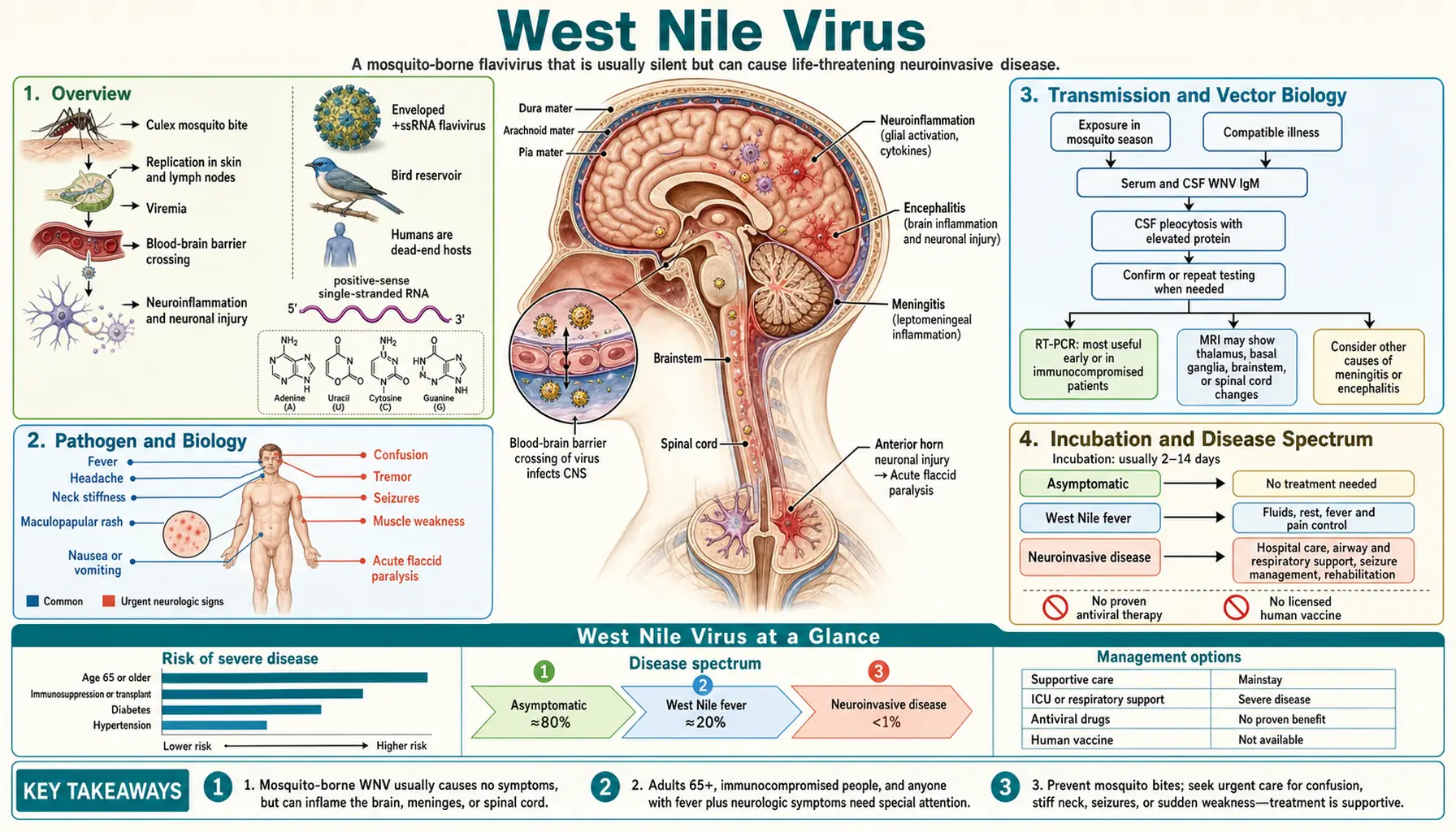
Table of Contents
- Overview
- Pathogen and Biology
- Transmission and Vector Biology
- Incubation and Disease Spectrum
- Symptoms and Clinical Presentation
- Diagnosis
- Treatment
- Home Care and Supportive Management
- Complications
- Prevention
- Key Research Papers
- PubMed Searches
- Connections
- Featured Videos
1. Overview
West Nile Virus (WNV) is a mosquito-borne flavivirus that has become the leading cause of domestically acquired arboviral encephalitis in the United States since it was introduced to North America in 1999. The virus was first isolated from a febrile woman in the West Nile district of Uganda in 1937, remained primarily an Old World pathogen for decades, and then emerged dramatically in New York City in the summer of 1999, rapidly spreading across the entire continental US by 2004.
The epidemiologic spectrum of WNV infection is heavily skewed: approximately 80% of infected people have no symptoms at all. About 20% develop a self-limited febrile illness called West Nile fever. Fewer than 1% develop West Nile neuroinvasive disease (WNND) — meningitis, encephalitis, or acute flaccid paralysis — which carries significant mortality and long-term morbidity.
From 1999 through 2022, the CDC reported over 56,000 cases of WNV disease in the United States, including more than 27,000 neuroinvasive cases and over 2,800 deaths. Because only symptomatic (and particularly severe) cases are typically reported, total infections are estimated in the millions over this period. There is no licensed vaccine or specific antiviral treatment for WNV in humans. Prevention depends entirely on mosquito control and personal protection.
2. Pathogen and Biology
West Nile Virus is a positive-sense, single-stranded RNA virus belonging to the genus Flavivirus, family Flaviviridae. It is closely related to other medically important flaviviruses including dengue virus, Zika virus, yellow fever virus, Japanese encephalitis virus, and St. Louis encephalitis virus.
Genome and Structure
The WNV genome is approximately 11 kilobases encoding a single polyprotein cleaved into 3 structural proteins (capsid C, membrane protein prM/M, envelope protein E) and 7 nonstructural proteins (NS1, NS2A, NS2B, NS3, NS4A, NS4B, NS5). The envelope (E) protein mediates receptor binding and membrane fusion and is the primary target of neutralizing antibodies. NS5 encodes the RNA-dependent RNA polymerase essential for viral replication and is a major drug target in flavivirus research.
Lineages
WNV exists in at least eight phylogenetic lineages. Lineage 1 (including the NY99 strain introduced in 1999 and the WN02 strain that replaced it) is responsible for virtually all North American disease. Lineage 2 has caused epidemic neuroinvasive disease in southern and eastern Europe since the mid-2000s. Lineage 1 strains show higher neuroinvasiveness and virulence in corvids (crows, ravens) — dead crow surveillance became an important early warning system in North America.
Neuroinvasion Mechanisms
WNV neuroinvasion involves several mechanisms: direct infection of endothelial cells of the blood-brain barrier (BBB) leading to transcellular or paracellular viral entry; “Trojan horse” transport inside infected monocytes/macrophages; peripheral nerve axonal transport; and olfactory nerve retrograde transport. Once inside the CNS, WNV infects neurons (particularly in the brainstem, anterior horn cells of the spinal cord, cerebellum, and cortex), triggering a neuroinflammatory cascade that causes both direct viral cytopathology and immune-mediated neuronal injury.
Risk Factors for Neuroinvasive Disease
Most people who develop severe disease have identifiable risk factors:
- Older age: The most important risk factor. Adults 60 and older account for the majority of WNV deaths and long-term neurologic sequelae. Risk increases steeply with each decade after age 50.
- Immunosuppression: Transplant recipients, HIV/AIDS patients, those on chemotherapy or chronic immunosuppressants — markedly elevated risk of neuroinvasive disease and death.
- Diabetes mellitus and hypertension: Associated with increased risk in multiple studies, possibly through impaired immune response or compromised BBB integrity.
- CCR5 32 deletion: Homozygous CCR5Δ32 mutation (found in approximately 1% of Europeans) confers natural HIV resistance but paradoxically increases susceptibility to WNV encephalitis, as CCR5 appears important for CNS viral clearance.
3. Transmission and Vector Biology
Culex Mosquito Vectors
WNV is transmitted principally by mosquitoes of the genus Culex in North America:
- Culex pipiens (northern house mosquito): Primary vector in the northeastern and north-central US; urban and suburban. Breeds in stagnant or slow-moving water: storm drains, catch basins, birdbaths, discarded tires, containers. Active from dusk to dawn.
- Culex quinquefasciatus (southern house mosquito): Primary vector in the southern US. Similar biology to C. pipiens; the two species hybridize in the transition zone.
- Culex tarsalis: Primary vector in the western US; rural and agricultural settings. Highly efficient amplifier of WNV in the West.
Over 65 mosquito species have been found infected with WNV in the US, but Culex species are the primary epidemiologic drivers.
The Bird Reservoir
Birds — particularly passerine (perching) birds — are the primary amplifying hosts for WNV. Infected birds develop high-level viremia, efficiently infecting mosquitoes that feed on them. The WNV transmission cycle is an enzootic cycle (bird↔mosquito↔bird) with humans as incidental “dead-end” hosts: human blood WNV titers are too low to efficiently infect a feeding mosquito, so humans do not meaningfully amplify virus back into the mosquito population.
Corvids (American crows, ravens, blue jays) are highly susceptible to WNV and die in large numbers during outbreak years — a pattern exploited for surveillance. Many other bird species (robins, house sparrows, house finches) develop high viremia without dying and serve as efficient reservoir hosts.
Non-Vector Transmission Routes
Though rare, WNV can be transmitted via:
- Blood transfusion: WNV entered the US blood supply in 2002, causing dozens of transfusion-associated infections. Nucleic acid testing (NAT) of all donated blood products for WNV has been standard since 2003 and has dramatically reduced but not eliminated this risk.
- Organ transplantation: WNV transmission through solid organ transplants from viremic donors has caused severe neuroinvasive disease in recipients.
- Vertical transmission (mother to infant): Documented but rare; breastfeeding transmission has been reported.
- Laboratory exposure: Occupational needle-stick cases reported.
- Not transmitted person-to-person: Casual contact, kissing, or sharing food/water does not transmit WNV.
4. Incubation and Disease Spectrum
Incubation Period
The incubation period is typically 2–14 days from mosquito bite, with most symptomatic cases developing illness 3–8 days after exposure. Immunocompromised individuals may have longer incubation periods (up to 21 days in transplant recipients).
Disease Spectrum
- Asymptomatic infection (~80%): Most people infected with WNV develop no symptoms. These individuals seroconvert and are immune to future WNV infection. They are identified through serologic surveys of blood donors, volunteer cohorts, and household contacts of symptomatic cases.
- West Nile fever (~20%): Acute febrile illness that is self-limited in nearly all cases. See Section 5 for details.
- West Nile neuroinvasive disease (<1%): Meningitis, encephalitis, or acute flaccid paralysis (WNV poliomyelitis). Potentially fatal or permanently disabling.
5. Symptoms and Clinical Presentation
West Nile Fever (Mild Disease)
Approximately 20% of infected individuals develop a febrile illness lasting 3–6 days, with complete recovery in most patients (though fatigue may persist for weeks):
- Fever (38–40°C), often abrupt in onset
- Headache (often frontal)
- Myalgias and arthralgias (muscle and joint aches)
- Fatigue, malaise, weakness
- Anorexia, nausea, vomiting, diarrhea (in a subset of patients)
- Maculopapular or roseolar rash on the trunk (reported in 20–50% of WNF cases) — non-pruritic, transient, spares the palms and soles
- Lymphadenopathy (mild)
- Retroorbital eye pain
West Nile fever is clinically indistinguishable from many other viral illnesses. Without epidemiologic context (residence or travel in endemic area during mosquito season), laboratory testing is not routinely indicated for mild illness.
West Nile Neuroinvasive Disease (WNND)
Fewer than 1% of infected individuals develop neuroinvasive disease, but this small fraction accounts for the serious morbidity and mortality associated with WNV. Three neurologic syndromes are recognized:
West Nile Meningitis
Milder of the three WNND syndromes. Presents like bacterial meningitis with fever, severe headache, photophobia, phonophobia, and meningismus (nuchal rigidity), but with a viral (aseptic) CSF profile. Most patients recover fully with supportive care. CSF shows lymphocytic pleocytosis (WBC typically 10–500 cells/mm³, predominantly lymphocytes), elevated protein (50–200 mg/dL), and normal or mildly low glucose.
West Nile Encephalitis
The most severe and most common form of WNND. Presents with altered level of consciousness (ranging from mild confusion to coma), seizures, focal neurologic deficits, and movement disorders. Distinctive features that may suggest WNV encephalitis over other viral encephalitides include:
- Tremors and parkinsonism: Coarse tremor (especially of the upper extremities), rigidity, bradykinesia, and postural instability — reflecting basal ganglia and substantia nigra involvement — are seen in approximately 75% of WNV encephalitis cases and are not typical of other viral encephalitides.
- Myoclonus
- Cranial nerve abnormalities
- MRI may show signal abnormalities in the basal ganglia, thalami, brainstem, cerebellum, and spinal cord.
West Nile Acute Flaccid Paralysis (WNV Poliomyelitis)
WNV can cause an asymmetric flaccid paralysis clinically identical to poliomyelitis, through direct anterior horn cell infection in the spinal cord. This syndrome presents as acute asymmetric limb weakness, often with rapid progression to complete flaccid paralysis of one or more limbs, absent deep tendon reflexes, and preserved sensation. Bulbar involvement and respiratory muscle paralysis can occur. EMG/nerve conduction studies show an axonal motor neuropathy pattern consistent with anterior horn cell disease. Prognosis for limb recovery is poor; permanent weakness is common. Some patients require long-term mechanical ventilation.
6. Diagnosis
Serology: IgM ELISA (Preferred Initial Test)
Detection of WNV-specific IgM antibodies in cerebrospinal fluid (CSF) or serum by enzyme-linked immunosorbent assay (ELISA) is the most commonly used diagnostic approach and is considered the standard of care for suspected WNND:
- CSF WNV IgM: Highly specific for neuroinvasive WNV infection (intrathecal IgM production confirms CNS involvement). Present in >90% of WNND patients within 8 days of illness onset.
- Serum WNV IgM: Sensitive and widely available. May remain positive for months after infection, limiting its utility for diagnosing recent acute infection in high-prevalence areas.
- Plaque reduction neutralization test (PRNT): Gold-standard confirmatory test to distinguish WNV from cross-reacting flaviviruses (dengue, Zika, St. Louis encephalitis, Japanese encephalitis, yellow fever vaccine). Required for confirmation when IgM is positive and another flavivirus infection is possible.
PCR
WNV RNA can be detected by RT-PCR in serum during the brief viremic phase (approximately 1–7 days after infection onset). By the time most patients present with neuroinvasive disease, viremia has cleared and RT-PCR on serum is typically negative. CSF PCR for WNV is also insensitive (<60%). Serology is therefore preferred over PCR for WNV diagnosis in the clinical setting. PCR is most useful in immunocompromised patients (who may not mount an adequate antibody response) and for blood donor screening.
CSF Analysis
Lumbar puncture is indicated for all patients with suspected WNV meningitis or encephalitis:
- Opening pressure: normal or mildly elevated
- Cell count: lymphocytic pleocytosis (typically 10–500 WBC/mm³; in rare cases, early in disease, neutrophils may predominate before lymphocytes take over)
- Protein: mildly to moderately elevated (50–200 mg/dL)
- Glucose: normal in viral meningitis (distinguishes from bacterial meningitis, which shows hypoglycorrhachia)
- WNV IgM: positive in >90% of WNND cases
MRI Brain
MRI findings in WNV encephalitis are variable and may be normal in many patients. When abnormal, signal changes (hyperintensities on T2/FLAIR) are most commonly seen in the basal ganglia, thalami, brainstem (especially substantia nigra), cerebellum, and spinal cord anterior horn regions — a pattern that can be suggestive. Enhancement after contrast may be seen in meningeal or parenchymal lesions.
7. Treatment
There is no specific antiviral treatment for West Nile Virus infection in humans. No antiviral drug has been approved or proven effective in clinical trials for WNV. Treatment is entirely supportive.
Investigational Antivirals
Several agents have been studied but not proven effective in clinical trials:
- Ribavirin: In vitro activity against WNV; no clinical benefit demonstrated in small trials.
- Interferon-alpha: Used empirically in severe cases; no controlled trial evidence of benefit.
- WNV-specific intravenous immunoglobulin (IVIG): Preparations with high WNV antibody titers (pooled from WNV-immune donors or from humanized anti-WNV monoclonal antibodies) have shown promise in animal models. Clinical trials in humans have been inconclusive to date.
- Corticosteroids: Not recommended for WNV encephalitis; potential harm by further suppressing antiviral immune responses.
Supportive Care for WNND
- ICU-level care for patients with encephalitis, altered consciousness, or respiratory compromise
- Seizure management with standard anticonvulsants
- Mechanical ventilation for respiratory failure (including bulbar or cervical cord paralysis)
- Physical and occupational therapy initiated early in recovery
- Careful monitoring for complications: SIADH, aspiration pneumonia, urinary tract infection, deep venous thrombosis
- Prevention of pressure ulcers and contractures in paralyzed patients
8. Home Care and Supportive Management
For the approximately 20% of WNV-infected people who develop West Nile fever (mild disease), recovery at home is appropriate with supportive care:
- Rest: Fatigue can be profound; avoid overexertion during the acute illness. Many patients report post-infectious fatigue lasting weeks to months even after mild WNF.
- Hydration: Drink plenty of fluids. Fever increases insensible fluid losses; dehydration worsens headache and malaise.
- Fever management: Acetaminophen or ibuprofen for fever and myalgias. Follow package dosing instructions.
- Avoid alcohol and sedating medications: These can mask worsening neurologic symptoms during the illness period.
When to seek emergency care from mild illness:
- Stiff neck, severe headache with fever (meningitis symptoms)
- Confusion, difficulty thinking clearly, or personality change
- Seizures
- Weakness or paralysis of any limb
- Difficulty swallowing or speaking
- Vision changes
- High fever (>39.5°C / 103°F) not responding to acetaminophen
Patients at high risk (age 60+, immunocompromised, diabetes) who develop fever during mosquito season in an endemic area should have a lower threshold for seeking medical evaluation even with initially mild symptoms, given the higher risk of progression to neuroinvasive disease.
9. Complications
Neurologic Sequelae in WNND Survivors
Long-term outcomes after WNND are often poor. Follow-up studies of WNND survivors show that many patients do not fully recover:
- Cognitive impairment: Memory problems, difficulty concentrating, slowed processing speed — reported in a majority of encephalitis survivors at one year follow-up
- Fatigue: Persistent, often debilitating fatigue lasting months to years is among the most common long-term complaints
- Depression and psychiatric symptoms
- Movement disorders: Tremor, parkinsonism, and gait abnormalities may persist or even progress after the acute illness in some patients
- Persistent limb weakness: Patients with WNV acute flaccid paralysis frequently have permanent weakness; some require long-term mechanical ventilation
- Seizure disorder: New-onset epilepsy in a subset of encephalitis survivors
Mortality
Among those with neuroinvasive disease, the overall case fatality rate is approximately 9%. Fatality is much higher in the elderly: approximately 17% in adults 70+. Encephalitis is more lethal than meningitis or flaccid paralysis in terms of acute mortality.
Long-Term Kidney Disease
Emerging evidence (particularly from a cohort study by Nolan et al.) suggests that WNV infection is associated with significantly increased risk of chronic kidney disease (CKD) and proteinuria in the years following infection, independent of other risk factors. The mechanism may involve persistent low-level renal inflammation or direct viral nephropathy.
Post-WNV Fatigue Syndrome
A substantial proportion of WNV fever patients (not just neuroinvasive cases) report persistent fatigue, cognitive difficulties, and myalgias months after the acute illness — a post-viral syndrome analogous to post-COVID or post-polio syndrome. This is an active area of research and is under-recognized in clinical practice.
10. Prevention
There is no licensed human vaccine for West Nile Virus (several veterinary vaccines are licensed for horses). Prevention relies on mosquito control at the community and personal level.
Personal Protection from Mosquitoes
- DEET repellent: DEET (N,N-diethyl-meta-toluamide) at concentrations of 20–30% is the most studied and effective mosquito repellent. EPA-registered. Apply to exposed skin, avoiding eyes and mouth. Duration of protection depends on concentration: 30% DEET provides approximately 6 hours of protection. DEET can be used safely on children over 2 months of age (concentrations up to 30%). Efficacy data from controlled field trials firmly support DEET as the gold standard.
- Picaridin (icaridin): Synthetic compound with efficacy comparable to DEET; less skin irritation; acceptable for use in young children. Available in 20% concentrations in the US.
- IR3535: Another effective synthetic repellent; used widely in Europe. Available in the US.
- Oil of lemon eucalyptus (OLE) / PMD: Plant-based; effective at concentrations of 30–40% for several hours. Not for use in children under 3 years.
- Permethrin-treated clothing: Apply 0.5% permethrin spray to clothing, boots, and gear (not skin). Highly effective against mosquitoes (kills on contact). Lasts through multiple washings.
- Clothing choices: Long sleeves, long pants during dusk and dawn peak mosquito activity hours. Light-colored clothing.
- Avoid peak mosquito hours: Culex mosquitoes (the primary WNV vectors) are most active from dusk to dawn. Limit outdoor activity during these hours, especially during WNV season (late spring through early fall in the US).
- Window screens and air conditioning: Ensure home window and door screens are in good repair. Air conditioning reduces need to open windows.
Eliminate Mosquito Breeding Sites
- Empty any standing water on property weekly: birdbaths, flowerpot saucers, clogged gutters, tarps, buckets, toys, unused containers
- Change water in birdbaths and pet water dishes weekly
- Drain water from outdoor furniture
- Keep swimming pools properly chlorinated and filtered
- Stock ornamental ponds with mosquito-eating fish (Gambusia affinis, fathead minnows)
- Use Bacillus thuringiensis israelensis (Bti) larvicide in standing water that cannot be eliminated (available as dunks or granules)
Community Mosquito Control
Municipal and county health departments conduct WNV surveillance (dead bird surveillance, mosquito trap monitoring, human case surveillance) and spray adulticide (e.g., pyrethrin-based insecticides) when threshold WNV activity is detected. Aerial and ground-based spraying can significantly reduce adult mosquito populations and WNV transmission during outbreaks.
Blood Supply Safety
Since 2003, all donated blood in the US is screened for WNV RNA by NAT. Minipool NAT testing (testing pools of 6–16 donations) is standard; individual NAT testing is activated in high-transmission regions during outbreak periods. This system has prevented thousands of transfusion-associated WNV infections annually.
11. Key Research Papers
- Nash D, Mostashari F, Fine A, et al. The outbreak of West Nile virus infection in the New York City area in 1999. N Engl J Med. 2001;344(24):1807–1814. PMID 11407341.
- Petersen LR, Brault AC, Nasci RS. West Nile virus: review of the literature. JAMA. 2013;310(3):308–315. PMID 23860989.
- Davis LE, DeBiasi R, Goade DE, et al. West Nile virus neuroinvasive disease. Ann Neurol. 2006;60(3):286–300. PMID 16983682.
- Hayes EB, Sejvar JJ, Zaki SR, Lanciotti RS, Bode AV, Campbell GL. Virology, pathology, and clinical manifestations of West Nile virus disease. Emerg Infect Dis. 2005;11(8):1174–1179. PMID 16102303.
- Petersen LR, Marfin AA. West Nile virus: a primer for the clinician. Ann Intern Med. 2002;137(3):173–179. PMID 12160365.
- Sejvar JJ, Haddad MB, Tierney BC, et al. Neurologic manifestations and outcome of West Nile virus infection. JAMA. 2003;290(4):511–515. PMID 12876094.
- Kleinschmidt-DeMasters BK, Marder BA, Levi ME, et al. Naturally acquired West Nile virus encephalomyelitis in transplant recipients: clinical, laboratory, diagnostic, and neuropathological features. Arch Neurol. 2004;61(8):1210–1220. PMID 15313840.
- Murray KO, Mertens E, Despres P. West Nile virus and its emergence in the United States of America. Vet Res. 2010;41(6):67. PMID 20828531.
- Glass WG, McDermott DH, Lim JK, et al. CCR5 deficiency increases risk of symptomatic West Nile virus infection. J Exp Med. 2006;203(1):35–40. PMID 16418398.
- Nolan MS, Podoll AS, Gardner AM, Murray KO, Khuwaja A, Chan W. Prevalence of chronic kidney disease and progression of disease over time among patients enrolled in the Houston West Nile virus cohort. PLoS One. 2012;7(7):e40827. PMID 22844414.
- Murray KO, Walker C, Gould E. The virology, epidemiology, and clinical impact of West Nile virus: a decade of advancements in research since its introduction into the Western Hemisphere. Epidemiol Infect. 2011;139(6):807–817. PMID 21306663.
- Petersen LR, Roehrig JT. West Nile virus: a reemerging global pathogen. Emerg Infect Dis. 2001;7(4):611–614. PMID 11585524.
PubMed Searches
Live PubMed queries for current peer-reviewed literature on West Nile Virus:
- West Nile virus review
- West Nile neuroinvasive disease
- West Nile virus encephalitis
- West Nile acute flaccid paralysis
- West Nile virus US epidemiology
- West Nile IgM serology diagnosis
- Culex mosquito West Nile transmission
- DEET mosquito repellent efficacy
- West Nile virus long-term outcomes
- West Nile virus blood transfusion safety
Connections
- Infectious Disease
- Meningitis
- Malaria
- Dengue Fever
- Sepsis
- Rocky Mountain Spotted Fever
- Lyme Disease
- HIV/AIDS
- Tuberculosis
- Hepatitis B
- Shingles
- Typhoid Fever
- Chickenpox
- Immune Boosting
- Parkinson's Disease
- Cellulitis
- Encephalitis — West Nile neuroinvasive disease most often presents as encephalitis.