Brain Aging: Mechanisms, Biomarkers, and Evidence-Based Strategies
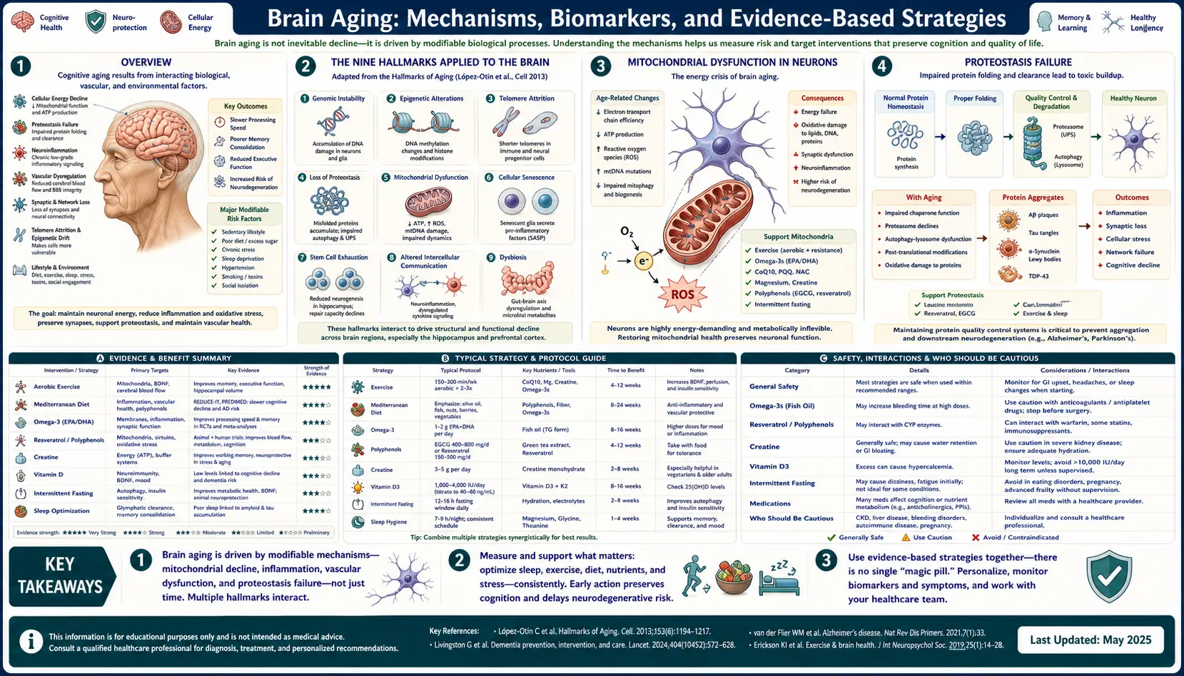
Table of Contents
- Overview
- The Nine Hallmarks Applied to the Brain
- Mitochondrial Dysfunction in Neurons
- Proteostasis Failure
- Glymphatic System and Sleep
- Neuroinflammation and Microglia Priming
- Vascular Contribution and Cerebral Small-Vessel Disease
- Insulin Resistance — "Type 3 Diabetes"
- Epigenetic Clocks Applied to Brain
- Sirtuins, NAD+, and Senescence
- Lifestyle Interventions With Strongest Evidence
- Targeted Supplements and Compounds
- Pharmacological Frontiers
- Biomarkers Worth Tracking
- Practical Synthesis
- Key Research Papers
- Connections
- Featured Videos
Overview
The phrase "brain aging" papers over two very different processes that happen to share a calendar. The first is biological brain aging — the slow accumulation of molecular and cellular damage in neurons, glia, and the vasculature, much of which proceeds silently for decades. The second is cognitive aging — the eventual appearance of subjective forgetfulness, reduced processing speed, mild executive slips, and (in the worst cases) frank dementia. The two are correlated but not identical: people with substantial Alzheimer's pathology at autopsy can have been cognitively normal in life (so-called "asymptomatic AD"), while others with relatively clean brains can decline catastrophically from vascular disease, depression, sleep apnea, alcohol, or polypharmacy.
The clinically useful split is between normal (healthy) aging and pathological decline. Healthy aging includes mildly slower recall of names, longer learning curves for new technical material, and a roughly linear shrinkage of total brain volume of about 0.2–0.5% per year after age 60. Pathological aging is defined by the accelerated loss of independence: difficulty managing finances, getting lost in familiar places, repeating questions within minutes, personality change, or motor parkinsonism. The transition zone — mild cognitive impairment (MCI) — is now the single most important intervention window, because the molecular damage driving Alzheimer's and Parkinson's begins 15–20 years before symptoms.
The encouraging finding of the past decade is that a meaningful fraction of late-life cognitive decline is modifiable. The 2024 Lancet Commission on dementia prevention concluded that addressing 14 risk factors across the lifespan could potentially prevent or delay up to 45% of dementia cases — including hearing loss, hypertension, smoking, obesity, depression, physical inactivity, diabetes, alcohol consumption, traumatic brain injury, air pollution, social isolation, low educational attainment, untreated visual impairment, and high LDL cholesterol. The biology that drives the modifiable fraction is the focus of this page: how the brain actually ages at a molecular level, which biomarkers reflect that process, and which interventions have human evidence rather than mouse-only or marketing-only support.
The unifying frame is the hallmarks of aging framework first articulated by López-Otín and colleagues in Cell in 2013 and updated in 2023. The brain is not exempt from the same nine (now twelve) hallmarks that drive aging in every other organ — it just expresses them through neuron-specific failures. Understanding which hallmark dominates which clinical syndrome (proteostasis in Alzheimer's, mitochondrial dysfunction in Parkinson's, vascular and inflammatory hallmarks in vascular dementia) is the bridge from longevity research to actionable prevention.
The Nine Hallmarks Applied to the Brain
López-Otín's original nine hallmarks of aging map onto neurons and glia with disease-relevant specificity. The framework was expanded to twelve hallmarks in 2023 (adding disabled macroautophagy, chronic inflammation, and dysbiosis), and every one of them has been demonstrated in human cortex. Reviewing them in order clarifies why "brain aging" is not one thing but a portfolio of failures that can be measured and, in many cases, slowed.
1. Genomic instability. Neurons are postmitotic and cannot dilute damaged DNA by dividing, so single-strand and double-strand breaks accumulate over decades. Oxidative damage to mitochondrial DNA is particularly important because mtDNA lacks histones and has limited repair capacity. Markers of DNA damage (gamma-H2AX foci, 8-oxoguanine) increase with age in pyramidal neurons. 2. Telomere attrition. Though neurons rarely divide, telomere shortening in peripheral leukocytes correlates with smaller hippocampal volume and faster cognitive decline; the relationship is bidirectional, because chronic stress accelerates telomere loss systemically.
3. Epigenetic alterations. DNA methylation drifts with age in ways measurable by the Horvath cortical clock; histone modifications and chromatin remodeling shift the transcriptome of aged neurons toward stress-response and inflammatory programs. 4. Loss of proteostasis. The capacity of the chaperone network (HSPs), the ubiquitin–proteasome system, and autophagy all decline with age, allowing aggregation-prone proteins (beta-amyloid, tau, alpha-synuclein, TDP-43) to accumulate. This is the central failure in classical neurodegeneration.
5. Deregulated nutrient sensing. Insulin/IGF-1 signaling, mTOR, AMPK, and sirtuins form an interconnected nutrient-sensing network. Hyperactive mTOR (from chronic overnutrition) suppresses autophagy and accelerates aging; conversely, calorie restriction, intermittent fasting, and rapamycin extend lifespan in every species tested. The brain is especially mTOR-sensitive because dendritic protein synthesis and synaptic plasticity depend on tightly regulated translation.
6. Mitochondrial dysfunction. Aged neurons show decreased Complex I and IV activity, increased ROS production, impaired calcium buffering, and accumulation of damaged mitochondria due to failing mitophagy. Because the brain uses 20% of resting energy expenditure for 2% of body mass, even modest bioenergetic deficits cascade into synaptic failure. 7. Cellular senescence. Senescent astrocytes and microglia accumulate in aged cortex and secrete the senescence-associated secretory phenotype (SASP) — IL-6, TNF-alpha, MMP-3, CCL2 — that drives chronic neuroinflammation. Senolytic compounds (dasatinib + quercetin, fisetin) selectively kill these cells in animal models with cognitive benefit.
8. Stem-cell exhaustion. Adult neurogenesis in the human hippocampal dentate gyrus has been documented through the ninth decade of life, but the rate falls steeply with age. Exercise, environmental enrichment, and BDNF preserve neurogenic capacity; chronic stress, cortisol, alcohol, and inflammation suppress it. 9. Altered intercellular communication. The aging brain shows "inflammaging" — rising baseline cytokines, complement-mediated synapse pruning by microglia, and loss of synchronized network oscillations. The 2023 expanded hallmarks add disabled macroautophagy (especially mitophagy), chronic inflammation, and dysbiosis (the gut microbiome's growing recognition as a regulator of neuroinflammation via the vagus nerve and bacterial metabolites).
Mitochondrial Dysfunction in Neurons
The brain is the most mitochondrially demanding organ in the body. A resting neuron consumes roughly 4.7 billion ATP molecules per second, almost all of it from oxidative phosphorylation, and the majority of that ATP fuels the Na+/K+ ATPase that maintains the resting membrane potential after each action potential. Even a 10–20% decline in oxidative capacity is enough to compromise synaptic transmission, particularly at the energy-hungry presynaptic terminals where vesicle recycling, calcium clearance, and neurotransmitter reuptake all draw on local ATP pools.
Aged neurons reliably show declines in Complex I and Complex IV activity, measurable both in postmortem cortex and in skin-derived fibroblasts from aged donors. Complex I (NADH:ubiquinone oxidoreductase) is the largest and most fragile of the respiratory complexes, and its decline drives a corresponding rise in electron leak and superoxide production. The resulting reactive oxygen species (ROS) damage mitochondrial lipids, proteins, and especially mtDNA, which sits unprotected adjacent to the inner membrane. Because mtDNA encodes 13 essential subunits of the respiratory complexes, oxidative mtDNA damage creates a self-amplifying spiral — the mitochondrial theory of aging in its modern molecular form.
Calcium handling is the second mitochondrial vulnerability. Mitochondria buffer cytosolic Ca2+ during high-frequency firing through the mitochondrial calcium uniporter (MCU), and matrix Ca2+ in turn stimulates the TCA cycle dehydrogenases. In aged neurons, the inner membrane is more permeable, the mitochondrial permeability transition pore opens at lower Ca2+ loads, and prolonged Ca2+ overload triggers cytochrome c release and apoptosis. This pathway is centrally implicated in selective vulnerability of substantia nigra dopaminergic neurons in Parkinson's disease, where Ca2+-driven pacemaking imposes an unusually high mitochondrial workload.
Mitophagy — the selective autophagic clearance of damaged mitochondria via the PINK1/Parkin pathway — declines with age. Loss-of-function mutations in PINK1 and PRKN cause early-onset Parkinson's; even in sporadic disease, mitophagy flux is reduced. Compounds that restore mitophagy (urolithin A, spermidine, NAD+ precursors) are an active area of clinical investigation. UCP2 (mitochondrial uncoupling protein 2), expressed in neurons of the arcuate nucleus and substantia nigra, modulates ROS production and calcium handling; its expression rises with caloric restriction and falls with chronic overnutrition, providing one mechanism by which diet directly influences brain bioenergetics.
Proteostasis Failure
The proteostasis network — chaperones, the ubiquitin–proteasome system, and autophagy — manages the lifecycle of every protein from synthesis through quality control to disposal. With age the network's capacity falls just as the demand on it rises: oxidized proteins accumulate, misfolded conformers escape chaperones, the 26S proteasome becomes less assembled, and the autophagic flux that should clear aggregates slows. The result is the visible pathology of late-life neurodegeneration — protein aggregates of various kinds in characteristic brain regions.
Beta-amyloid (Abeta). Sequential cleavage of the amyloid precursor protein (APP) by beta-secretase (BACE1) and gamma-secretase generates Abeta peptides of 38–43 residues. The longer, more hydrophobic Abeta42 is highly aggregation-prone, forming soluble oligomers (the most synaptotoxic species), protofibrils, and ultimately the extracellular plaques that define Alzheimer's disease. The "amyloid cascade hypothesis" — that Abeta accumulation is upstream of tau pathology, neuroinflammation, and neurodegeneration — was clinically validated by the modest but real benefit of the monoclonal antibodies lecanemab and donanemab, which clear amyloid and slow decline by ~25–35% over 18 months in early disease.
Tau. Tau is a microtubule-stabilizing protein in axons. Hyperphosphorylation (especially at sites recognized by the AT8 antibody) detaches tau from microtubules and allows it to aggregate into paired helical filaments — the neurofibrillary tangles of Alzheimer's. Crucially, tau pathology correlates with cognitive decline far better than amyloid burden; plasma p-tau217 has emerged as the single most useful blood biomarker for early AD, performing at the level of CSF assays and amyloid PET in some cohorts. Tau also drives the primary tauopathies (progressive supranuclear palsy, corticobasal degeneration, frontotemporal lobar degeneration with tau pathology).
Alpha-synuclein aggregates into Lewy bodies in Parkinson's disease and Lewy body dementia; its prion-like spread from gut and olfactory bulb to brainstem to cortex (the Braak staging hypothesis) explains the long prodrome of constipation, anosmia, and REM sleep behavior disorder that precedes motor symptoms by 10–20 years. TDP-43 aggregates drive amyotrophic lateral sclerosis and the majority of frontotemporal dementia, and are also found in a large fraction of aged hippocampi as the "LATE" (limbic-predominant age-related TDP-43 encephalopathy) phenotype, often misdiagnosed as Alzheimer's.
The common thread is chaperone exhaustion and autophagy failure. Heat-shock proteins (HSP70, HSP90, HSP104 in yeast) fold and refold client proteins; the unfolded protein response (UPR) in the endoplasmic reticulum diverts translation when misfolded proteins accumulate. Both systems become less effective with age. Macroautophagy — especially the chaperone-mediated and mitophagy subsets — declines steeply, allowing aggregates that would have been cleared in youth to persist and seed further misfolding. Interventions that boost autophagy (caloric restriction, rapamycin, spermidine) are a leading anti-aging strategy precisely because they target proteostasis at its source.
Glymphatic System and Sleep
In 2012–2013, Maiken Nedergaard and colleagues at Rochester described the glymphatic system — a brain-wide network that uses cerebrospinal fluid (CSF) flowing along periarterial spaces, through the parenchyma via astrocytic aquaporin-4 (AQP4) channels, and out along perivenous routes to clear interstitial solutes including amyloid-beta and tau. The discovery filled a long-standing puzzle: the brain has no conventional lymphatic system, yet must dispose of metabolic waste from a tissue with the highest metabolic rate in the body.
The defining finding from Nedergaard's group, published in Science in 2013, was that glymphatic clearance is dramatically more active during sleep than during wake. During slow-wave sleep the brain's interstitial space expands by about 60%, CSF-interstitial fluid exchange roughly doubles, and the clearance of injected radiolabeled Abeta accelerates correspondingly. The phenomenon depends on AQP4 polarization to astrocytic end-feet around blood vessels — a polarization that is lost in the aged brain and in AD, producing a measurable "glymphatic failure" that is itself an aging hallmark of the central nervous system.
The clinical implications are enormous. Chronic short sleep, fragmented sleep, and especially obstructive sleep apnea compromise glymphatic clearance and are now established risk factors for cognitive decline and dementia. Sleep apnea drives intermittent hypoxia, sympathetic activation, blood–brain barrier disruption, and direct impairment of slow-wave sleep architecture. Treatment with CPAP improves cognition in moderate-to-severe OSA in randomized trials, and screening with home sleep studies has become an essential part of any modern cognitive-decline workup.
Practical glymphatic-friendly sleep hygiene includes prioritizing seven to nine hours of opportunity for sleep, sleeping on the side (lateral position appears to optimize glymphatic flow in animal models), maintaining strict circadian regularity, treating sleep apnea aggressively, restricting alcohol (which suppresses slow-wave sleep), and limiting late-evening light exposure. Magnesium-L-threonate and apigenin (from chamomile) have modest evidence for improving sleep architecture; melatonin at low doses (0.3–1 mg) may help phase-shift the circadian clock in older adults. The single most powerful glymphatic intervention, however, is simply consistent, deep, undisrupted sleep.
Neuroinflammation and Microglia Priming
Microglia are the brain's resident immune cells, derived embryologically from yolk-sac progenitors and self-renewing locally throughout life. In youth they survey the neuropil with motile processes, prune underutilized synapses during development, and clear debris and pathogens. With age they undergo a profound shift toward a "primed" phenotype: shorter, less motile processes; elevated baseline production of IL-1beta, IL-6, and TNF-alpha; exaggerated response to subsequent inflammatory stimuli; and accumulation of dystrophic, senescent forms that secrete the senescence-associated secretory phenotype.
The NLRP3 inflammasome is a central driver of microglial inflammaging. NLRP3 assembles in response to a variety of damage-associated molecular patterns (DAMPs) including Abeta fibrils, mitochondrial ROS, and extracellular ATP, recruiting caspase-1 to cleave pro-IL-1beta and pro-IL-18 into their active forms. Chronic NLRP3 activation produces a self-sustaining loop of microglial activation, synaptic dysfunction, and neuronal death. NLRP3 inhibitors (the small molecule MCC950 and structurally related compounds) reduce pathology in AD animal models and are in early human trials.
The complement cascade — specifically C1q, C3, and C3a — tags synapses for engulfment by microglia. Beth Stevens and colleagues showed that complement-mediated synaptic pruning, an essential developmental process, is inappropriately reactivated in the aging brain and dramatically so in AD models. Blocking C1q rescues synapse density and cognitive function in mice, and anti-C1q antibodies are entering human trials. This synaptic-pruning mechanism is one of the most direct molecular links between systemic inflammation and cognitive decline.
Imaging of neuroinflammation in living humans is now possible with TSPO PET (translocator protein, an outer mitochondrial membrane protein upregulated in activated microglia). TSPO signal is elevated in mild cognitive impairment, increases over time in AD, and correlates with tau pathology and clinical decline. Plasma GFAP (glial fibrillary acidic protein, an astrocyte intermediate filament) tracks astrocyte reactivity and has joined plasma p-tau217 and NfL as a standard biomarker triplet for early AD and other neurodegenerative diseases. Targeting senescent microglia with senolytics (dasatinib + quercetin, fisetin) is one of the most actively pursued strategies for reversing the neuroinflammatory component of brain aging.
Vascular Contribution and Cerebral Small-Vessel Disease
For most of the past century the vascular contribution to dementia was treated as a separate disease — "multi-infarct dementia" caused by discrete strokes. The modern picture is far more inclusive: cerebral small-vessel disease (cSVD), driven by endothelial dysfunction and chronic hypertension, contributes to the majority of late-life cognitive impairment and synergizes with Alzheimer's pathology in mixed dementia, which is in fact the most common pathological substrate found at autopsy in patients clinically diagnosed with AD.
The hallmark MRI findings of cSVD are white matter hyperintensities (WMH), lacunar infarcts, cerebral microbleeds, enlarged perivascular spaces, and cortical superficial siderosis. WMH burden — quantifiable as a Fazekas score on FLAIR MRI — is one of the strongest imaging predictors of future cognitive decline and stroke. WMH reflects chronic ischemic damage to the deep penetrating arterioles that supply the white matter, with axonal demyelination and gliosis the histological substrate. The Rotterdam, Framingham, and ARIC cohorts have repeatedly demonstrated that midlife hypertension (especially systolic blood pressure in the 50s and 60s) drives WMH expansion measured 20 years later.
Enlarged perivascular spaces (EPVS) in the basal ganglia and centrum semiovale, visible as small CSF-intensity tubules on T2 MRI, are now recognized as a marker of impaired glymphatic clearance. They are not benign: high EPVS burden predicts cognitive decline independent of WMH and amyloid load. Cerebral microbleeds (small hemosiderin deposits) reflect either hypertensive arteriolar damage (deep distribution) or cerebral amyloid angiopathy (lobar distribution); the latter is increasingly recognized as a risk factor for amyloid-related imaging abnormalities (ARIA) during anti-amyloid antibody therapy.
Berislav Zlokovic's work has highlighted the role of blood–brain barrier (BBB) breakdown in early aging and AD, demonstrable with dynamic contrast-enhanced MRI and CSF/plasma albumin ratios. BBB leakage is detectable in the hippocampus before measurable cognitive change and before amyloid PET positivity in some APOE4 carriers, suggesting that vascular dysfunction may sometimes precede protein aggregation. The clinical lever is straightforward: aggressive treatment of midlife and late-life hypertension (target SBP <130 mmHg per SPRINT-MIND), control of diabetes, smoking cessation, regular aerobic exercise to maintain endothelial function, and treatment of atrial fibrillation to prevent embolic infarcts and silent strokes.
Insulin Resistance — "Type 3 Diabetes"
Suzanne de la Monte's coining of "Type 3 diabetes" for Alzheimer's disease in 2008 was provocative but biologically apt. The brain expresses insulin receptors densely in the hippocampus, entorhinal cortex, and hypothalamus; central insulin signaling regulates synaptic plasticity, glucose uptake by astrocytes, neuronal survival, and the trafficking of neurotransmitter receptors at the synapse. In AD brain, insulin and IGF-1 signaling are profoundly impaired — insulin receptor expression is reduced, downstream IRS-1 is hyperphosphorylated at inhibitory serine sites, and PI3K/Akt signaling is suppressed. The molecular picture closely parallels that of insulin-resistant skeletal muscle in type 2 diabetes.
One particularly elegant mechanism links peripheral and central insulin resistance through insulin-degrading enzyme (IDE). IDE is a metalloprotease that degrades both insulin and Abeta, and the two substrates compete for the enzyme. Chronic hyperinsulinemia saturates IDE with insulin, slowing Abeta clearance and tilting the brain toward amyloid accumulation. This is one molecular explanation for the epidemiologic finding that type 2 diabetes roughly doubles dementia risk independent of cerebrovascular disease.
The insulin/IGF-1 axis also overlaps with longevity biology in striking ways. Reduced IGF-1 signaling extends lifespan in worms (daf-2), flies (chico), and mice (Igf1r heterozygotes); centenarians more frequently carry IGF-1 receptor variants associated with reduced signaling. The brain-specific lesson is not that IGF-1 should be minimized (low IGF-1 in elderly humans is associated with frailty and worse outcomes) but that the brain needs responsiveness rather than chronic high signaling. Intermittent fasting, exercise, and metformin all sensitize the brain to insulin in animal models and improve cognitive outcomes in humans with prediabetes or type 2 diabetes.
Clinical levers include treating diabetes to HbA1c targets that avoid hypoglycemia (severe hypoglycemia itself accelerates cognitive decline), reducing visceral adiposity, intermittent fasting or time-restricted eating (16:8 or 14:10), aerobic exercise (which acutely improves insulin sensitivity for 24–48 hours), resistance training (which improves long-term glycemic control by increasing muscle glucose disposal), and reducing dietary refined carbohydrates. GLP-1 receptor agonists (semaglutide, tirzepatide) are now in Phase 3 trials for AD prevention, with strong biological rationale and encouraging early signals.
Epigenetic Clocks Applied to Brain
The discovery in 2013 by Steve Horvath of an epigenetic clock — a weighted sum of DNA methylation at ~353 CpG sites that predicts chronological age across nearly all human tissues with a median error under 4 years — transformed aging research from a phenotype-driven enterprise to a quantitative one. The clock works because DNA methylation drifts in stereotyped patterns across the genome with age, and that drift is largely tissue-independent. Horvath subsequently developed a cortical-specific clock trained on prefrontal cortex samples that performs better in brain tissue and reveals the brain age gap — the difference between predicted methylation age and chronological age — as an independent predictor of cognitive decline and AD risk.
Second-generation clocks have moved beyond simply predicting chronological age to predicting biological age and mortality. PhenoAge (Levine et al., 2018) is trained against a composite of nine clinical markers (CRP, glucose, albumin, etc.) and predicts mortality, cancer risk, and disability better than the original Horvath clock. GrimAge (Lu et al., 2019) is trained against plasma proteins and smoking pack-years, and predicts time-to-death and time-to-dementia more accurately than any prior clock. DunedinPACE (Belsky et al., 2022) measures the current pace of aging from a single blood draw, calibrated against a longitudinal cohort followed from birth.
The MRI-based brain age gap uses machine learning on T1-weighted structural MRI to predict chronological age from gray matter morphology. The residual (predicted minus actual) is the brain age gap; positive values (brain appears older than chronological age) predict accelerated cognitive decline, AD progression, mortality, and a host of psychiatric outcomes. The metric is now routinely included in research-grade neuroimaging pipelines and is starting to appear in commercial direct-to-consumer reports.
The crucial caveat is that epigenetic clocks are biomarkers, not biological mechanisms. Slowing the clock per se is not the goal — reducing the underlying damage that drives clock acceleration is. Interventions documented to slow epigenetic clocks in human trials include caloric restriction (CALERIE trial: DunedinPACE slowed by ~2%), the Fahy et al. growth-hormone/metformin/DHEA combination (TRIIM trial: ~2.5 years of biological age reversed), and the Mediterranean diet plus exercise (modest GrimAge slowing). Whether these represent true rejuvenation or merely epigenetic correlate change remains a live research question.
Sirtuins, NAD+, and Senescence
The seven mammalian sirtuins (SIRT1–7) are NAD+-dependent deacylases originally discovered as longevity factors in yeast (Sir2). They sit at the intersection of nutrient sensing, mitochondrial biogenesis, DNA repair, and cellular senescence, and their activity falls with age primarily because of declining NAD+ availability. David Sinclair's laboratory and many others have shown that restoring NAD+ via the precursors nicotinamide riboside (NR) and nicotinamide mononucleotide (NMN) reverses aspects of mitochondrial decline, vascular function, and (in some models) cognitive aging in mice.
The age-related fall in tissue NAD+ has several causes. CD38, an NADase expressed on immune cells and vascular endothelium, increases dramatically with age and consumes NAD+ at a rate that outpaces synthesis. CD38 inhibitors (78c, apigenin at high doses) raise tissue NAD+ in rodents. NAMPT, the rate-limiting enzyme of the NAD+ salvage pathway, declines with age in muscle, liver, and brain. The net effect is that tissue NAD+ in 60-year-old humans is roughly half that of 30-year-olds, with corresponding decreases in SIRT1 and SIRT3 activity.
One particularly relevant brain-specific NAD+ pathway involves NMNAT2, a chaperone-like enzyme that synthesizes NAD+ locally in axons and is essential for axon survival. Loss of NMNAT2 triggers Wallerian degeneration — the active, self-destruction program of distal axons after injury — through activation of the NADase SARM1. SARM1 inhibitors are now in clinical trials for diabetic peripheral neuropathy and chemotherapy-induced neuropathy, and the same pathway is implicated in the axonal pathology of glaucoma, traumatic brain injury, and possibly Alzheimer's. Preserving axonal NAD+ may be one of the more underappreciated strategies for brain aging.
The human evidence for NAD+ precursors is mixed. NR and NMN reliably raise blood NAD+ levels (NMN somewhat more efficiently in some studies), and small trials have shown improvements in walking speed, blood pressure, and aerobic capacity in older adults. Larger, longer trials have not yet demonstrated meaningful cognitive benefit. The most rigorous brain-specific data come from a 2022 Maryland trial of NR (1 g/day for 10 weeks) in mild cognitive impairment, which showed measurable increases in brain NAD+ on phosphorus MR spectroscopy without significant cognitive change at that timescale. NAD+ supplementation is reasonable and well tolerated, but should be regarded as a foundation rather than a magic bullet, and combined with exercise and CD38-inhibiting compounds (apigenin from parsley and chamomile, quercetin) for synergy.
Lifestyle Interventions With Strongest Evidence
The single intervention with the strongest evidence base for slowing brain aging is aerobic exercise. Erickson and colleagues' landmark 2011 trial published in PNAS randomized 120 older adults to either one year of moderate-intensity aerobic exercise (40-minute walks 3x/week) or stretching control, and demonstrated a 2% increase in hippocampal volume in the exercise arm versus 1.4% decline in controls — effectively reversing two years of normal age-related hippocampal atrophy. The effect was mediated in part by exercise-induced brain-derived neurotrophic factor (BDNF), a master regulator of synaptic plasticity, neurogenesis, and neuronal survival. Subsequent meta-analyses have confirmed that aerobic exercise produces meaningful improvements in executive function, attention, and memory in older adults, and reduces incident dementia by 15–30% in long-term cohorts.
The dose-response relationship favors moderate-to-vigorous aerobic exercise for 150–300 minutes per week, including some interval-style work that produces a clear lactate response (which itself crosses the blood–brain barrier and acts as a neuronal fuel and signaling molecule). Resistance training adds independent benefit through preservation of muscle mass, improved insulin sensitivity, and stimulation of IGF-1 and myokines that cross the BBB. The combination of aerobic and resistance work is superior to either alone in head-to-head trials in older adults with MCI.
The Mediterranean diet and its variant the MIND diet (Mediterranean–DASH Intervention for Neurodegenerative Delay) have the strongest dietary evidence. Morris and colleagues' 2015 development and validation of the MIND diet in Alzheimer's & Dementia showed that high adherence reduced incident AD by 53% over five years in the Rush Memory and Aging Project. The MIND diet emphasizes ten brain-healthy food groups (leafy greens, other vegetables, berries, nuts, beans, whole grains, fish, poultry, olive oil, modest wine) and five to limit (red meat, butter/margarine, cheese, pastries/sweets, fried/fast food). Subsequent randomized trials (FINGER, MIND-CHINA) have replicated benefit on multidomain cognitive composites.
Sleep architecture is the third pillar. As discussed under glymphatic clearance, slow-wave sleep is when the brain washes itself; chronic short sleep (<6 hours), fragmented sleep, and untreated sleep apnea are all independent dementia risk factors. The fourth pillar is cognitive and social engagement. Sustained intellectual activity (learning a new language, musical instrument, complex hobby) builds "cognitive reserve" — the brain's ability to maintain function despite accumulating pathology. Social isolation, conversely, accelerates cognitive decline and is associated with a 50% higher risk of dementia in meta-analyses. The Lancet Commission lists social isolation among its 14 modifiable risk factors precisely because the effect size is so large and the intervention so straightforward.
Targeted Supplements and Compounds
The supplement landscape for brain aging is enormous and the evidence quality varies dramatically. The compounds with the strongest human data for slowing cognitive decline or modifying meaningful biomarkers are concentrated in a small number of categories: omega-3 fatty acids, B-vitamins for homocysteine, magnesium, lithium at microdoses, creatine, urolithin A, and methylene blue.
Omega-3 EPA and DHA. DHA constitutes roughly 30% of the brain's phospholipid fatty acids, particularly enriched in synaptic membranes where it modulates membrane fluidity, ion channel function, and the resolution of neuroinflammation through specialized pro-resolving mediators (resolvins, protectins, maresins). Higher dietary omega-3 intake and higher red blood cell omega-3 indices are associated with slower cognitive decline and lower dementia risk in epidemiological cohorts. Randomized trials are less impressive: the largest (AREDS2, VITAL-COG) showed modest or null effects on cognition in unselected older adults. The probable explanation is that high baseline omega-3 status (from fish-eating populations) blunts the response. APOE4 carriers and individuals with low baseline omega-3 status appear to benefit most. Reasonable dose: 1–2 g/day of combined EPA+DHA, with food, ideally from a high-quality fish oil or algae source.
B12, folate, and B6 for homocysteine. Elevated plasma homocysteine is a strong independent predictor of brain atrophy, white matter hyperintensities, and dementia incidence. The VITACOG trial (Smith et al., Oxford) randomized older adults with MCI and homocysteine >11 µmol/L to high-dose B12 (500 µg), folate (800 µg), and B6 (20 mg) versus placebo for two years, and demonstrated a 53% reduction in brain atrophy rate by MRI in the active arm, with the largest effects in those with the highest baseline homocysteine. The benefit was concentrated in the temporal lobe regions vulnerable in AD. Subsequent analyses showed cognitive benefit specifically when omega-3 status was also adequate — suggesting that homocysteine-lowering and omega-3 sufficiency are synergistic. Check serum B12, holotranscobalamin, methylmalonic acid, and homocysteine; treat to homocysteine <9 µmol/L.
Magnesium-L-threonate is a specific magnesium chelate developed by Liu and colleagues that crosses the blood–brain barrier more efficiently than other magnesium forms and raises brain magnesium levels in animal models. A small 2016 trial showed cognitive improvement in older adults with subjective memory complaints. Magnesium is also important for sleep architecture, NMDA receptor regulation, and vascular function. Lithium orotate at microdoses (1–5 mg of elemental lithium) is supported by epidemiologic data showing that drinking water lithium levels correlate inversely with dementia incidence in Danish and Texan populations, and by a small Brazilian trial showing slowed cognitive decline in MCI. Mechanism likely involves GSK-3-beta inhibition (which reduces tau hyperphosphorylation) and BDNF induction. Microdoses are vastly below the pharmacologic doses used for bipolar disorder and have a wide safety margin.
Creatine (3–5 g/day) is best known for muscle performance but crosses the BBB modestly and supports brain bioenergetics; meta-analyses show small improvements in working memory and processing speed in older adults and vegetarians. Urolithin A (Mitopure) is a gut-microbiome metabolite of ellagitannins from pomegranates and walnuts; it induces mitophagy in muscle and brain in animal models and has shown muscle-function benefit in human trials. Direct cognitive trial data are still preliminary. Methylene blue at low dose (0.5–4 mg/kg, or in the 10–50 mg range for adults) is an old dye and antimalarial with mitochondrial-enhancing and tau-aggregation-inhibiting effects in preclinical models; small Phase 2 trials of the modified leuco-methylthioninium derivative (LMTM) in tauopathies have been mixed. Methylene blue is interesting but should be used cautiously and not combined with SSRIs (serotonin syndrome risk).
Pharmacological Frontiers
Several prescription compounds are now being repurposed for brain aging and AD prevention, alongside the first generation of disease-modifying anti-amyloid antibodies. The pace of clinical development is unprecedented, and the next decade is likely to redefine what "brain aging" treatment looks like.
Rapamycin and rapalogs. Rapamycin (sirolimus) is the most consistently lifespan-extending intervention across model organisms and the leading candidate small-molecule geroprotector. It works by inhibiting mTOR Complex 1 (mTORC1), thereby upregulating autophagy, reducing senescence burden, and improving immune function. In animal models of AD, rapamycin reduces amyloid and tau pathology and preserves cognition. Human trials in healthy older adults (PEARL trial, Kaeberlein group; ongoing rapamycin observational cohorts) suggest that intermittent dosing (5–10 mg weekly) is reasonably well tolerated, with stomatitis and lipid elevations as the main side effects. The FDA-approved rapalog everolimus has been studied as an immunomodulator in older adults and produced improved influenza vaccine responses, a marker of immune rejuvenation.
Senolytics. The dasatinib + quercetin combination, developed by James Kirkland's group at Mayo Clinic, selectively kills senescent cells in mouse models and produced measurable reductions in senescent cell markers and improvements in physical function in early human trials. Fisetin, a flavonoid found in strawberries and onions, is a milder senolytic with a benign safety profile and is being tested in older adults at doses of 20 mg/kg administered for 2–3 consecutive days monthly. Senolytic strategies for the brain are particularly attractive because senescent microglia and astrocytes drive a large fraction of neuroinflammation; preclinical work shows that senolytics reduce neuroinflammation and improve cognition in aged and AD-model mice.
GLP-1 receptor agonists. Semaglutide, liraglutide, and tirzepatide are revolutionizing obesity and diabetes treatment and are now in Phase 3 prevention trials for Alzheimer's (EVOKE, EVOKE+). The rationale is multifold: GLP-1 receptors are expressed in brain regions affected by AD, GLP-1 signaling improves insulin sensitivity centrally, and GLP-1 reduces neuroinflammation in preclinical models. Early secondary analyses of diabetes trials suggested 30–40% reductions in dementia incidence, though confounding from improved metabolic control complicates interpretation. Results of the dedicated AD prevention trials are expected in 2026.
Lecanemab and donanemab are the first anti-amyloid antibodies to show clinically meaningful disease modification, with ~25–35% reductions in cognitive decline rate over 18 months in early symptomatic AD. They define a new clinical reality: AD is now a treatable disease in its earliest stages, and the value of early biomarker-based diagnosis (plasma p-tau217, amyloid PET) has correspondingly skyrocketed. ARIA — amyloid-related imaging abnormalities (edema and microhemorrhages) — remains the main safety issue, particularly in APOE4 homozygotes. The clinical implication for prevention is profound: identifying preclinical AD via plasma biomarkers and intervening before symptoms become manifest is now the central focus of the field.
Biomarkers Worth Tracking
The biomarker landscape for brain aging has transformed in the past five years, with plasma assays now approaching the accuracy of CSF and PET for AD-relevant pathology. The currently useful panel for tracking brain aging in research or aggressive personal-medicine contexts includes the following:
Plasma p-tau217 is the single most useful blood biomarker for early Alzheimer's. The PrecivityAD2 assay and others have demonstrated AUCs above 0.90 for distinguishing amyloid-positive from amyloid-negative individuals, and the Lilly p-tau217 assay is approaching FDA approval as an AD diagnostic. Plasma p-tau217 rises 15–20 years before symptom onset in autosomal dominant AD and is highly responsive to anti-amyloid therapy. Plasma GFAP tracks astrocyte reactivity and is elevated in preclinical AD, MCI, and a range of other neurodegenerative conditions. Plasma neurofilament light (NfL) reflects axonal injury and is elevated in essentially all neurodegenerative diseases, traumatic brain injury, MS, and stroke; its non-specificity actually makes it useful as a general "brain damage" screen and treatment response marker.
APOE genotype remains the single most important common genetic risk factor for late-onset AD. APOE4 heterozygotes have ~3x AD risk, homozygotes ~12x; APOE2 carriers are protected. APOE4 homozygotes can now be reframed as "stage 0" AD because their lifetime amyloid PET conversion approaches 100% by their seventies. Knowledge of APOE status materially changes the calculus for early biomarker screening and lifestyle prioritization. Cognitive composites (the Preclinical Alzheimer Cognitive Composite, PACC; the Cogstate battery; remote cognitive testing platforms) detect subtle decline years before clinical diagnosis. Annual cognitive testing in at-risk individuals is increasingly recommended.
MRI volumetry — hippocampal volume, entorhinal cortex thickness, brain age gap — provides a structural correlate of trajectory. The NeuroQuant and Volbrain pipelines now provide normative comparisons accessible to clinicians. Quantitative WMH burden (Fazekas score, or volumetric measurement) tracks the vascular contribution to cognitive aging. Sleep tracking via WHOOP, Oura, Apple Watch, or polysomnography quantifies the duration and quality of slow-wave sleep, and screens for sleep apnea via overnight oximetry or home sleep studies. A single overnight home sleep study should be in the workup of any older adult with subjective cognitive complaints.
Other increasingly used markers include homocysteine (treatable, see B-vitamin discussion), hs-CRP (systemic inflammation), HbA1c and fasting insulin (metabolic), omega-3 index, ferritin and transferrin saturation (iron metabolism), and the epigenetic clocks GrimAge or DunedinPACE (commercially available through TruDiagnostic, Elysium, and others). The pace of biomarker development is brisk; expect plasma alpha-synuclein assays, plasma microglial markers, and digital phenotyping from smartphone use to enter routine practice within five years.
Practical Synthesis
Compressing the science into an actionable framework: brain aging interventions divide into three tiers, and the higher tiers should not substitute for the foundational ones. The order matters because the highest-impact interventions are also the cheapest and most accessible, and pharmacological interventions tested on top of bad foundations consistently disappoint.
Foundation (free or near-free, do these first). Aerobic exercise 150–300 minutes per week with some intervals plus resistance training twice weekly. Seven to nine hours sleep opportunity nightly, side-sleeping when possible, strict circadian regularity, treat sleep apnea aggressively. Mediterranean or MIND-style eating pattern with leafy greens daily, berries multiple times weekly, fish two to three times weekly, olive oil as primary fat, minimal ultra-processed food and added sugar. Active social and cognitive engagement — learn new things, maintain relationships, treat depression and hearing loss. Stop smoking, moderate alcohol (ideally <7 drinks/week), correct visual impairment.
Pillar 2 (labs and correction, low-cost). Test and treat: blood pressure to <130/80, LDL cholesterol per current cardiovascular guidelines (lower is generally better for brain), HbA1c <5.7 ideally, homocysteine <9 µmol/L (treat with B12, folate, B6), 25-OH vitamin D 40–80 ng/mL, omega-3 index >8%, TSH and thyroid panel, B12 with methylmalonic acid, hs-CRP. Screen for sleep apnea, obstructive uropathy disrupting sleep, atrial fibrillation. Consider APOE genotyping in those with family history. Plasma p-tau217 and GFAP are reasonable in those over 60 with subjective cognitive complaints or strong family history.
Pillar 3 (targeted compounds, moderate cost). Omega-3 1–2 g/day if dietary intake is insufficient. Magnesium-L-threonate 1–2 g at night. Vitamin D3 to target 25-OH levels. B-complex if homocysteine is elevated. Creatine 3–5 g/day. Consider lithium orotate microdoses (1–5 mg elemental), spermidine (from wheat germ or supplement), and NAD+ precursors (NR or NMN at 250–500 mg) in those with established commitment to the foundations. Urolithin A (Mitopure) at 500 mg/day has reasonable mitophagy evidence.
Pillar 4 (pharmacological, requires clinician). In those with metabolic syndrome or obesity, GLP-1 receptor agonists likely have brain-protective effects beyond weight loss. Rapamycin at 5–10 mg weekly under physician supervision in those willing to accept some uncertainty. Senolytic protocols (D+Q or fisetin) periodically as the evidence base matures. Anti-amyloid antibodies (lecanemab, donanemab) for those with biomarker-confirmed preclinical or early symptomatic AD, weighed against ARIA risk and APOE status.
What to avoid. Polypharmacy — anticholinergics (diphenhydramine, oxybutynin, many tricyclics) are particularly damaging; benzodiazepines and Z-drugs for chronic sleep; proton pump inhibitors at chronic high doses (may worsen B12 status); long-term opioids. Heavy alcohol. Chronic short sleep. Untreated depression. Untreated hearing loss (hearing aids reduce dementia incidence in randomized trials). Sedentary lifestyle. Social isolation. When to escalate. Subjective cognitive decline progressing over months, getting lost in familiar places, family concern, repeated medication errors, or any functional decline warrants a formal cognitive assessment, MRI brain, and plasma biomarker panel rather than reassurance.
Key Research Papers
- López-Otín C, Blasco MA, Partridge L, Serrano M, Kroemer G. The hallmarks of aging. Cell. 2013;153(6):1194-1217. — Search PubMed · doi:10.1016/j.cell.2013.05.039
- López-Otín C, Blasco MA, Partridge L, Serrano M, Kroemer G. Hallmarks of aging: an expanding universe. Cell. 2023;186(2):243-278. — Search PubMed · doi:10.1016/j.cell.2022.11.001
- Xie L, Kang H, Xu Q, et al. Sleep drives metabolite clearance from the adult brain. Science. 2013;342(6156):373-377. — PubMed PMID: 24136970 · doi:10.1126/science.1241224
- Iliff JJ, Wang M, Liao Y, et al. A paravascular pathway facilitates CSF flow through the brain parenchyma and the clearance of interstitial solutes, including amyloid beta. Sci Transl Med. 2012;4(147):147ra111. — PubMed PMID: 22896675 · doi:10.1126/scitranslmed.3003748
- Sweeney MD, Sagare AP, Zlokovic BV. Blood-brain barrier breakdown in Alzheimer disease and other neurodegenerative disorders. Nat Rev Neurol. 2018;14(3):133-150. — PubMed PMID: 29377008 · doi:10.1038/nrneurol.2017.188
- Horvath S. DNA methylation age of human tissues and cell types. Genome Biol. 2013;14(10):R115. — PubMed PMID: 24138928 · doi:10.1186/gb-2013-14-10-r115
- Lu AT, Quach A, Wilson JG, et al. DNA methylation GrimAge strongly predicts lifespan and healthspan. Aging (Albany NY). 2019;11(2):303-327. — PubMed PMID: 30669119 · doi:10.18632/aging.101684
- Erickson KI, Voss MW, Prakash RS, et al. Exercise training increases size of hippocampus and improves memory. Proc Natl Acad Sci USA. 2011;108(7):3017-3022. — PubMed PMID: 21282661 · doi:10.1073/pnas.1015950108
- Morris MC, Tangney CC, Wang Y, Sacks FM, Bennett DA, Aggarwal NT. MIND diet associated with reduced incidence of Alzheimer's disease. Alzheimers Dement. 2015;11(9):1007-1014. — PubMed PMID: 25681666 · doi:10.1016/j.jalz.2014.11.009
- Mattson MP, Moehl K, Ghena N, Schmaedick M, Cheng A. Intermittent metabolic switching, neuroplasticity and brain health. Nat Rev Neurosci. 2018;19(2):63-80. — PubMed PMID: 29321682 · doi:10.1038/nrn.2017.156
- Smith AD, Smith SM, de Jager CA, et al. Homocysteine-lowering by B vitamins slows the rate of accelerated brain atrophy in mild cognitive impairment: a randomized controlled trial. PLoS One. 2010;5(9):e12244. — PubMed PMID: 20838622 · doi:10.1371/journal.pone.0012244
- de la Monte SM, Wands JR. Alzheimer's disease is type 3 diabetes-evidence reviewed. J Diabetes Sci Technol. 2008;2(6):1101-1113. — PubMed PMID: 19885299 · doi:10.1177/193229680800200619
- Imai S, Guarente L. NAD+ and sirtuins in aging and disease. Trends Cell Biol. 2014;24(8):464-471. — PubMed PMID: 24786309 · doi:10.1016/j.tcb.2014.04.002
- Hou Y, Dan X, Babbar M, et al. Ageing as a risk factor for neurodegenerative disease. Nat Rev Neurol. 2019;15(10):565-581. — PubMed PMID: 31501588 · doi:10.1038/s41582-019-0244-7
- Madeo F, Eisenberg T, Pietrocola F, Kroemer G. Spermidine in health and disease. Science. 2018;359(6374):eaan2788. — Search PubMed · doi:10.1126/science.aan2788
- Livingston G, Huntley J, Liu KY, et al. Dementia prevention, intervention, and care: 2024 report of the Lancet standing Commission. Lancet. 2024;404(10452):572-628. — PubMed PMID: 39096926 · doi:10.1016/S0140-6736(24)01296-0
Connections
- All Remedies
- Menin Protein & Brain Aging
- Rapamycin
- Longevity Protocols
- NAD+ / NMN
- Gut–Brain Axis
- Alzheimer's Disease
- Parkinson's Disease
- Vitamin D3
- Vitamin B12
- Vitamin E
- Magnesium
- Zinc
- Selenium
- Berberine
- Morley Robbins
- Homocysteine
- Blackburn, Greider & Szostak — telomeres, telomerase, and what telomere length does and does not tell you