Forssmann, Cournand & Richards: The Man Who Catheterised His Own Heart
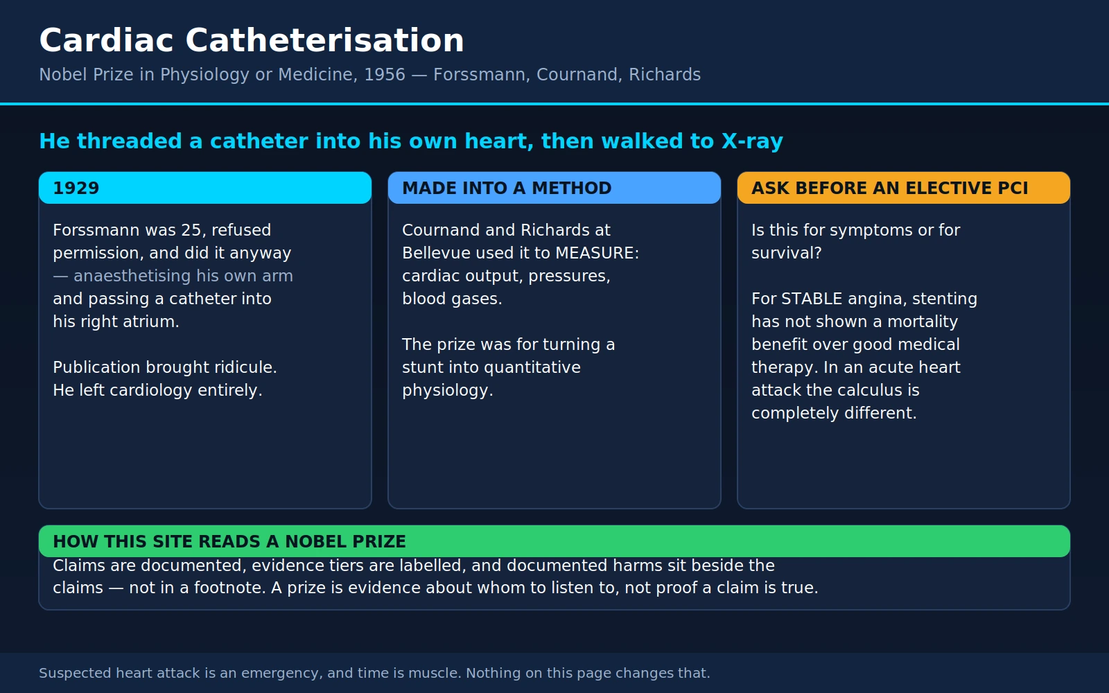
In the summer of 1929, a 25-year-old German surgical trainee threaded a thin rubber tube up a vein in his own arm, pushed it about two feet into his chest until it reached the right upper chamber of his own heart, and then walked downstairs to the X-ray department to prove he had done it. Almost everyone who heard about it thought he was a lunatic. He was told, in effect, to take his trick to a circus. He gave up heart research entirely and spent most of his working life as a urologist in provincial German towns.
Twenty-seven years later he shared a Nobel Prize with two men in New York who had read his paper, believed it, and spent a decade turning his stunt into a measuring instrument. Nearly a century on, the direct descendant of that rubber tube is how a blocked coronary artery is opened during a heart attack, how a narrowed aortic valve is replaced without opening the chest, and how pulmonary arterial hypertension is definitively diagnosed. This page tells that story accurately — including the parts that are uncomfortable, and the parts where the evidence later complicated the enthusiasm.
Table of Contents
- The Prize and the Three Men
- Summer 1929: The Self-Experiment
- What Happened Next: Ridicule, Not Acclaim
- Forssmann's Nazi Party Membership
- Cournand and Richards Make It a Method
- What Catheterisation Actually Lets You See
- The Line from 1929 to Today
- What a Patient Actually Experiences
- Where the Evidence Complicated the Enthusiasm
- What This Means If You Are Offered a Procedure
- Self-Experimentation in Medicine
- Where Mainstream Medicine Agrees — and What Remains Debated
- Key Research Papers
- Connections
- Featured Videos
1. The Prize and the Three Men
The 1956 Nobel Prize in Physiology or Medicine was awarded jointly to Werner Forssmann, André Frédéric Cournand and Dickinson W. Richards "for their discoveries concerning heart catheterization and pathological changes in the circulatory system."
It is one of the odder groupings in the history of the prize, because the three men had never worked together, were separated by an ocean and a world war, and contributed things of very different kinds. One had the nerve. Two had the patience.
Werner Forssmann (1904–1979)
Born in Berlin, Forssmann qualified in medicine in 1929 and immediately took a surgical training post at the Auguste Viktoria Home, a small hospital in Eberswalde, a town northeast of Berlin. He was 25 years old and had no research position, no funding, no laboratory and no supervisor's blessing. What he had was an idea taken from a nineteenth-century physiology textbook and a conviction that everyone else's objection to it was an assumption rather than a fact.
After the 1929 experiment he spent almost no further time in cardiac research. He became a urologist. When the Nobel telephone call came in October 1956, he was working in a small-town practice and reportedly said he felt like a village pastor who had just been told he was to be made a bishop. He had not published on the heart in a quarter of a century.
André Cournand (1895–1988)
Born in Paris, Cournand studied medicine there, served as a battalion surgeon in the French army in the First World War — he was decorated for it — and moved to New York in 1930 for what was meant to be a one-year residency at Bellevue Hospital. He stayed for the rest of his life, becoming an American citizen in 1941. He was the physiologist of the group: methodical, obsessive about calibration, interested less in the drama of getting a catheter into a heart than in what number you could trust once it was there.
Dickinson Richards (1895–1973)
Born in Orange, New Jersey, Richards trained at Columbia University's College of Physicians and Surgeons and worked at Bellevue's chest service. He was Cournand's senior colleague and, in the early years, his mentor; the two began collaborating in 1932 on the physiology of the lung, long before either of them touched a heart catheter. Richards was the one who framed the questions. Their partnership lasted more than forty years and is one of the most productive in twentieth-century clinical science.
The essential point about the 1956 prize: it was not awarded for a daring act. It was awarded because a daring act was converted into a reliable, repeatable, quantitative method for measuring what the human circulation is doing while the patient is alive and awake. Forssmann proved the thing was survivable. Cournand and Richards proved it was useful, which is a much harder and much less glamorous thing to prove.
2. Summer 1929: The Self-Experiment
Here is what Forssmann was reasoning about. Physicians of the era needed a way to deliver drugs directly to the heart in an emergency, and a way to measure what was happening inside its chambers. Injecting adrenaline through the chest wall with a long needle was being done, and it was dangerous — you could puncture a lung or a coronary artery on the way in. Forssmann's idea was that you could reach the heart from the inside, along a vein, without piercing anything.
The idea was not without precedent in animals. Claude Bernard had passed catheters into the hearts of horses and dogs in the mid-nineteenth century and had coined the term cardiac catheterisation; the physiology of it was, in animals, an old technique. What nobody had done was try it in a living human being, because of a belief — universal, confidently held, and never actually tested — that an instrument touching the inside of a human heart would cause a fatal arrhythmia or fatal shock.
What is well documented
These elements are supported by the contemporary record rather than by later recollection:
- In the summer of 1929, working at the hospital in Eberswalde, Forssmann anaesthetised the crook of his own arm, opened a vein, and passed a flexible ureteric catheter — a urological instrument, the only thing available that was long, thin and flexible enough — up the vein of his arm and into the right side of his own heart.
- He documented the result radiographically. The X-ray showing the catheter tip in his own right atrium was published with the paper and is the single most important piece of evidence in the whole story, because it is not a claim; it is an image.
- He published the account in Klinische Wochenschrift in November 1929, under the title Die Sondierung des rechten Herzens — "The probing of the right heart."
- He did not die, faint, or develop a dangerous rhythm. The central assumption of the era was simply wrong, and one experiment demonstrated it.
What rests on his own later memoir
The vivid, frequently retold details of the story come almost entirely from Forssmann's autobiography, published in 1974 — forty-five years after the event and eighteen years after the Nobel Prize. They should be read as his recollection, not as documentation, and they have accumulated embellishments in retelling. His account is that:
- His chief, Richard Schneider, refused him permission to attempt it on a patient and refused to let him attempt it on himself.
- He needed access to the sterile instruments, which were controlled by the operating-theatre nurse, Gerda Ditzen. He talked her round — by his own telling, partly by persuasion and partly by deception — and she agreed on one condition: that he perform the experiment on her, not on himself.
- He appeared to accept this. He had her lie on the table and strapped her down. Then he anaesthetised his own arm and did it to himself, out of her line of sight, while she was still restrained.
- With the catheter in place, he freed her and the two of them walked or were wheeled down to the X-ray department in the basement, where a nurse held a mirror in front of the fluoroscope screen so that Forssmann could watch his own catheter advance and push it the last distance into the right atrium.
- A colleague named Peter Romeis, seeing what was happening, tried to pull the catheter out of his arm.
Some retellings compress or alter these events — putting the mirror in Forssmann's own hand, moving the confrontation with Romeis to a different attempt, or turning Ditzen's role into pure victimhood or pure collaboration. Historians who have looked at the primary sources have noted that Forssmann's own accounts of how many times he catheterised himself, and in what sequence, are not entirely consistent between tellings.
The honest summary is this: the experiment happened, the X-ray exists, the publication is real, and the details of how he got hold of the equipment come from a memoir written by a man recalling his own youth after he had become famous for it. That does not make the memoir false. It makes it a different kind of evidence, and it is worth saying so plainly rather than repeating the theatrical version as though it were a laboratory notebook.
One detail deserves comment on its own terms. Whatever the precise sequence, Forssmann's own account describes physically restraining a colleague under false pretences in order to conduct an unapproved experiment. That is not a charming anecdote. It is a description of deceiving a co-worker, and it belongs in the story as a fact about how the experiment was obtained rather than as a joke about a bold young doctor.
3. What Happened Next: Ridicule, Not Acclaim
Publication did not make Forssmann's career. It came close to ending it.
The response in German academic medicine ranged from indifference to open contempt. The experiment was read not as a demonstration but as a stunt — self-promotion by an unqualified junior who had bypassed his chief. A priority dispute followed almost immediately, with the Berlin surgeon Ernst Unger claiming earlier work of a similar kind, which further muddied the reception. Forssmann was briefly taken on at the Charité in Berlin under the celebrated surgeon Ferdinand Sauerbruch and was dismissed. The line most often quoted from that dismissal — that one might lecture on such tricks in a circus but not in a respectable German clinic — comes from Forssmann's own memoir, and like the rest of that book should be read as his recollection of the tone rather than as a verified transcript. The tone, at least, is not in dispute.
He went back to Eberswalde, did a small amount of further work — including attempts to inject contrast medium through a catheter to visualise the heart chambers, again partly on himself — and then stopped. He retrained in urology, the specialty where his ureteric catheter had come from in the first place, and practised it for the rest of his working life.
What the profession got wrong, and why it matters
The belief that blocked cardiac catheterisation for decades was that touching the inside of the heart with an instrument would kill the patient. It was held confidently. It was taught. It shaped what experiments people thought were worth attempting. And it had never been tested in a human being, because the belief itself was the reason nobody tested it — a closed loop that is much more common in medicine than the profession likes to admit.
It was also not entirely baseless. The heart is irritable; a catheter in the right ventricle can and does provoke ectopic beats and, rarely, sustained ventricular arrhythmias. The error was not in noticing the risk. The error was in treating an unquantified risk as a certainty, and a certainty as a reason not to look.
This is the pattern that recurs on this site — in Barry Marshall's fight over whether bacteria could live in the stomach, in the long dismissal of experimental physiology as irrelevant to bedside medicine, in the reception of Einthoven's string galvanometer as an unwieldy laboratory toy. The consensus is not usually wrong because it is stupid. It is wrong because a plausible assumption has been sitting unexamined for so long that it has stopped feeling like an assumption.
4. Forssmann's Nazi Party Membership
This site does not launder biographical records, so the facts are stated here rather than omitted or smoothed over.
Werner Forssmann joined the Nazi Party in 1932, before the party took power. During the Second World War he served as a surgeon in the German army, reaching the rank of major in the medical corps, and was taken prisoner by American forces near the end of the war. He was released in 1945.
Two further facts belong beside that one, and both are matters of record rather than opinion:
- He was not among the physicians implicated in the medical atrocities prosecuted at Nuremberg. He was not a defendant in the Doctors' Trial and has not been linked by historians to the human experimentation, the killing of disabled patients, or the camp medicine that those proceedings addressed. His war service was as a front-line and hospital surgeon.
- His party membership had consequences for him after the war. Under denazification he was barred from medical practice for a period, worked at one point as a lumberjack, and the family was supported substantially by his wife, Elsbeth Forssmann, who was herself a physician. He later resumed practice as a urologist, eventually at Bad Kreuznach.
In his memoir he characterised the decision to join as a young man's political naivety. Readers may weigh that for themselves; it is his account of his own motives, which is exactly the kind of evidence that deserves the least automatic deference.
What this page will not do is either of the two easy moves. It will not use his decades of professional obscurity as a kind of retroactive punishment that squares the account — his obscurity had nothing to do with his politics and everything to do with his profession's reaction to a catheter. And it will not use the 1929 experiment, or the Nobel Prize, as a reason to leave the membership out. Both things are true about the same person, and a reader is entitled to know both.
5. Cournand and Richards Make It a Method
At Bellevue Hospital in New York, Cournand and Richards had spent the 1930s on a different problem: how the lung actually works as a gas exchanger in disease, particularly in chronic lung disease and emphysema. That work kept running into the same wall. To know how much oxygen the body is consuming and how efficiently blood is being loaded with it, you need blood that has come back from the whole body and mixed thoroughly — mixed venous blood. You cannot get that from a vein in the arm, because blood from an arm is not representative of blood from the gut, the legs, the brain and the kidneys. The only place mixed venous blood exists is inside the right heart and the pulmonary artery.
The 70-year-old formula that nobody could use
This mattered because of a piece of arithmetic published by Adolf Fick in 1870. The Fick principle says that the amount of oxygen the body consumes per minute must equal the blood flow per minute multiplied by the amount of oxygen each unit of blood picks up as it crosses the lungs. Rearranged:
Cardiac output = oxygen consumption ÷ (arterial oxygen content − mixed venous oxygen content)
That is a complete method for measuring how many litres of blood the heart pumps per minute in a living person. It had been sitting in the literature for seventy years, essentially unusable, because one of its three terms could not be obtained. Forssmann's catheter was the missing instrument.
What they actually did
Cournand and Richards began working towards human right-heart catheterisation in the mid-1930s, first in animals and cadavers, establishing that a catheter could sit in the right atrium for hours without provoking arrhythmias, clotting or infection. The first human right-heart catheterisation at Bellevue was performed in 1941, and Cournand and Hilmert Ranges published it that year in the Proceedings of the Society for Experimental Biology and Medicine as "Catheterization of the right auricle in man" — a paper that predates PubMed's indexing and so has no modern record.
Then came the part that earned the prize. Over the following years the Bellevue group used the catheter to measure, systematically and reproducibly:
- Cardiac output by the Fick principle, published in full in the Journal of Clinical Investigation in 1945 — the first robust method for measuring the output of a living human heart.
- Pressures in the right atrium, right ventricle and pulmonary artery, establishing the normal ranges against which every abnormal tracing since has been compared.
- Blood gases at each stage of the circuit, which is what makes it possible to detect and locate a shunt between the left and right sides of the heart.
- The effect of disease on all of the above — in congenital heart disease, valve disease, chronic lung disease, and heart failure. This is the "pathological changes in the circulatory system" half of the Nobel citation.
The war work, which saved lives directly
During the Second World War, Richards chaired national work on shock and blood substitutes for the American war effort, and the Bellevue catheterisation programme was turned onto the problem of traumatic and haemorrhagic shock — the commonest way a wounded soldier died before reaching definitive surgery.
The prevailing management of shock was confused, partly because the underlying physiology was disputed. Some held that shock was primarily a failure of the blood vessels' tone; others that it was a toxin released from damaged tissue. Catheterisation settled it by measurement. Richards' 1944 account, The Circulation in Traumatic Shock in Man, showed that the dominant derangement was a fall in circulating blood volume and therefore in cardiac output, and that the treatment that corrected it was replacing volume — whole blood and plasma — rather than the vasoconstrictor drugs and warming measures that had been used.
That is an unusually direct line from a physiology laboratory to lives saved on a battlefield, and it is the least famous part of the story. It is also why the 1956 prize is properly described as a prize for turning a stunt into quantitative physiology: not because measurement is intellectually superior to daring, but because a number that can be repeated in another hospital by another doctor is what changes practice.
6. What Catheterisation Actually Lets You See
A cardiac catheter is, at bottom, a way of putting a pressure sensor and a sampling straw inside a chamber of the heart while the person is awake. Two kinds of information come out of it, and understanding them makes the rest of modern cardiology much easier to follow.
Pressure tracings
As the catheter is advanced, the pressure at its tip is recorded continuously and drawn as a waveform. Each chamber has a characteristic shape and a characteristic range, so the operator can tell where the catheter is from the tracing alone, and a pressure outside the normal range is a direct measurement of a problem rather than an inference about one. As broad orientation:
- Right atrium — a low mean pressure, typically a few millimetres of mercury. Elevated when the right heart is failing or the pericardium is constricting it, and this is the number a clinician is estimating from the neck veins at the bedside.
- Right ventricle — a pumping chamber, so a high systolic and a very low diastolic pressure.
- Pulmonary artery — the same systolic pressure as the right ventricle if the pulmonary valve is normal, with a higher diastolic pressure. Its mean is the number that defines pulmonary hypertension.
- Pulmonary artery wedge pressure — obtained by advancing a small balloon until it occludes a branch of the pulmonary artery. With flow stopped, the tip is effectively reading back through the lung capillaries to the left atrium. It is the standard estimate of left-heart filling pressure, and it is how you distinguish fluid in the lungs caused by a failing left heart from fluid in the lungs caused by lung injury itself.
The oxygen saturation "run"
The second procedure is a series of blood samples drawn at successive points as the catheter travels: from the veins returning to the heart, then the right atrium, then the right ventricle, then the pulmonary artery. Each sample's oxygen saturation is measured.
In a normal heart these values are all low and all similar, because it is all the same de-oxygenated blood on its way to the lungs. If the saturation suddenly steps up at one point, oxygen-rich blood from the left side of the heart is leaking into the right side at that level — and the location of the step tells you where the hole is. A step-up at the atrium indicates an atrial septal defect; at the ventricle, a ventricular septal defect; at the pulmonary artery, a patent ductus arteriosus. The size of the step lets you calculate how much blood is being shunted, which is what determines whether the defect needs closing.
This is Cournand and Richards' contribution in its purest form. A congenital heart defect that had previously been a matter of listening to a murmur and guessing became a defect you could locate and quantify before deciding whether to operate.
Valve gradients
If a valve is narrowed, pressure must be higher on the upstream side to drive blood through it. Measuring the pressure on both sides simultaneously gives the gradient, and combining the gradient with cardiac output gives an estimate of the valve area. Echocardiography now does this non-invasively in most patients, and does it well; catheterisation is reserved for cases where the ultrasound findings are unclear or conflict with the patient's symptoms. See aortic stenosis and valvular heart disease for the conditions this applies to.
Pulmonary hypertension: the one diagnosis that still requires a catheter
This is worth stating clearly, because it is the clearest surviving example of Forssmann's technique being irreplaceable.
An echocardiogram can estimate pulmonary artery pressure from the velocity of a regurgitant jet across the tricuspid valve. That estimate is good enough for screening and it is how most patients are first flagged. It is not good enough to diagnose, because it is an estimate derived from a velocity and an assumption, and it can be substantially wrong in either direction.
Right-heart catheterisation remains the only way to definitively diagnose pulmonary arterial hypertension. Under the current European Society of Cardiology / European Respiratory Society guidelines, pulmonary hypertension is defined as a mean pulmonary arterial pressure above 20 mmHg measured at rest by right-heart catheterisation; distinguishing pre-capillary disease (a problem in the pulmonary arteries themselves) from pressure backed up from a failing left heart additionally requires a wedge pressure of 15 mmHg or less together with a raised pulmonary vascular resistance. Those three numbers cannot be obtained any other way, and the distinction between them decides the entire treatment pathway — the drugs used for pulmonary arterial hypertension are ineffective or harmful when the real problem is left-sided heart disease.
Nobody catheterises a heart out of nostalgia. It is done here because a wrong answer sends the patient down the wrong road for years. See pulmonary arterial hypertension for the condition itself.
Filling pressures in heart failure
In advanced heart failure, catheterisation answers questions that the bedside examination answers only approximately: how congested is this patient really, how much is the heart actually pumping, and is the low blood pressure due to an empty circulation or a failing pump? Those are opposite problems with opposite treatments. As section 9 describes, however, being able to measure something and being able to improve outcomes with the measurement turned out to be two different questions — and that distinction is one of the most instructive episodes in modern cardiology.
7. The Line from 1929 to Today
Forssmann proved a catheter could reach the heart. Cournand and Richards proved it could measure. Everything since has been about what else you can send down the same tube.
1958: Mason Sones and a very lucky accident
Injecting contrast dye into the heart chambers was established by the 1950s. Injecting it directly into a coronary artery was considered unthinkable, for the same class of reason that catheterisation itself had been unthinkable: everyone assumed that filling a coronary artery with dye would stop the heart.
On 30 October 1958 at the Cleveland Clinic, F. Mason Sones was performing an aortic root injection in a young man with rheumatic heart disease. The catheter tip slipped, unnoticed, into the opening of the right coronary artery, and a large bolus of contrast — accounts vary, but on the order of 30 to 40 millilitres — went straight down the artery.
Sones expected to watch the patient die. The heart stopped briefly. By the accounts of those present, Sones shouted at the patient to cough, the mechanical effect of coughing restored a rhythm, and the patient recovered fully.
The right lesson was drawn immediately, and it is the same lesson as 1929: the catastrophe everyone feared did not happen, so the assumption behind the fear was wrong. Sones set about doing deliberately what he had just done by accident, developing selective coronary arteriography over the next few years and publishing the technique with Earl Shirey in 1962. For the first time, a living patient's coronary anatomy could be seen. Everything downstream — bypass surgery, angioplasty, stents — depends on being able to see where the blockage is.
1964 and 1977: Dotter, Grüntzig and the balloon
In 1964, Charles Dotter and Melvin Judkins in Oregon showed that a narrowed leg artery could be opened by passing progressively larger catheters through it — treatment, not just diagnosis, delivered through the catheter. The idea was received in the United States with roughly the enthusiasm Forssmann had received in Germany.
It was picked up in Europe by Andreas Grüntzig, a German physician working in Zurich, who improved on it decisively by replacing the sequence of dilating catheters with an inflatable balloon on the catheter tip. He developed the early balloons largely at home, with his wife's help, on his own kitchen table — a detail that is well attested and not merely folklore. He applied it to leg arteries from 1974.
On 16 September 1977, in Zurich, Grüntzig performed the first coronary balloon angioplasty in a human being, dilating a narrowing in the left anterior descending artery of a 38-year-old man who was awake throughout. He reported the case as a brief letter to the Lancet in 1978, and the first series of 50 patients in the New England Journal of Medicine in 1979. Grüntzig died in a light-aircraft crash in 1985, aged 46.
1986: Stents
Balloon angioplasty had two serious problems. The vessel could collapse abruptly after the balloon came down, which was an emergency requiring immediate bypass surgery; and in a large minority of patients the vessel re-narrowed over the following months.
The answer was a small expandable metal scaffold left behind in the artery to hold it open. The first coronary stents were implanted in human patients in 1986 by Jacques Puel in Toulouse and Ulrich Sigwart in Lausanne, and reported in the New England Journal of Medicine in 1987. Bare-metal stents largely abolished abrupt closure and cut re-narrowing. Drug-eluting stents, introduced from the early 2000s, coat the scaffold with a drug that suppresses the scar-tissue overgrowth that caused the remaining re-narrowing, and are now standard.
Primary PCI: the catheter as an emergency treatment
The most consequential use of all of this is in an acute heart attack. When a coronary artery is completely blocked by a clot, heart muscle downstream begins dying within minutes, and the amount of muscle lost — and therefore the patient's long-term heart function and survival — depends heavily on how fast flow is restored.
Clot-dissolving drugs came first. Then trials compared them against going straight to the catheter laboratory and mechanically opening the artery: primary percutaneous coronary intervention. A 2003 pooled analysis of 23 randomised trials found primary angioplasty better than thrombolysis on essentially every outcome that matters — short-term death 7% versus 9%, re-infarction 3% versus 7%, stroke 1% versus 2%, and the combination of death, re-infarction and stroke 8% versus 14%.
Because the benefit is so time-dependent, health systems began measuring door-to-balloon time — the interval from a patient's arrival at hospital to the balloon inflating in the blocked artery — and treating 90 minutes as the target. It became one of the most-tracked quality metrics in hospital medicine, and it worked: national door-to-balloon times fell dramatically. Attention has since shifted towards total ischaemic time, measured from the onset of symptoms rather than from arrival at the hospital door, because the largest remaining delay in most systems is now the time before the patient calls for help. That has a direct implication for readers, and it appears again in section 10.
TAVI: replacing a heart valve without opening the chest
In April 2002, in Rouen, Alain Cribier compressed a prosthetic aortic valve onto a balloon catheter, threaded it up from a vein and across into the heart of a man dying of aortic stenosis who had been refused surgery, and expanded it inside the diseased valve. It worked. He reported the first human case in Circulation later that year.
Transcatheter aortic valve implantation — TAVI, or TAVR in American usage — began as a last resort for patients too frail for open surgery. It has since worked its way steadily down the risk scale: by 2019 randomised trials were showing it to be at least as good as surgery in patients at low surgical risk. For many older patients with severe aortic stenosis it now means a hospital stay of a day or two instead of a sternotomy and a long convalescence. The durability of the valves over two and three decades in younger patients is still being established, which is the main reason surgery has not simply been abandoned.
Ablation: burning or freezing the source of an arrhythmia
An electrode on a catheter tip can record the heart's electrical activity from inside the chamber, map where an abnormal rhythm is originating, and then destroy that tissue with radiofrequency heat or a cryoablation freeze. The field was transformed in 1998, when Michel Haïssaguerre's group in Bordeaux showed that most paroxysmal atrial fibrillation is triggered by ectopic beats arising in the pulmonary veins — which made electrical isolation of those veins a rational and now routine target. Supraventricular tachycardias are frequently cured outright by ablation, which is a word cardiology rarely gets to use.
Structural closure devices
Holes and appendages can be plugged through a catheter: atrial septal defects and patent foramen ovale closed with a double-disc device; the left atrial appendage occluded in patients with atrial fibrillation who cannot take anticoagulants; a leaking mitral valve reduced with a clip that holds its leaflets together. Each of these was, within living memory, an open-heart operation or nothing.
The scale of it
Take the whole list together — diagnosis of coronary disease, opening arteries in a heart attack, elective stenting, valve replacement, valve repair, cure of arrhythmias, closure of congenital defects, measurement of pulmonary pressures, evaluation for transplantation — and a very large fraction of everything modern cardiology does to a patient, it does through a catheter. The chest is not opened. The patient is usually awake. The entry wound is a puncture in a wrist. That is the direct descendant of a rubber ureteric catheter in a 25-year-old's arm in a small hospital in Eberswalde.
8. What a Patient Actually Experiences
This section is deliberately practical. If you or someone close to you has been booked for a coronary angiogram or a right-heart catheterisation, this is roughly what happens.
Where they go in: wrist versus groin
There are two standard access points.
- Radial access — the radial artery at the wrist, the one your pulse is taken from. Now the preferred default in most centres for coronary work.
- Femoral access — the femoral artery in the groin. Still used when the wrist arteries are too small or tortuous, when a large-bore device is needed (as in TAVI or mechanical support), or when the radial route has already failed.
The shift to the wrist happened because of two large randomised trials. RIVAL randomised 7,021 patients with acute coronary syndromes and found no difference in the main combined outcome of death, heart attack, stroke or major bleeding — 3.7% with radial versus 4.0% with femoral. What it did find was a marked reduction in access-site problems: large haematomas occurred in 42 of 3,507 radial patients versus 106 of 3,514 femoral patients, and pseudoaneurysms needing repair in 7 versus 23. The larger MATRIX trial randomised 8,404 patients and found radial access reduced the combined rate of adverse clinical events (9.8% versus 11.7%), driven by less major bleeding (1.6% versus 2.3%) and, in that trial, lower all-cause mortality (1.6% versus 2.2%).
The practical translation: the wrist bleeds less, and bleeding after a cardiac procedure is not a trivial complication — it is associated with worse outcomes, partly through the transfusions and the interruption of antiplatelet drugs that follow it. It also means you can sit up straight afterwards instead of lying flat for hours. It is entirely reasonable to ask which route your operator plans to use and why.
Awake, not asleep
A diagnostic catheterisation is done under local anaesthetic plus conscious sedation, not general anaesthesia. You are given something to make you relaxed and drowsy — you may remember little of it — but you are breathing on your own, you can talk to the team, and you will be asked at points to hold your breath, take a deep breath, or cough. Coughing is not an emergency signal; it is used deliberately to clear contrast and to help maintain blood pressure. General anaesthesia is reserved for the bigger structural procedures such as TAVI and some complex ablations.
The local anaesthetic sting at the wrist or groin is the sharpest sensation most people report. The catheter itself moving through the arteries is not painful — the inside of a blood vessel has no sensory nerves of the kind that would register it.
The heat
When contrast is injected, particularly a larger injection into the left ventricle for a ventriculogram, most people feel a sudden intense wave of warmth spreading through the chest and down through the pelvis and legs, lasting perhaps 10 to 20 seconds. It very commonly comes with the strong and convincing sensation of having wet yourself. You have not. Every catheter laboratory team warns patients about this in advance for exactly that reason, and it passes quickly.
How long it takes
A straightforward diagnostic coronary angiogram typically takes 20 to 40 minutes of procedure time, with rather more time either side for preparation and recovery. If it proceeds to stenting in the same sitting, add roughly another 30 to 60 minutes, more for complex or multi-vessel work. A right-heart catheterisation alone is often quicker. Expect the whole hospital visit to take most of a day even when everything is routine.
Afterwards
With radial access, a firm compression band goes on the wrist and is gradually deflated over roughly one to three hours. You can sit up and eat immediately. Diagnostic cases are frequently discharged the same day. You will be told not to lift heavy things with that arm for a few days, and the wrist will be bruised and tender.
With femoral access, the sheath is removed and pressure is applied, or a closure device is deployed. You then lie flat, keeping the leg straight, for something in the range of two to six hours depending on the device and the drugs used. Lying flat is the part patients find most trying, particularly with a bad back. Bruising in the groin and down the thigh is common and can be dramatic-looking without being dangerous.
The honest risk figures
All numbers below are approximate, and every one of them depends heavily on how sick the patient is and how experienced the operator and centre are. A healthy 55-year-old having an elective diagnostic angiogram and an 85-year-old in cardiogenic shock are not in the same risk category, and quoting them a single number would be dishonest.
For a diagnostic catheterisation in experienced hands, serious complications — death, stroke, heart attack, a vessel injury needing surgery, a major bleed — occur in well under 1% of procedures in aggregate. The classic large-scale benchmark is the Society for Cardiac Angiography and Interventions registry, which reported on 71,916 patients across 63 laboratories in 1990 and found a mortality rate for diagnostic procedures of 0.11% — about one death per 900 procedures, in an older and sicker population than earlier registries, and notably stable across decades of reporting. Equipment, catheters, contrast agents and antiplatelet drugs have all improved substantially since. Minor complications — bruising, a small haematoma, a transient arrhythmia, a vasovagal faint, an allergic reaction to the contrast — are considerably more common than serious ones.
Interventional procedures carry higher risk than diagnostic ones, because a stent is being deployed in a diseased artery rather than dye being squirted past it. That risk is still low for elective single-vessel work and rises with the complexity of the anatomy and the instability of the patient.
Contrast and the kidneys: who is actually at risk
Iodinated contrast is filtered by the kidneys, and a rise in creatinine after angiography is called contrast-associated acute kidney injury. This deserves a careful treatment, because it is a real phenomenon that has also been substantially over-attributed.
The problem with the older literature is that most of it lacked a control group. Hospitalised patients who are unwell — dehydrated, on diuretics, on antibiotics, septic, in heart failure — have creatinine rises all the time without going anywhere near a contrast injection. Studies that compared patients who received contrast against matched patients who did not found much smaller attributable effects than the uncontrolled studies had implied. The modern review literature reflects that correction: contrast is a genuine but modest nephrotoxin whose importance was inflated by study design.
Where the risk concentrates is not in doubt:
- Pre-existing chronic kidney disease is the dominant risk factor, and risk rises steeply as kidney function falls.
- Diabetes with kidney impairment raises it further; diabetes with entirely normal kidneys adds relatively little.
- Contrast volume matters — which is why operators track it and why a diagnostic study is lower risk than a long, complex intervention.
- Dehydration, heart failure, low blood pressure, and concurrent nephrotoxic drugs all add to it.
What actually helps is unglamorous: intravenous fluid before and after with isotonic saline, using the smallest contrast volume that answers the question, and pausing nephrotoxic drugs. What does not help, on good evidence, is the two remedies that were used for years on the strength of small early trials: a large randomised trial published in 2018 tested intravenous sodium bicarbonate against ordinary saline and oral N-acetylcysteine against placebo in high-risk patients undergoing angiography, and found no benefit from either for death, dialysis, persistent loss of kidney function or contrast-associated kidney injury.
If you have known kidney disease, the questions worth asking are what your current eGFR is, whether you will be given fluids beforehand, and whether any of your regular medicines should be paused around the procedure.
Radiation, in context
Catheterisation is guided by X-ray fluoroscopy, so it involves a real radiation dose. Typical figures, again approximate and highly variable with body size and procedure length: a diagnostic coronary angiogram is on the order of 3 to 10 millisieverts, and a PCI commonly 10 to 20 millisieverts or more for prolonged complex cases.
For scale: natural background radiation is roughly 3 millisieverts per year in most of the developed world; a chest CT is in the same general range as an angiogram; a plain chest X-ray is a small fraction of one millisievert. So a single angiogram is roughly comparable to one to a few years of background exposure. That is not nothing, and it is a genuine consideration for young patients and for anyone facing repeated procedures. It is also a small consideration next to an untreated blocked artery. The people for whom cumulative dose matters most are, in practice, the interventional cardiologists and radiographers standing beside the table all day, which is why they wear lead.
9. Where the Evidence Complicated the Enthusiasm
This is the section that makes this page worth reading, and it is the one most often left out of admiring accounts of catheterisation.
A technique that lets you measure something previously unmeasurable, or open something previously unopenable, generates enormous and entirely understandable enthusiasm. Enthusiasm is not evidence. In two separate areas, when catheter-based practice was finally tested against the question "does the patient end up better off?", the answer came back more complicated than anyone expected.
9.1 Right-heart catheterisation in the critically ill
From the 1970s, the balloon-tipped pulmonary artery catheter — the Swan-Ganz catheter, a direct descendant of the Bellevue work — became routine in intensive care. It gave continuous, precise numbers on filling pressures and cardiac output in the sickest patients in the hospital. At its peak, well over a million were being placed each year in the United States. It felt self-evidently useful. How could knowing more about a shocked patient's circulation not help?
In 1996, Connors and colleagues published an observational study of 5,735 critically ill patients across five teaching hospitals. Using propensity matching to compare like with like, patients who received a right-heart catheter in their first 24 hours in intensive care had higher 30-day mortality (odds ratio 1.24, 95% confidence interval 1.03–1.49), longer stays and higher costs. No subgroup showed benefit. The authors were careful about what this could and could not prove — an observational study cannot exclude the possibility that sicker patients were selected for catheterisation in ways the propensity score failed to capture — and they explicitly called for randomised trials.
They got them. The most relevant to this site's readers is ESCAPE, published in 2005, which randomised 433 patients hospitalised with severe heart failure to therapy guided by a pulmonary artery catheter plus clinical assessment, or by clinical assessment alone. Both groups improved substantially. But the catheter made no difference to the primary outcome — days alive and out of hospital over six months, 133 versus 135 — and none to mortality (10% versus 9%) or days hospitalised. In-hospital adverse events were more common in the catheter group (21.9% versus 11.5%).
Routine use collapsed over the following decade, and appropriately so.
The lesson here is precise, and it is easy to get wrong in both directions. This is not evidence that right-heart catheterisation is useless, and it is not evidence that Cournand and Richards were mistaken. The measurements are accurate; they are exactly what the Bellevue group established them to be. The finding is that a measurement only improves outcomes if it changes a decision, and that decision changes an outcome. In an average intensive-care patient, the catheter's numbers were either already adequately estimated at the bedside, or led to treatment changes — more inotropes, more aggressive diuresis — that carried their own harms. Meanwhile the catheter itself carried a small but real risk. Right-heart catheterisation retains a clear role where the number genuinely decides something: diagnosing pulmonary hypertension, assessment for transplantation or mechanical circulatory support, and untangling shock whose cause is genuinely unclear.
9.2 Stents for stable angina
This is one of the most important and most misunderstood findings in modern cardiology, and it needs to be stated carefully rather than sensationally.
The setting matters absolutely. Everything in this subsection concerns stable coronary disease — a person with predictable exertional angina, whose symptoms come on with a known amount of activity and settle with rest, who is not having a heart attack. It says nothing whatever about acute presentations. That distinction is spelled out in 9.3, and if you read only one part of this section, read that one.
The intuition behind stenting stable angina is powerful and almost visually irresistible: there is a narrowing, you can see it on the angiogram, you open it, blood flows again. Both symptom relief and prevention of future heart attacks seem to follow obviously.
On survival and heart attacks, two major randomised trials tested that intuition against optimal medical therapy — meaning aggressive treatment with statins, antiplatelet drugs, blood pressure control, and lifestyle change.
- COURAGE (2007) randomised 2,287 patients with stable coronary disease and objective ischaemia to PCI plus optimal medical therapy, or optimal medical therapy alone. Over a median of 4.6 years, death or non-fatal heart attack occurred in 19.0% of the PCI group and 18.5% of the medical therapy group — hazard ratio 1.05, p=0.62. No difference.
- ISCHEMIA (2020) was designed to answer the main objection to COURAGE by enrolling only patients with moderate or severe ischaemia on stress testing — the group most expected to benefit. It randomised 5,179 patients. Over a median of 3.2 years there was no reduction in ischaemic cardiovascular events or death. The pattern over time is worth noting because it is honest and rarely quoted: at 6 months the invasive group had more events (5.3% versus 3.4%), reflecting procedure-related infarctions; at 5 years it had slightly fewer (16.4% versus 18.2%, a difference of 1.8 percentage points with a confidence interval crossing zero). Deaths were 145 versus 144.
On symptoms, the picture is genuinely different, and this is where the story is most frequently mangled in both directions.
ISCHEMIA's companion health-status analysis found that patients assigned to the invasive strategy did have better angina-related quality of life — and, crucially, that the benefit was concentrated where you would expect it. Among patients who had daily or weekly angina at baseline the improvement was substantial; among the 35% who had no angina at all in the month before enrolment it was essentially zero. Stenting an asymptomatic narrowing does not make an asymptomatic person feel better, because there is nothing to feel better about.
Then came ORBITA (2018), which asked the harder question. Every previous trial compared stenting against not-stenting, with everyone knowing which they had received. ORBITA compared PCI against a placebo procedure — patients were sedated, taken to the catheter laboratory, had a catheter placed and pressure wires passed, and then either did or did not receive a stent, with patients and assessors blinded. Two hundred patients with severe single-vessel narrowings (mean area stenosis 84%) were randomised after six weeks of medication optimisation.
The primary endpoint was the improvement in treadmill exercise time. The difference between real PCI and the placebo procedure was 16.6 seconds, with a confidence interval running from −8.9 to +42.0 seconds — not statistically significant (p=0.200).
This landed hard, and it was widely over-interpreted as showing that stents "don't work". They were also over-defended. The fair reading is narrower and more interesting: in patients already on optimised anti-anginal medication, adding a stent produced no clearly demonstrable exercise-capacity gain beyond that of undergoing the procedure itself. A meaningful part of what patients and doctors had attributed to the metal was attributable to the ritual, the expectation, and the drugs.
The story did not stop there, and honesty requires reporting the sequel. ORBITA-2 (2023) asked a different and arguably more realistic question: what if the stent is compared against placebo in patients who are not taking anti-anginal drugs? Three hundred and one patients stopped their anti-anginal medication, then were randomised to PCI or a placebo procedure and followed for 12 weeks. This time PCI clearly won: mean angina symptom score 2.9 with PCI versus 5.6 with placebo (odds ratio 2.21, 95% CI 1.41–3.47, p<0.001).
Putting ORBITA and ORBITA-2 together gives the accurate picture, and it is not the picture either camp wanted:
- Stenting a stable narrowing does genuinely relieve angina compared with a placebo procedure. ORBITA-2 established that against the most rigorous possible comparator.
- Much of that same relief is also achievable with medication. When patients are already on good anti-anginal therapy, the additional benefit of a stent is small enough that ORBITA could not distinguish it from placebo.
- Some of the apparent benefit of any invasive procedure is placebo and expectation effect, which is real, is not fraudulent, and is precisely why blinded comparison was necessary to find out what the metal itself contributes.
- Neither trial found a survival benefit, and neither was designed to. For stable disease, that question was answered by COURAGE and ISCHEMIA, and the answer was no.
So a patient with stable angina faces a genuine choice with two reasonable options, not one right answer: medication first, with stenting held in reserve if symptoms persist or are intolerable; or stenting earlier, for faster symptom relief at the cost of a procedure with a small risk and no survival advantage. Which is preferable depends on how bad the symptoms are, how the patient feels about taking daily medication, and what they want. That is a preference-sensitive decision, and it should be made as one.
9.3 None of this applies to a heart attack
This must be unambiguous, because the sections above are exactly the kind of material that gets clipped out of context and repeated as "studies show stents don't work".
In an acute heart attack, opening the blocked artery with a catheter is one of the most effective treatments in all of medicine, and it is time-critical. The 23-trial analysis cited in section 7 found primary angioplasty reduced short-term death from 9% to 7%, re-infarction from 7% to 3%, stroke from 2% to 1%, and the combination of all three from 14% to 8%, against the best available drug alternative. This is a large benefit, and heart muscle is dying while the decision is being made.
The distinction is mechanical and easy to state. In stable angina, the narrowing has been there for years, collateral vessels have developed, and no muscle is dying — the treatment is for symptoms, and there is time to think. In a heart attack, an artery has abruptly and completely occluded, muscle downstream is dying now, and the treatment is to save that muscle and the patient. Same technique, same equipment, entirely different calculus.
Similarly, patients with unstable symptoms or a non-ST-elevation heart attack who are at higher risk do benefit from an early invasive strategy. The trials described above concern stable disease only.
If you have symptoms suggesting a heart attack — chest pressure, tightness or pain lasting more than a few minutes, especially with breathlessness, sweating, nausea, or pain spreading to the arm, neck or jaw — call emergency services immediately. Do not drive yourself. Do not wait to see whether it settles. See heart attack for the full symptom picture, including the presentations that are less classic in women and in people with diabetes.
10. What This Means If You Are Offered a Procedure
If a cardiologist has recommended an angiogram or a stent for a stable problem — and only for a stable problem — these are the questions that separate a genuinely informed decision from a nod.
The three questions that matter most
- "Is this being done to help me feel better, or to help me live longer?"
This is the single most clarifying question you can ask, and the evidence in section 9 is why. For stable coronary disease the honest answer is usually "to help you feel better." That is a perfectly good reason for a procedure — symptoms matter, and being unable to walk uphill is a real loss. But it is a different conversation from one about survival, and many patients leave the clinic believing they have been offered the second when they have been offered the first. Studies of patient understanding have repeatedly found that most people who receive an elective stent believe it will prevent a heart attack or extend their life. - "What does the evidence show for someone in my situation specifically?"
Not for coronary disease in general — for your degree of symptoms, your ischaemia on testing, your anatomy, your kidney function, your age. The answer for a person with daily angina despite three drugs is genuinely different from the answer for a person with a narrowing found incidentally who has no symptoms at all. - "What is the alternative, and what happens if I try that first?"
For stable angina the alternative is optimal medical therapy, which is not "doing nothing" — it is a specific, active, evidence-based regimen. Ask what it consists of, how long a fair trial would be, and what would trigger reconsidering the procedure. Reserving a stent is not refusing one.
Worth asking as well
- "Will you use the wrist or the groin, and why?" Radial access is now the default for most coronary work for the bleeding reasons in section 8.
- "How many of these does this operator and this centre do a year?" Volume correlates with outcome across interventional cardiology. This is an entirely legitimate question and a good team will not take offence.
- "If you find a narrowing, will you stent it in the same sitting, or stop and discuss it with me?" This matters. If you want the decision to stent to be a separate, considered one, say so before the procedure, when you are not sedated on a table.
- "What will I need to take afterwards, and for how long?" A stent commits you to dual antiplatelet therapy for a defined period. That has real implications for bleeding risk, for any surgery or dental work planned in that window, and for anyone already on an anticoagulant.
- "What is my kidney function, and what are you doing about the contrast?" See section 8.
- "Would a second opinion be reasonable here?" For an elective, preference-sensitive procedure, yes, and a confident clinician will say so.
The exception, stated plainly
Everything above concerns elective, planned procedures for stable symptoms, where you have days or weeks to think, gather information and get another opinion.
A suspected heart attack is not that situation, and the calculus is entirely different. There, the delay caused by deliberation is itself the main harm. Heart muscle dies at a rate measured in minutes, the benefit of opening the artery quickly is large and well established, and the correct action is to call emergency services at once. Nothing on this page — and nothing in the careful, important, genuinely surprising evidence about elective stenting — should ever be used as a reason to hesitate in that situation.
11. Self-Experimentation in Medicine
Forssmann belongs to a small and strange tradition: researchers who tested a hypothesis on themselves because they could not get permission, could not find a volunteer, or did not think it right to ask anyone else to take a risk they would not take.
Werner Forssmann, 1929. A catheter into his own right atrium, to test whether an untested universal assumption was true. It was not.
Barry Marshall, 1984. Told that Helicobacter pylori could not possibly cause peptic ulcers because nothing could survive in stomach acid, and unable to reproduce the disease in animals, Marshall drank a culture of the organism himself. He developed gastritis, documented it endoscopically, and treated it. He shared the 2005 Nobel Prize with Robin Warren, and peptic ulcer disease went from a chronic condition managed with acid suppression and surgery to an infection curable with a week of antibiotics.
The Haldanes. John Scott Haldane repeatedly exposed himself to carbon monoxide and mine gases to work out their physiological effects and the safe limits — work that produced the canary-in-the-mine method and much of what is known about gas poisoning. His son J.B.S. Haldane ran decompression and gas-mixture experiments on himself and a small group of colleagues during the Second World War, to establish what submarine escape and diving crews could survive. He recorded perforated eardrums, crushed vertebrae and convulsions among the results.
Others. Max von Pettenkofer drank a culture of cholera to argue against germ theory, and survived, which he took as vindication and which was in fact luck. Jesse Lazear, working with Walter Reed's yellow fever commission, allowed himself to be bitten by infected mosquitoes and died of the disease he had helped prove was mosquito-borne.
Two things are true at once
The first is that self-experimentation produced real, durable, life-saving discoveries. Cardiac catheterisation. The bacterial cause of ulcers. The physiology of decompression. Not marginal findings — foundational ones. And it did so partly because the researcher took the risk personally: it is much easier to run an experiment you sincerely believe is safe when you are the one on the table, and much harder to fool yourself about the risk.
The second is that none of these experiments would be approved by a research ethics committee today, and they should not be. Modern research ethics — informed consent, independent review, favourable risk-benefit assessment, the ability to withdraw — exists because of the Nuremberg Doctors' Trial, because of Tuskegee, and because of a long record of researchers deciding on their own authority that the knowledge was worth someone else's risk. A researcher's own body does not sit outside that framework as cleanly as it first appears. Junior researchers can be pressured into "volunteering". An investigator cannot give truly independent consent to their own protocol. Forssmann's own account involves deceiving and physically restraining a colleague to obtain equipment — which is not self-experimentation at all, but experimentation on someone else's professional standing and safety.
There is also a survivorship problem that no admiring retelling of these stories can be allowed to hide. We know the names of the self-experimenters who lived and were right. Lazear died. Pettenkofer survived and was wrong, and drew exactly the wrong conclusion from his survival. The ones who took a risk, were mistaken, and quietly harmed themselves for nothing did not get memoirs or prizes. The tradition looks braver and more successful than it was, because the failures are invisible.
Both facts stand. The discoveries were real, and the method is not one to romanticise or revive. Forssmann's experiment was correct, brave, and unrepeatable — and the reason it is unrepeatable is a genuine improvement in medicine, not a loss of nerve.
12. Where Mainstream Medicine Agrees — and What Remains Debated
Settled
- Cardiac catheterisation is safe enough to be routine. Forssmann's assumption-breaking result has been confirmed several hundred million times over.
- Right-heart catheterisation is required to diagnose pulmonary arterial hypertension. Echocardiography screens; it does not diagnose.
- Primary PCI is the preferred treatment for ST-elevation heart attack where it can be delivered promptly, and speed matters enormously.
- Radial (wrist) access reduces bleeding and access-site complications compared with femoral access, and is the default for coronary work in most centres.
- For stable coronary disease, PCI does not reduce death or heart attack compared with optimal medical therapy. COURAGE and ISCHEMIA agree, and ISCHEMIA specifically tested the group thought most likely to benefit.
- PCI does relieve angina compared with a placebo procedure in patients not on anti-anginal drugs (ORBITA-2), and the relief is concentrated in patients who actually have symptoms to relieve.
- Routine pulmonary artery catheterisation in critically ill and heart failure patients does not improve outcomes and has largely been abandoned as routine practice, while retaining specific indications.
- Bicarbonate and N-acetylcysteine do not prevent contrast-associated kidney injury. Fluids and contrast-volume restraint are what is left.
Genuinely debated
- How much of ORBITA's null result reflects its design. ORBITA was small, single-vessel, six weeks long, and used exercise time as its endpoint. Critics argue this under-powered and under-sensitive design missed a real effect; defenders point out that the same objections were not raised about unblinded trials showing benefit. ORBITA-2 addressed several of the criticisms and found a positive result, which supports the critics on symptoms while leaving the underlying methodological point about placebo control fully intact.
- Whether ISCHEMIA's late curve-crossing signals a real long-term benefit. The event curves cross at around two years, and the 5-year difference favoured the invasive strategy without reaching significance. Whether longer follow-up would show a genuine benefit, and whether the definition of procedural heart attack used disadvantaged the invasive arm early on, are both live arguments. The trial's own authors noted that the primary outcome was sensitive to which infarction definition was used.
- How much stable disease should be revascularised at all, and how much of current practice is driven by the visual persuasiveness of an angiogram — the "oculostenotic reflex", the pull to fix a narrowing simply because you are looking at it. Physiological guidance (measuring whether a narrowing actually restricts flow, rather than judging it by appearance) has moderated this, but the debate is not settled.
- The long-term durability of transcatheter valves in younger, lower-risk patients, and where the crossover point with surgical valves lies. This is the main open question in structural cardiology and will take another decade of follow-up to answer.
- The true magnitude of contrast-associated kidney injury in patients with moderate kidney impairment, once the control-group problem is properly accounted for.
- How Forssmann's 1929 experiment should be narrated. Historians differ on how much of his memoir to take at face value, and on how prominently his party membership belongs in accounts of his science. This page's position is stated in section 4: state it, contain it, do not use it to erase the work and do not use the work to erase it.
13. Key Research Papers
- Forssmann W. Die Sondierung des rechten Herzens ["The probing of the right heart"]. Klinische Wochenschrift, 1929. The original self-experiment report, in German, published before the era covered by modern bibliographic indexing — it has no PubMed record. Search PubMed for later accounts of Forssmann's experiment
- Cournand A, Riley RL, Breed ES, et al. Measurement of cardiac output in man using the technique of catheterization of the right auricle or ventricle. J Clin Invest 1945;24(1):106-16 — the Fick principle finally made usable in living patients; Richards is a co-author.
- Richards DW. The Circulation in Traumatic Shock in Man. Bull N Y Acad Med 1944;20(7):363-93 — the wartime shock work, which changed resuscitation practice directly.
- Cournand A. Cardiac catheterization; development of the technique, its contributions to experimental medicine, and its initial applications in man. Acta Med Scand Suppl 1975;579:3-32 — Cournand's own historical account of the field, written near the end of his career.
- Sones FM Jr, Shirey EK. Cine coronary arteriography. Mod Concepts Cardiovasc Dis 1962;31:735-8 — the technique that followed from the 1958 accident, and the foundation of every coronary intervention since.
- Grüntzig AR, Senning A, Siegenthaler WE. Nonoperative dilatation of coronary-artery stenosis: percutaneous transluminal coronary angioplasty. N Engl J Med 1979;301(2):61-8 — the first published series of coronary balloon angioplasty.
- Connors AF Jr, Speroff T, Dawson NV, et al. The effectiveness of right heart catheterization in the initial care of critically ill patients. JAMA 1996;276(11):889-97 — the observational study that broke the consensus on routine pulmonary artery catheterisation.
- Binanay C, Califf RM, Hasselblad V, et al. Evaluation study of congestive heart failure and pulmonary artery catheterization effectiveness: the ESCAPE trial. JAMA 2005;294(13):1625-33 — the randomised trial that confirmed it in heart failure.
- Boden WE, O'Rourke RA, Teo KK, et al. Optimal medical therapy with or without PCI for stable coronary disease. N Engl J Med 2007;356(15):1503-16 — the COURAGE trial.
- Al-Lamee R, Thompson D, Dehbi HM, et al. Percutaneous coronary intervention in stable angina (ORBITA): a double-blind, randomised controlled trial. Lancet 2018;391(10115):31-40 — the first placebo-controlled trial of a coronary intervention.
- Maron DJ, Hochman JS, Reynolds HR, et al. Initial Invasive or Conservative Strategy for Stable Coronary Disease. N Engl J Med 2020;382(15):1395-1407 — the ISCHEMIA trial.
- Rajkumar CA, Foley MJ, Ahmed-Jushuf F, et al. A Placebo-Controlled Trial of Percutaneous Coronary Intervention for Stable Angina. N Engl J Med 2023;389(25):2319-2330 — ORBITA-2, which found PCI superior to a placebo procedure for angina in patients off anti-anginal medication.
- Jolly SS, Yusuf S, Cairns J, et al. Radial versus femoral access for coronary angiography and intervention in patients with acute coronary syndromes (RIVAL): a randomised, parallel group, multicentre trial. Lancet 2011;377(9775):1409-20
- Mehran R, Dangas GD, Weisbord SD. Contrast-Associated Acute Kidney Injury. N Engl J Med 2019;380(22):2146-2155 — the modern review, including why the older literature overstated the risk.
Live PubMed Searches
- Cardiac catheterization history
- Percutaneous coronary intervention in stable angina
- Radial versus femoral access
- Right heart catheterization outcomes
- Contrast-associated acute kidney injury
14. Connections
- All Notable Doctors
- Nobel Prize in Physiology or Medicine — the full roll of laureates, and where the 1956 prize sits in it
- Willem Einthoven — the electrocardiogram: the other instrument that made the living heart legible, and also dismissed at first as a laboratory curiosity
- Barry Marshall — the site's other famous self-experimenter, who drank Helicobacter pylori to prove a point nobody would let him prove otherwise
- Goldstein & Brown — the LDL receptor, and the mechanism behind the statins that make up half of "optimal medical therapy"
- John Vane — how aspirin works, and therefore why antiplatelet therapy sits alongside every stent
- Furchgott, Ignarro & Murad — nitric oxide and the endothelium, which is why nitrates relieve angina
- Cardiology — the full index of heart and circulation conditions on this site
- Heart Attack — the emergency in which catheterisation is unambiguously life-saving, and the symptoms that should prompt a call for help
- Angina — stable chest pain: what it is, how it is treated with drugs, and where a stent fits
- Coronary Artery Disease — the underlying condition that an angiogram is looking for
- Atherosclerosis — how the narrowings form in the first place
- Heart Failure — where filling pressures matter, and where ESCAPE tested whether measuring them helps
- Aortic Stenosis — the valve disease that TAVI transformed
- Atrial Fibrillation — and catheter ablation of the pulmonary veins
- Pulmonary Arterial Hypertension — the one diagnosis that still cannot be made without a right-heart catheter
- Cardiac Troponin — the blood test that says heart muscle is dying, and often the trigger for the catheter laboratory
- Coronary Calcium Score — the non-invasive scan that has taken over much of the risk-stratification work angiography once did