Iodine for Thyroid Function
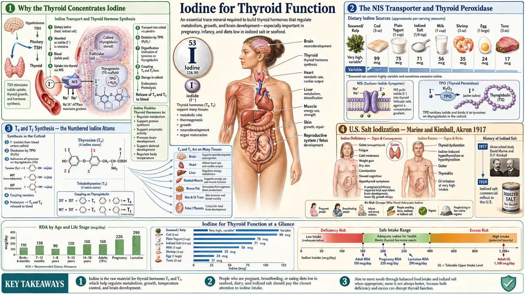
The thyroid gland is the dominant iodine-consuming organ in the body, and the synthesis of thyroxine (T4, four iodine atoms) and triiodothyronine (T3, three iodine atoms) is the best-established use of iodine in human biology. Without adequate iodide arriving at the sodium-iodide symporter (NIS) on the basolateral membrane of the thyroid follicular cell, the entire downstream cascade — thyroid peroxidase (TPO) iodination of tyrosine, coupling into iodothyronines, storage in thyroglobulin colloid, release into the bloodstream, peripheral conversion to active T3, and binding to nuclear thyroid hormone receptors in every tissue — collapses. This page walks through that cascade, the history of U.S. salt iodization (Marine and Kimball in Akron, Ohio, 1917), the Wolff-Chaikoff effect and its escape and what the research reports about iodine-induced thyroid disease, the high-dose iodine protocol described by Guy Abraham and David Brownstein set beside that research, the pharmacology of Lugol's solution, and the research on iodide versus molecular iodine in different tissues.
Table of Contents
- Why the Thyroid Concentrates Iodine
- The NIS Transporter and Thyroid Peroxidase
- T4 and T3 Synthesis — the Numbered Iodine Atoms
- U.S. Salt Iodization — Marine and Kimball, Akron 1917
- The Wolff-Chaikoff Effect and Escape
- Iodine Deficiency, Goiter, and Hypothyroidism
- Brownstein and Abraham High-Dose Iodine Protocols
- Lugol's Solution and Iodide-vs-Molecular-Iodine
- Selenium and the Thyroid
- Hashimoto's and the Iodine Controversy
- Key Research Papers
- Connections
- Featured Videos
Why the Thyroid Concentrates Iodine
The human thyroid gland weighs only 15-25 grams in an adult but contains approximately 70-80% of the body's total iodine pool — about 15-20 milligrams of iodine in an iodine-replete adult. The reason is biochemical necessity. Thyroid hormones are unique in human biology because they contain a halogen (iodine) at three or four specific positions on a coupled-tyrosine backbone, and there is no synthesis pathway that does not require dietary iodine as the input atom.
No other nutrient can substitute for iodine in that synthesis. The resulting conditions — hypothyroidism, goiter, cretinism, and the wider spectrum that the Australian researcher Basil Hetzel proposed in 1983 to call "iodine deficiency disorders" (IDD) — are corrected by iodine itself; Hetzel described iodized salt and iodized oil as suitable for correcting iodine deficiency on a mass scale (Hetzel 1983).
Iodine is unevenly distributed on Earth: glaciation, flooding and leaching of soils left it concentrated mostly in coastal areas, and seaweed and other seafood are the most common dietary sources (Leung 2012). Seaweed iodine varies enormously — in one analysis of 12 species, from 16 µg/g in nori to over 8,165 µg/g in one sample of processed kelp granules (Teas 2004). Before salt iodization, the U.S. "goiter belt" of the Great Lakes, Appalachians and Northwest had clinically apparent goiter in 26-70% of children (Leung 2012); Switzerland was another historic goiter region where salt iodization began in the 1920s (Leung 2012).
The NIS Transporter and Thyroid Peroxidase
The sodium-iodide symporter (NIS) is a 13-transmembrane glycoprotein encoded by the SLC5A5 gene and expressed on the basolateral membrane of thyroid follicular cells; it mediates the active transport of iodine from the circulation into the thyroid (Leung and Braverman 2014). Its cDNA was cloned in 1996 (Dai 1996). NIS uses the sodium gradient maintained by Na+/K+ ATPase to co-transport iodide into the cell against its concentration gradient, and TSH stimulates iodide uptake.
Several environmental anions compete with iodide for NIS uptake. In cells expressing human NIS, perchlorate was 15, 30 and 240 times as potent as thiocyanate, iodide and nitrate, respectively, at inhibiting iodide uptake on a molar basis, and the anions acted by simple competition with no evidence of synergism (Tonacchera 2004). The main competitors:
- Perchlorate (ClO4⁻) — a rocket-fuel oxidizer found in some U.S. drinking-water supplies (Tonacchera 2004), and also used as a drug to block thyroid iodide uptake, for example in one form of amiodarone-induced thyrotoxicosis (Leung and Braverman 2014)
- Thiocyanate (SCN⁻) — a goitrogen formed from cyanide in cigarette smoke and from glucosinolate breakdown in cruciferous vegetables
- Nitrate (NO3⁻) — the weakest of the three competitors tested (Tonacchera 2004), relevant to agricultural runoff contamination of drinking water
Once inside the follicular cell, iodide is transported across the apical membrane into the colloid lumen by the pendrin transporter (encoded by SLC26A4; mutations cause Pendred syndrome, a combined deafness-goiter syndrome). At the apical surface, thyroid peroxidase (TPO) — a heme-containing enzyme — oxidizes iodide to a reactive iodinating species (iodinium ion or iodine radical) using hydrogen peroxide (H2O2) generated by the dual oxidase enzymes DUOX1 and DUOX2. The activated iodine then attaches to specific tyrosine residues on the giant 660 kDa thyroglobulin glycoprotein stored in the colloid.
T4 and T3 Synthesis — the Numbered Iodine Atoms
The thyroid hormone names are not arbitrary — they describe the molecular structure with precision. Thyroxine (T4) contains four iodine atoms; triiodothyronine (T3) contains three. The "T" stands for "thyronine," the coupled-tyrosine backbone. The "4" or "3" is literally the count of iodine atoms attached to that backbone at the 3, 5, 3', and 5' positions of the two aromatic rings.
The synthesis sequence on thyroglobulin tyrosine residues is:
- A single tyrosine residue gets one iodine attached → monoiodotyrosine (MIT) — one iodine atom
- A different tyrosine residue gets two iodines attached → diiodotyrosine (DIT) — two iodine atoms
- Two DIT residues couple (TPO-catalyzed phenol coupling) → thyroxine (T4) — 2 + 2 = four iodine atoms
- A DIT plus an MIT couple → triiodothyronine (T3) — 2 + 1 = three iodine atoms
The thyroid produces about 80% T4 and 20% T3 directly. T4 is the major circulating form (half-life ~7 days) and functions essentially as a prohormone. The biological activity is in T3, which is generated peripherally by deiodinase enzymes that remove one iodine from T4. The 5'-deiodinase (type 1 and type 2, both selenium-dependent) converts T4 to active T3 by removing the outer-ring iodine; the type 3 deiodinase removes the inner-ring iodine to generate reverse T3 (rT3), an inactive isomer.
Because T3 carries one fewer iodine atom than T4, a thyroid short of iodine shifts toward making relatively more T3; serum T4 can fall while T3 remains in the normal range before frank hypothyroidism appears.
U.S. Salt Iodization — Marine and Kimball, Akron 1917
The history of iodine prophylaxis in the United States is one of public health's most successful and least-remembered interventions. In 1917 David Marine, a physician in Ohio, and his colleagues — chiefly Oliver Kimball — began an iodine prophylaxis program in more than 2,100 schoolgirls in Akron, Ohio, in the Great Lakes "goiter belt" (Leung 2012).
Over the next few years they published a series of papers, among them a 1920 report in the Archives of Internal Medicine (Marine and Kimball 1920), reporting a markedly lower frequency of goiter in the children given iodine (0.2%) than in children who did not receive it (more than 25%) (Leung 2012). Those reports, together with the work of the Michigan pediatrician David Cowie, spurred U.S. salt iodization (Leung 2012).
Iodized table salt first went on sale in Michigan on 1 May 1924, initially fortified at 100 mg of iodine per kg of salt (Leung 2012). U.S. salt iodization has remained voluntary: iodized salt is now fortified at about 45 mg iodide per kg, a survey found 47 of 88 table-salt brands below the FDA's recommended range of 46-76 mg iodide/kg, and the share of U.S. households using only iodized salt has stayed at 70-76% since the 1950s (Leung 2012). Worldwide, programs of universal salt iodization (USI), recommended by the WHO and the ICCIDD Global Network (now the Iodine Global Network), have led the progress against iodine deficiency, and about 120 countries have made iodization of all food-grade salt mandatory (Leung 2012).
Several later changes are discussed in the research:
- The majority of U.S. salt consumption is assumed to come from processed foods, which are made primarily with non-iodized salt (Leung 2012)
- Iodate dough conditioners now add iodine to U.S. bread only occasionally (Leung 2012); in a 2001-2002 Boston survey, three bread varieties contained more than 300 µg of iodine per slice while the rest averaged about 10 µg per slice (Pearce 2004). Proponents of high-dose iodine add that commercial baking replaced iodate with bromate, which they describe as competing with iodine
- Median urinary iodine in the U.S. National Health and Nutrition Examination Survey (NHANES) fell by more than 50% between 1971-1974 (32.0 µg/dL) and 1988-1994 (14.5 µg/dL), while still indicating adequate intake for the population as a whole (Hollowell 1998). The decline then stabilized: the median was 164 µg/L in both 2005-2006 and 2007-2008, though pregnant women surveyed in 2005-2008 had a median of 125 µg/L (Caldwell 2011)
Proponents of high-dose iodine also describe fluoride as another halide that competes with iodine in the thyroid; the research on fluoride and the thyroid is covered on the Fluoride page.
The Wolff-Chaikoff Effect and Escape
The acute Wolff-Chaikoff effect was described in 1948 by Jan Wolff and Israel Lyon Chaikoff at the University of California, Berkeley: in rats given large amounts of iodide, organic binding of iodide in the thyroid fell when plasma iodide levels were raised, a transient reduction (lasting about 24 hours) in the synthesis of thyroid hormones (Wolff and Chaikoff 1948; Eng 1999; Leung and Braverman 2014). The mechanism is not completely understood. It is thought to be explained at least partly by iodine-containing substances formed in the gland (iodolactones, iodoaldehydes and/or iodolipids) that inhibit thyroid peroxidase, and reduced deiodinase activity may also contribute (Leung and Braverman 2014). Markou and colleagues describe the effect as an effective means by which the thyroid rejects large quantities of iodide and avoids synthesizing large quantities of hormone (Markou 2001).
The same 1948 work reported that the gland adapted, or escaped, in about two days despite continuing high plasma iodide (Eng 1999). Later rat studies showed that the escape is caused by a fall in iodide transport into the thyroid, which lowers the intrathyroidal iodine content below the inhibitory threshold so that organification resumes; Eng and colleagues found that NIS messenger RNA and protein both decreased after iodide excess, and that serum T4 and T3 fell after 1 day of high iodide in drinking water and returned to normal by 6 days (Eng 1999). In most individuals the fall in hormone production is only transient (Leung and Braverman 2014). Guy Abraham argues that the rat finding was over-extrapolated to humans and produced what he calls "medical iodophobia"; the human research summarized below describes who fails to escape.
What the research reports in people:
- Who is at risk. Intakes above the 150 µg/day adult recommendation are generally well tolerated, but in susceptible individuals — those with pre-existing thyroid disease, the elderly, fetuses and newborns — excess iodine can cause hypothyroidism or hyperthyroidism, either subclinical or overt, and the source of the excess may not be readily apparent (Leung and Braverman 2014). Iodides may rarely cause either condition in people with completely normal thyroid function (Vagenakis and Braverman 1975).
- Iodine-induced hypothyroidism (failure to escape). Susceptible groups include people with autoimmune thyroid disease such as Hashimoto's thyroiditis; Graves' disease previously treated with surgery, radioactive iodine or antithyroid drugs; past subacute or postpartum thyroiditis; hemithyroidectomy; interferon-alpha therapy; and lithium use; failure to escape may also be more likely in fetuses and newborns (Leung and Braverman 2014; Markou 2001). Markou and colleagues report that the hypothyroidism is transient, with thyroid function returning to normal 2-3 weeks after iodide withdrawal, but that many of these patients later develop permanent hypothyroidism (Markou 2001). In rats, excess iodide also lowered TPO messenger RNA, which Eng and colleagues suggested may contribute to the iodine-induced hypothyroidism that is common in patients with Hashimoto's thyroiditis (Eng 1999).
- Iodine-induced hyperthyroidism (Jod-Basedow phenomenon). First described in the early 1800s, when thyrotoxicosis was more common among patients with endemic goiter treated with iodine; it can be transient or permanent, and risk factors include nontoxic or diffuse nodular goiter, latent Graves' disease and long-standing iodine deficiency. It has also been reported in people with nodular goiter in iodine-sufficient areas when supplementation is excessive (Leung and Braverman 2014). Autonomous nodules are another setting in which iodides may induce hyperthyroidism (Vagenakis and Braverman 1975).
- Deliberate medical use. Lugol's solution or a saturated solution of potassium iodide (SSKI) is used in thyroid storm and before thyroidectomy in Graves' disease, together with antithyroid drugs (Leung and Braverman 2014; Calissendorff and Falhammar 2017); most of the regimens collated by Calissendorff and Falhammar gave iodide for about 10 days before surgery. In a randomized trial of 36 patients with Graves' disease, preoperative Lugol's solution lowered thyroid blood flow, microvessel density and intraoperative blood loss (Erbil 2007).
Iodine Deficiency, Goiter, and Hypothyroidism
The clinical sequence of iodine deficiency is well-characterized. Initially the thyroid compensates by upregulating NIS expression, increasing its iodine-trapping efficiency, and shifting hormone production toward T3 (which requires one fewer iodine atom than T4). Serum TSH rises as the pituitary senses falling T4. Under sustained TSH stimulation, the thyroid hypertrophies — the follicular cells enlarge, multiply, and increase colloid storage capacity, producing visible gland enlargement (goiter).
Goiter is the classic clinical sign of iodine deficiency, visible as a swelling at the front of the neck. In children and adolescents, the goiter is typically diffuse and smooth. In adults with long-standing deficiency, the gland often becomes nodular — multinodular goiter — with discrete nodules that may become functionally autonomous (producing thyroid hormone independent of TSH regulation, a setting for iodine-induced hyperthyroidism if iodine intake suddenly increases).
Hypothyroidism develops when the thyroid's compensatory hypertrophy can no longer keep up with the demand for hormone production. Clinical features include:
- Fatigue, cold intolerance, sluggish thinking, depressed mood
- Constipation, dry skin, brittle hair, thinning of the outer third of the eyebrows ("Queen Anne sign")
- Weight gain (usually modest; severe weight gain is rarely attributable to hypothyroidism alone)
- Bradycardia, diastolic hypertension, elevated LDL cholesterol
- Menstrual irregularities (heavy periods, infertility, miscarriage)
- In severe long-standing untreated cases, myxedema — the puffy, doughy skin and tongue thickening from glycosaminoglycan accumulation
The related conditions are covered on the Hashimoto's Thyroiditis and Thyroid Disorders pages, and iodine, thyroid hormone and energy use in the Metabolism & Energy deep-dive.
Brownstein and Abraham High-Dose Iodine Protocols
The U.S. Recommended Dietary Allowance (RDA) for iodine, set by the Institute of Medicine, is 150 µg/day for adults, 220 µg/day in pregnancy and 290 µg/day in lactation (Leung 2012), and the adult Tolerable Upper Intake Level (UL) is 1,100 µg/day (Leung and Braverman 2014). The RDA is defined as the intake sufficient to meet the needs of nearly all (97-98%) healthy people. For populations, a median urinary iodine above 10 µg/dL is the criterion for iodine sufficiency (Hollowell 1998).
A group of practitioners — the physician Guy Abraham, Jorge Flechas, and David Brownstein, who popularized the approach in clinical practice — argue that the RDA is far too low: in their view it is the minimum that prevents visible thyroid disease, not the optimum for every iodine-concentrating tissue in the body. These are their own arguments. They point to:
- Extra-thyroidal uptake — they note that the breast, salivary glands, stomach lining and other tissues also take up iodine, and argue that these tissues were not considered when the goiter-prevention RDA was set. Whether iodine has roles in other tissues beyond making thyroid hormone is an open research question (Aceves 2013).
- Japanese dietary iodine intake — Japanese intake, largely from seaweed, has been estimated at 1,000-3,000 µg/day (Zava 2011), and high seaweed iodine intake has been associated with the low incidence of benign and cancerous breast and prostate disease in Japan (Aceves 2013). The research also reports thyroid effects of high intake there: among people without thyroid antibodies, those from coastal areas of Japan where iodine-rich seaweed is abundant had a higher prevalence of hypothyroidism than those from non-coastal areas (12.1% versus 2.3%), and in a small study seaweed consumption was associated with papillary thyroid cancer in postmenopausal women (Leung and Braverman 2014).
- Iodine loading test — Abraham developed a test in which a 50 mg oral dose of iodine/iodide is given and 24-hour urinary iodine is measured. Its premise, as Abraham described it, is that an iodine-sufficient body excretes about 90% or more of the dose and a depleted body retains more. It is not among the assessment methods described in the major reviews — urinary iodine, goiter, newborn TSH and blood thyroglobulin (Zimmermann 2009) — and validation of its 90% threshold against an independent measure of body iodine was not found for this page.
- Bromide and halide displacement — they argue that milligram doses of iodine displace stored bromide and fluoride, and report increases in urinary bromide excretion in the weeks after starting iodine. Their evidence consists largely of their own urine-excretion reports; controlled trials testing the idea were not found for this page.
The protocol as Brownstein describes it uses 12.5-50 mg of iodine per day (combined iodide plus molecular iodine, as in Lugol's solution drops or tablet versions of the same mixture), with selenium (200-400 µg), magnesium, vitamin C and unrefined salt described as companion nutrients, and with TSH, free T4, free T3, TPO antibodies and thyroglobulin antibodies checked at baseline and every 3-6 months. Those doses are far above the adult UL of 1,100 µg (1.1 mg) per day.
Brownstein and colleagues describe a favorable safety record in their own case series, reported mainly in books and in The Original Internist; those reports are not indexed in PubMed, and no large randomized trials of the protocol were found for this page. The concerns raised in the endocrinology literature include:
- Iodine-induced hyperthyroidism (Jod-Basedow) in people with nodular goiter or autonomous nodules (Leung and Braverman 2014; Vagenakis and Braverman 1975)
- Iodine-induced hypothyroidism and thyroid autoimmunity: in a five-year Chinese cohort, more-than-adequate or excessive iodine intake was associated with more hypothyroidism and autoimmune thyroiditis (Teng 2006). Brownstein argues that this reflects inadequate selenium rather than iodine itself
- The lack of large prospective randomized trials
Practitioners who use the protocol describe reserving it for people with documented low iodine status or for conditions such as fibrocystic breast disease, recurrent ovarian cysts and prostate enlargement, combining it with the companion nutrients and laboratory follow-up, and starting at 3-6 mg/day before increasing over weeks. The milligram-dose trials found for this page studied breast pain: 0.07-0.09 mg/kg of molecular iodine in the controlled study reviewed by Ghent and colleagues (Ghent 1993), and 1.5, 3.0 or 6.0 mg/day of molecular iodine for 6 months in a randomized placebo-controlled trial of 111 women with cyclic breast pain, in which pain improved with dose and no dose-related increase in adverse events was observed (Kessler 2004).
Lugol's Solution and Iodide-vs-Molecular-Iodine
Lugol's solution was developed in 1829 by the French physician Jean Lugol, initially as a cure for tuberculosis; it is a solution of elemental iodine (5%) and potassium iodide (10%) in distilled water (Calissendorff and Falhammar 2017). Publications collated by Calissendorff and Falhammar list it as supplying 5-8 mg of iodide per drop. The solution contains both forms of iodine:
- Iodide (I⁻) — the form transported by NIS into the thyroid (and into the salivary glands, stomach lining and lactating breast). The thyroid's native substrate for hormone synthesis.
- Molecular iodine (I₂) — a research group at the National Autonomous University of Mexico (UNAM; Aceves and colleagues) has reviewed animal and human studies in which molecular iodine supplementation suppressed the development and size of benign and cancerous growths, with laboratory evidence for mechanisms that include mitochondria-mediated apoptosis and the formation of iodolipids (iodolactones) that activate PPAR-γ (Aceves 2013). Proponents describe the non-lactating breast and the prostate as preferring molecular iodine.
On that basis, proponents of the high-dose protocol argue that potassium iodide (KI) tablets — the form used for thyroid blocking in radiation emergencies — deliver iodide only and may serve extra-thyroidal tissues less well, and they favor combined iodine-plus-iodide preparations such as Lugol's solution or tablet versions of it. Sea vegetables (kelp, dulse, nori) supply iodine in amounts that vary widely by species and processing (Teas 2004).
The fibrocystic-breast studies reviewed by Ghent and colleagues compared forms: 70% of 233 women given sodium iodide improved clinically, but the rate of side effects was high; 40% of 588 given protein-bound iodide improved; and 74% of those switched to molecular iodine improved. In the controlled, double-blind study (23 women on molecular iodine, 33 on a placebo mixture), 65% of the treatment group improved subjectively and objectively. The authors concluded that the fibrocystic breast reacts differently to the three forms and that molecular iodine was the most beneficial (Ghent 1993).
Selenium and the Thyroid
Selenium and iodine are closely linked in thyroid biology. The thyroid uses hydrogen peroxide (H2O2) generated by DUOX enzymes as the oxidant required by TPO to iodinate tyrosine residues. Excess H2O2 not consumed by TPO is potentially cytotoxic. The selenium-dependent glutathione peroxidases (including GPx1 and GPx3) help neutralize it, and the iodothyronine deiodinases that convert T4 to T3 are also selenoproteins. All of these enzymes carry selenium as an active-site selenocysteine residue.
Gärtner and colleagues noted that areas with severe selenium deficiency have a higher incidence of thyroiditis, which they attributed to decreased selenium-dependent glutathione peroxidase activity within thyroid cells, and suggested that even mild selenium deficiency may contribute to autoimmune thyroid disease (Gärtner 2002). One proposed mechanism is that in selenium deficiency H2O2 accumulates, damages thyroid follicular cells, and exposes intracellular antigens (notably TPO itself and thyroglobulin) to circulating lymphocytes.
This is the basis for a practice described by proponents of the high-dose protocol: giving selenium before or alongside iodine. Their descriptions include:
- 200-400 µg/day of selenium as selenomethionine or a mixed selenium product (the adult Tolerable Upper Intake Level for selenium, set by the Institute of Medicine, is 400 µg/day)
- Brazil nuts as a food source of selenium
- Selenium for at least 2-4 weeks before high-dose iodine is introduced
Selenium's role in thyroid biology is covered on the Selenium page.
Hashimoto's and the Iodine Controversy
Hashimoto's thyroiditis is the autoimmune destruction of the thyroid gland, accompanied by anti-TPO and anti-thyroglobulin antibodies. It is the most common cause of hypothyroidism in iodine-sufficient countries. Concern about iodine in Hashimoto's rests on clinical and population findings: iodine-induced hypothyroidism is common in Hashimoto's thyroiditis (Eng 1999; Markou 2001); in a five-year Chinese cohort of 3,018 people, more-than-adequate or excessive iodine intake was associated with more hypothyroidism and autoimmune thyroiditis, and among people with thyroid antibodies but normal thyroid function at baseline, raised TSH developed more often with higher iodine intake (Teng 2006); and in Sri Lanka, schoolgirls surveyed after salt iodization became compulsory in 1993 had a markedly raised prevalence of thyroglobulin antibodies (14.3% at age 11 to 69.7% at age 16), which the authors attributed most likely to excessive iodination of thyroglobulin making it more immunogenic (Premawardhana 2000).
The integrative counter-position, advanced by Brownstein, Flechas, Abraham, and others, is that the observed autoimmunity increase is not caused by iodine itself but by the failure to provide adequate selenium at the same time. Their mechanistic argument is that suddenly increasing iodine in a selenium-deficient population overwhelms the thyroid with H2O2 it cannot safely neutralize, damages thyroid cells, and exposes the autoantigens that drive the autoimmune cascade. This is their clinical position, not a trial finding.
What the selenium trials and practitioner descriptions report:
- In a blinded, placebo-controlled trial in 70 women with autoimmune thyroiditis who were taking levothyroxine, 200 µg/day of sodium selenite for 3 months lowered mean TPO antibody concentrations to 63.6% of baseline, against 88% with placebo; TSH and free thyroid hormone levels did not change (Gärtner 2002)
- A Cochrane review found four trials (463 participants) at unclear to high risk of bias; selenomethionine 200 µg/day lowered TPO antibody levels compared with placebo in two of them, but the clinical relevance was unclear, and the authors judged the evidence to support or refute selenium in Hashimoto's thyroiditis incomplete (van Zuuren 2013)
- Practitioners who use iodine in Hashimoto's describe starting with selenium alone (200-400 µg/day) for 6-12 weeks and rechecking antibodies, then introducing iodine at a few hundred micrograms a day rather than the 12.5-50 mg of the high-dose protocol, following TSH, free T4, free T3 and thyroid antibodies every 3 months, and reducing iodine if antibody titers rise. This approach has not been tested in trials found for this page
- They also describe people with low iodine status on urinary testing, including the loading test, as the most likely to benefit; as noted above, the loading test has not been validated against an independent measure of body iodine
Key Research Papers
- Marine D, Kimball OP (1920). Prevention of simple goiter in man. Archives of Internal Medicine 25:661 — doi:10.1001/archinte.1920.00090350088005
- Wolff J, Chaikoff IL (1948). Plasma inorganic iodide as a homeostatic regulator of thyroid function. Journal of Biological Chemistry 174:555-564 — PubMed PMID: 18865621
- Dai G, Levy O, Carrasco N (1996). Cloning and characterization of the thyroid iodide transporter. Nature 379(6564):458-460 — PubMed PMID: 8559252
- Hetzel BS (1983). Iodine deficiency disorders (IDD) and their eradication. The Lancet 2(8359):1126-1129 — PubMed PMID: 6138653
- Abraham GE (2005). The historical background of the iodine project. The Original Internist. The author's own account of the high-dose iodine project; not indexed in PubMed or Crossref.
- Brownstein D. Iodine: Why You Need It, Why You Can't Live Without It (book). The author's own descriptions of clinical experience with high-dose iodine; not indexed in PubMed.
- Gärtner R, Gasnier BC, Dietrich JW, Krebs B, Angstwurm MW (2002). Selenium supplementation in patients with autoimmune thyroiditis decreases thyroid peroxidase antibodies concentrations. Journal of Clinical Endocrinology & Metabolism 87(4):1687-1691 — PubMed PMID: 11932302
- Zimmermann MB (2009). Iodine deficiency. Endocrine Reviews 30(4):376-408 — PubMed PMID: 19460960
- Pearce EN, Pino S, He X, Bazrafshan HR, Lee SL, Braverman LE (2004). Sources of dietary iodine: bread, cows' milk, and infant formula in the Boston area. Journal of Clinical Endocrinology & Metabolism 89(7):3421-3424 — PubMed PMID: 15240625
- Caldwell KL, Makhmudov A, Ely E, Jones RL, Wang RY (2011). Iodine status of the U.S. population, National Health and Nutrition Examination Survey, 2005-2006 and 2007-2008. Thyroid 21(4):419-427 — PubMed PMID: 21323596
- Markou K, Georgopoulos N, Kyriazopoulou V, Vagenakis AG (2001). Iodine-induced hypothyroidism. Thyroid 11(5):501-510 — PubMed PMID: 11396709
- Vagenakis AG, Braverman LE (1975). Adverse effects of iodides on thyroid function. Medical Clinics of North America 59(5):1075-1088 — PubMed PMID: 1099355
- Eng PH, Cardona GR, Fang SL, Previti M, Alex S, Carrasco N, Chin WW, Braverman LE (1999). Escape from the acute Wolff-Chaikoff effect is associated with a decrease in thyroid sodium/iodide symporter messenger ribonucleic acid and protein. Endocrinology 140(8):3404-3410 — PubMed PMID: 10433193
- Leung AM, Braverman LE (2014). Consequences of excess iodine. Nature Reviews Endocrinology 10(3):136-142 — PubMed PMID: 24342882
- Leung AM, Braverman LE, Pearce EN (2012). History of U.S. iodine fortification and supplementation. Nutrients 4(11):1740-1746 — PubMed PMID: 23201844
- Tonacchera M, Pinchera A, Dimida A, Ferrarini E, Agretti P, Vitti P, Santini F, Crump K, Gibbs J (2004). Relative potencies and additivity of perchlorate, thiocyanate, nitrate, and iodide on the inhibition of radioactive iodide uptake by the human sodium iodide symporter. Thyroid 14(12):1012-1019 — PubMed PMID: 15650353
- Hollowell JG, Staehling NW, Hannon WH, Flanders DW, et al. (1998). Iodine nutrition in the United States. Trends and public health implications: iodine excretion data from National Health and Nutrition Examination Surveys I and III (1971-1974 and 1988-1994). Journal of Clinical Endocrinology & Metabolism 83(10):3401-3408 — PubMed PMID: 9768638
- Teng W, Shan Z, Teng X, Guan H, et al. (2006). Effect of iodine intake on thyroid diseases in China. New England Journal of Medicine 354(26):2783-2793 — PubMed PMID: 16807415
- Premawardhana LD, Parkes AB, Smyth PP, Wijeyaratne CN, et al. (2000). Increased prevalence of thyroglobulin antibodies in Sri Lankan schoolgirls — is iodine the cause? European Journal of Endocrinology 143(2):185-188 — PubMed PMID: 10913936
- Erbil Y, Ozluk Y, Giriş M, Salmaslioglu A, et al. (2007). Effect of Lugol solution on thyroid gland blood flow and microvessel density in the patients with Graves' disease. Journal of Clinical Endocrinology & Metabolism 92(6):2182-2189 — PubMed PMID: 17389702
- Calissendorff J, Falhammar H (2017). Lugol's solution and other iodide preparations: perspectives and research directions in Graves' disease. Endocrine 58(3):467-473 — PubMed PMID: 29075974
- Aceves C, Anguiano B, Delgado G (2013). The extrathyronine actions of iodine as antioxidant, apoptotic, and differentiation factor in various tissues. Thyroid 23(8):938-946 — PubMed PMID: 23607319
- Zava TT, Zava DT (2011). Assessment of Japanese iodine intake based on seaweed consumption in Japan: a literature-based analysis. Thyroid Research 4:14 — PubMed PMID: 21975053
- Ghent WR, Eskin BA, Low DA, Hill LP (1993). Iodine replacement in fibrocystic disease of the breast. Canadian Journal of Surgery 36(5):453-460 — PubMed PMID: 8221402
- Kessler JH (2004). The effect of supraphysiologic levels of iodine on patients with cyclic mastalgia. Breast Journal 10(4):328-336 — PubMed PMID: 15239792
- van Zuuren EJ, Albusta AY, Fedorowicz Z, Carter B, Pijl H (2013). Selenium supplementation for Hashimoto's thyroiditis. Cochrane Database of Systematic Reviews (6):CD010223 — PubMed PMID: 23744563
- Teas J, Pino S, Critchley A, Braverman LE (2004). Variability of iodine content in common commercially available edible seaweeds. Thyroid 14(10):836-841 — PubMed PMID: 15588380
PubMed Topic Searches
Connections
- All Minerals
- Iodine Benefits Hub
- Iodine Overview
- Iodine for Brain Development
- Iodine for Breast Health
- Iodine for Metabolism & Energy
- Selenium (TPO cofactor)
- Tyrosine (T4/T3 backbone)
- Zinc
- Hashimoto's Thyroiditis
- Thyroid Disorders
- Thyroid Cancer
- Fluoride (NIS competitor)
- Heavy Metals
- Fatigue
- Deficiency
- Toxicity
- History